pISSN 2233-8233 · eISSN 2233-8241 Clin Exp Reprod Med 2020;47(3):180-185
Localization (and profiles) of
tyrosine-phosphorylated proteins in female reproductive
organs of adult rats
Sudtida Bunsueb1, Nareelak Tangsrisakda1,2, Alexander T.H. Wu3,4, Sitthichai Iamsaard1,2
1Department of Anatomy, Faculty of Medicine, Khon Kaen University, Khon Kaen; 2Research Institute for Human High Performance and Health
Promotion (HHP & HP), Khon Kaen University, Khon Kaen, Thailand; 3PhD Program for Translational Medicine, College of Medical Science and
Technology, Taipei Medical University and Academia Sinica, Taipei; 4Graduate Institute of Medical Sciences, National Defense Medical Center, Taipei,
Taiwan
Objective: Tyrosine phosphorylation is an essential process in many biological systems, including the male reproductive system. The presence of tyrosine-phosphorylated (TyrPho) proteins has been well documented in male reproductive organs, but research in fertile females is still lim-ited.
Methods: The ovary, oviduct, and uterus of adult female Sprague-Dawley rats in the estrus phase were used to localize TyrPho proteins using an immunohistochemical technique. These proteins were separated and their expression patterns were examined by sodium dodecyl sulfate-polyacrylamide gel electrophoresis and Western blot analysis, respectively.
Results: TyrPho proteins were localized in the cytoplasm of the oocyte except the antral fluid; in the granulosa cells, theca cells, and stromal cells of the ovary; at the apical surface of oviductal epithelial cells; and in the basal epithelium and submucosa of the uterine wall. Moreover, we found that 72-, 43-, and 28-kDa TyrPho proteins were localized in the ovary, while 170-, 55-, and 43-kDa proteins were localized in the oviduct. In the uterus, we detected four major bands, corresponding to 61-, 55-, 54-, and 43-kDa TyrPho proteins.
Conclusion: Given that these TyrPho proteins were found in major reproductive organs in the estrus phase, these proteins may play important roles in female fertility.
Keywords: Ovary; Oviduct, Phosphorylation; Rats; Uterus
Introduction
Tyrosine phosphorylation is a post-transcriptional process that is essential to the regulation of cell proliferation, division, growth, and
differentiation [1-4]. In the male reproductive system, tyrosine-phos-phorylated (TyrPho) proteins have been found to be localized in the Sertoli cells [5,6], spermatogonia, spermatocytes, and Leydig cells, as well as the spermatids of rat testes [6], the epididymis epithelium [7], and the seminal epithelium and fluid [8]. Previous reports have dem-onstrated that the patterns of expression of testicular TyrPho proteins can be altered by drugs or other chemical agents [9-15]. Recently, Tongpan et al. [8] showed that TyrPho proteins were localized in the seminal epithelium and present in the seminal fluid and blood plas-ma. Moreover, it was recently demonstrated that the expression of a TyrPho protein was altered in the seminal vesicles of diabetic mice [16].
Received: February 5, 2020 ∙ Revised: March 13, 2020 ∙ Accepted: April 1, 2020 Corresponding author: Sitthichai Iamsaard
Department of Anatomy, Faculty of Medicine, Khon Kaen University, 123 Mitrapap Road, Amphoe Muang Khon Kaen 40002, Thailand Tel: +66-4336-3212 Fax: +66-4336-3212 E-mail: [email protected]
This is an Open Access article distributed under the terms of the Creative Commons Attribution Non-Commercial License (http://creativecommons.org/licenses/by-nc/4.0/) which permits unrestricted non-commercial use, distribution, and reproduction in any medium, provided the original work is properly cited.
In the female system, TyrPho proteins were found to be localized in the pig oocyte and were assumed to be involved in the formation of chromatin during metaphase [17]. Richards et al. [18] also demon-strated the localization of TyrPho proteins in the uterine protein ly-sate of ovariectomized mice, an endogenous estrogen-deficient model. It has also been revealed that estrogen affects the tyrosine phosphorylation protein profiles in the uterine cycles of the ovipa-rous lizard Lampropholis guichenoti and the vivipaovipa-rous lizard Eulam-prus tympanum [19]. Although tyrosine phosphorylation has been investigated in certain female reproductive organs of both mammals and non-mammalian animals, the specific localization patterns of TyrPho proteins have never been assessed in organs such as the ova-ry, oviduct, and uterus in adult female rats. To better understand the fundamentals of tyrosine phosphorylation in the female reproduc-tive system, this study aimed to investigate the localization and ex-pression of TyrPho proteins in the ovary, oviduct, and uterus of adult fertile rats.
Methods
1. Animals
Four adult female Sprague-Dawley rats (mass, 200–250 g) with nat-ural estrus were selected from six animals without hormonal induc-tion as confirmed by a vaginal smear. The rats were obtained from the animal unit of the Khon Kaen University Faculty of Medicine in Khon Kaen, Thailand. The animals were kept in plastic cages under condi-tions of a 12-hour light/dark cycle, temperature of 23°C±2°C, relative humidity of 30%–60%, sound level of <85 decibels, and light intensi-ty of 350–400 lux in the Laboratory Animal Unit of the Khon Kaen University Faculty of Medicine. The animals received commercial pel-let food and water ad libitum. This study was approved by the Animal Research Ethics Committee of the Khon Kaen University Faculty of Medicine (No. AEMDKKU 011/2019), in accordance with the Ethical Principles and Guidelines for the Use of Animals for Scientific Purposes published by the National Research Council of Thailand.
2. Immunohistochemical analysis
After euthanasia, the ovary, oviduct, and uterus of each animal were routinely processed into paraffin blocks. Then, the paraffinized female reproductive tissue sections were placed on gelatin-coated glass slides and dried in a hot air oven (60°C). All tissue sections were deparaffinized with xylene and rehydrated before antigen retrieval by soaking in citrate buffer (pH 6.0) and heating using a microwave at 80°C–95°C for 2 minutes. Then, the antigen-retrieved slides were cooled and washed with phosphate-buffered saline (PBS). Endoge-nous peroxidase activity was blocked with 30% H2O2 in PBS for 30
minutes, after which the blocking of nonspecific proteins was
in-duced by incubation with 0.3% bovine serum albumin (MilliporeSig-ma, Burlington, MA, USA) in PBS in a moist chamber overnight. The sections of tissue were further incubated with mouse phosphotyro-sine monoclonal antibody (MilliporeSigma) diluted in PBS (1:200 [v/ v]) at 4°C overnight, whereas the negative control slides were not treated with this primary antibody. After washing with PBS for 5 min-utes to remove the excess antibody, all sections were incubated with horseradish peroxidase-conjugated goat anti-mouse immunoglobu-lin G (dilution 1:300 [v/v]; Invitrogen, Carlsbad, CA, USA) at 25°C for 2 hours. Subsequently, the sections were incubated with the 3,3’-di-aminobenzidine (DAB) substrate (Vector Laboratories, Burlingame, CA, USA) and counterstained with hematoxylin for 5 minutes. Then, all sections were dehydrated and cleared with xylene before mount-ing with dibutyl phthalate polystyrene xylene (BDH Laboratories, Poole, UK). Images of positive immunoreactivity of the tissue sec-tions were captured using a Nikon Light ECLIPSE E200 light micro-scope equipped with a DXM1200 digital camera (Nikon, Tokyo, Ja-pan).
3. Western blot analysis
To investigate the levels of localization of TyrPho proteins in the ovary, oviduct, and uterus, the total proteins of each tissue lysate were loaded at 100 µg/lane and separated via 10% sodium dodecyl sulfate -polyacrylamide gel electrophoresis (SDS-PAGE). Then, the separated proteins on the SDS gel were transferred onto a nitrocellu-lose membrane. Subsequently, all transferred protein membranes were incubated with 5% skim milk in 0.1% TBST (0.1% Tween-20 with tris-buffered saline, pH 7.4) for 1 hour to block nonspecific pro-tein binding. All membranes of the ovary, oviduct, and uterus propro-tein lysates were incubated with primary anti-phosphotyrosine antibody (1:2,000 dilution, catalog no. 05-321; MilliporeSigma) at 4°C over-night followed by washing and incubating with secondary anti-mouse antibody (1:2,000 dilution, TM catalog number G-21234; Invi-trogen) in 0.1% TBST for 1 hour at room temperature. Epidermal growth factor (MilliporeSigma) was used as a positive control with regard to tyrosine phosphorylation, while bovine serum albumin (MilliporeSigma) was used as a negative control. An enhanced che-miluminescence substrate reagent kit (GE Healthcare Life Sciences, Chicago, IL, USA) was used for incubation prior to visualization under gel documentation protocol 4 (ImageQuant 400; GE Healthcare Life Sciences) at the Vejawichakarn building of the Khon Kaen University Faculty of Medicine. For quantitative analysis, the relative intensity of protein expression was measured and analyzed using the ImageJ software program version 1.49 (National Institutes of Health, Bethes-da, MD, USA).
Results
The immunolocalization of TyrPho proteins in female reproductive organs during the estrus phase is shown in Figure 1. Compared to the negative control (Figure 1D-F), the results revealed that TyrPho proteins were localized in the stroma of the ovary and in all develop-ing ovarian follicles, especially mature follicles (Figure 1A). TyrPho proteins were observed to be localized in the cytoplasm of the oo-cyte and in granulosa cells, theca cells, and stroma cells; however, these proteins were not detected in the antrum (Figure 1B).
More-over, compared to the negative control, TyrPho proteins were found to be localized at the apical surface of oviductal epithelial cells (Figure 1) as well as in the basal cytoplasm and submucosa of the uterine tube epithelium (Figure 1C). The negative control was not treated with primary antibody and was counterstained with hematoxylin as shown in Figure 1D-F.
In addition, the levels of TyrPho proteins in the ovary, oviduct, and uterus are demonstrated in Figure 2. The protein profiles of each or-gan were revealed by SDS-PAGE with Coomassie blue staining (Fig-ure 2). Fig(Fig-ure 2A shows that 72-, 43-, and 28-kDa TyrPho proteins
Figure 1. Immunohistochemical micrographs stained with 3,3’-diaminobenzidine (DAB) demonstrating the localization of
anti-phosphotyro-sine antibody in the female reproductive organs (the mature follicles of the ovary, oviduct, and uterus; A-C). The negative control (D-F) was not treated with primary antibody. CyO, cytoplasm of the oocyte; GC, granulosa cell; ThC, theca cell; Ost, ovarian stroma cell; Epi, epithelial cell; Sub, submucosa. A B C D E F Ovary Nega tiv e c on trol An ti-phosphot yr osine an tibody Oviduct Uterus 100 µm 100 µm 100 µm 100 µm 100 µm 100 µm
Figure 2. Protein profiles (100 mg/lane) of the ovary (OVR; A), oviduct (OVD; B), and uterus (UTR; C). Proteins were separated by sodium
dodec-yl sulfate-polyacrdodec-ylamide gel electrophoresis with Coomassie blue staining and Western blotting with anti-phosphotyrosine antibody. EGF, epi-dermal growth factor (used as a positive control for tyrosine phosphorylation); BSA, bovine serum albumin (used as a negative control).
Ost Ost ThC ThC ThC Epi Epi Epi Epi Epi Epi Epi Epi Sub Sub Sub CyO CyO Antrum Antrum GC GC GC GC OVR EGF 170 170 130 130 95 95 55 55 72 72 72 43 43 43 34 34 26 26 28 17 17 10 10 BSA OVR A 170 170 170 55 43 130 130 95 95 55 55 72 72 43 43 34 34 26 26
OVD EGF BSA OVD
B 170 170 130 130 95 95 55 55 61 55 54 43 72 72 43 43 34 34 26 26
UTR EGF BSA UTR
were localized in the adult rat ovary. In the oviduct, 170-, 55-, and 43-kDa TyrPho proteins were clearly localized (Figure 2B). Moreover, in the total protein lysate of the uterus, four major bands were ob-served, corresponding to the expression of 61-, 55-, 54-, and 43-kDa TyrPho proteins (Figure 2C).
Discussion
Tyrosine phosphorylation is known to play important roles in the internal fertilization processes of mammals, especially in capacitation and the acrosome reaction. Additionally, TyrPho proteins have been shown to be localized in the testis [9-15], epididymis [7], and seminal vesicle [8] by using anti-TyrPho antibodies (clone 4G10; catalog no. 05-321; MilliporeSigma). Such localization patterns indicate the im-portance of TyrPho proteins in the facilitation of primary male fertility via roles in spermatogenesis, androgen production, functional sperm maturation, and seminal fluid production. Regarding the female sys-tem, the localization patterns of TyrPho proteins are summarized in Figure 3. Previously, a 42-kDa TyrPho protein was found in the cyto-plasm of ovulated secondary oocytes in pigs [17]; however, the pres-ent study was the first to demonstrate the presence of multiple Tyr-Pho proteins in adult rat ovarian tissue (Figure 3). Miyano et al. [17] suggested that the 42-kDa TyrPho protein detected in the pig oocyte may be involved in the formation of chromatin during metaphase.
Many studies have reported that TyrPho proteins in capacitated sperm induced in the female reproductive tracts, particularly the ovi-duct, are associated with male fertility [20-22]. However, the expres-sion and localization patterns of TyrPho proteins in the rat oviduct have never been reported. Our study was the first to report the
pres-ence of multiple TyrPho proteins in the apical epithelium of the ovi-duct (Figure 1). Moreover, the oviovi-ductal TyrPho proteins detected in this study had masses of 170, 55, and 43 kDa, as summarized in Fig-ure 3. These oviductal TyrPho proteins may play roles in sperm ca-pacitation and the acrosome reaction when sperm travel into the oviduct to fertilize the ovulated egg. In an ovariectomized mouse model, Richards et al. [18] revealed that TyrPho proteins are also lo-calized in the uterus; specifically, they identified the presence of 180- and 170-kDa TyrPho proteins in uterine epithelial cells. Their results suggested that the expression of those TyrPho proteins is regulated by estrogen [18]. In addition, 61-, 52-, and 48-kDa TyrPho proteins have been demonstrated to be localized in the uterus of the ovipa-rous lizard L. guichenoti and the vivipaovipa-rous lizard E. tympanum [19]. As demonstrated by Richards et al. [18] in the mouse uterus, in the present study, we found a 170-kDa TyrPho protein in rat uterine tis-sue (Figure 2). Moreover, this study showed that additional TyrPho proteins (with masses of 70, 55, and 43 kDa) are localized in the adult rat uterus and localized in the epithelial cells and the submucosa and basal cytoplasm of the epithelium (Figures 2 and 3). In contrast, the expression patterns of uterine TyrPho proteins demonstrated in the present study are entirely different from those previously observed in vertebrate lizards [19]. These findings suggest that a 170-kDa pro-tein may be conserved for some uterine functions in mammalian ani-mals, especially the mouse and rat, while lower vertebrates may use different TyrPho proteins to maintain their uterine physiology. To dis-cover and better understand the functions of TyrPho proteins found in the ovary, oviduct, and uterus in our study, these proteins must be identified and further characterized using proteomics.
In this study, we report that TyrPho proteins are localized in the
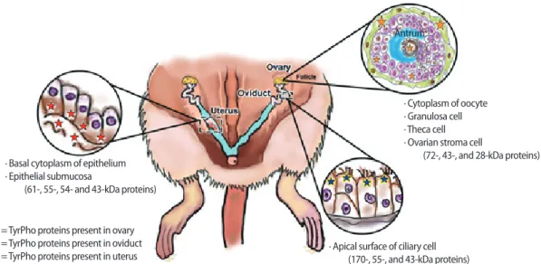
Figure 3. Summary of the localization of tyrosine-phosphorylated (TyrPho) proteins in the ovary, especially in the mature follicle (72-, 43-, and
28-kDa proteins), oviduct (170-, 55-, and 43-kDa proteins), and uterus (61-, 55-, 54-, and 43-kDa proteins), of adult female rats in the estrus phase.
∙ Basal cytoplasm of epithelium ∙ Epithelial submucosa
(61-, 55-, 54- and 43-kDa proteins)
∙ Apical surface of ciliary cell (170-, 55-, and 43-kDa proteins) =TyrPho proteins present in ovary
=TyrPho proteins present in oviduct =TyrPho proteins present in uterus
∙ Cytoplasm of oocyte ∙ Granulosa cell ∙ Theca cell ∙ Ovarian stroma cell
(72-, 43-, and 28-kDa proteins) Antrum
ovary, oviduct, and uterus of adult female rats in the estrus phase. Those proteins can be separated and clearly detected in the tissue ly-sate, indicating that TyrPho proteins may play important roles in fe-male fertility.
Conflict of interest
No potential conflict of interest relevant to this article was reported.
Acknowledgments
We would like to thank the Khon Kaen University Faculty of Medi-cine for kindly providing financial support via an Invitation Research Grant (IN 62336) to Miss Sudtida Bunsueb and Associate Professor Dr. Sitthichai Iamsaard.
ORCID
Sudtida Bunsueb https://orcid.org/0000-0001-9067-1403 Nareelak Tangsrisakda https://orcid.org/0000-0003-2444-8478 Alexander T.H. Wu https://orcid.org/0000-0002-0178-6530 Sitthichai Iamsaard https://orcid.org/0000-0002-6793-2879
Author contributions
Conceptualization: SI, ATHW. Data curation: SB. Formal analysis: SB, NT. Funding acquisition: SI. Methodology: SB, NT. Project administra-tion: SI. Visualizaadministra-tion: NT, SI. Writing–original draft: SI, SB. Writing–re-view & editing: SI, ATHW.
References
1. Keller N, Ozmadenci D, Ichim G, Stupack D. Caspase-8 function, and phosphorylation, in cell migration. Semin Cell Dev Biol 2018;82:105-17.
2. Grindheim AK, Saraste J, Vedeler A. Protein phosphorylation and its role in the regulation of Annexin A2 function. Biochim Bio-phys Acta Gen Subj 2017;1861(11 Pt A):2515-29.
3. Hanks SK, Quinn AM, Hunter T. The protein kinase family: con-served features and deduced phylogeny of the catalytic do-mains. Science 1988;241:42-52.
4. Ullrich A, Schlessinger J. Signal transduction by receptors with tyrosine kinase activity. Cell 1990;61:203-12.
5. Arad-Dann H, Beller U, Haimovitch R, Gavrieli Y, Ben-Sasson SA. Immunohistochemistry of phosphotyrosine residues: identifica-tion of distinct intracellular patterns in epithelial and steroido-genic tissues. J Histochem Cytochem 1993;41:513-9.
6. Chaichun A, Arun S, Burawat J, Kanla P, Iamsaard S. Localization and identification of tyrosine phosphorylated proteins in adult Sprague-Dawley rat testis. Int J Morphol 2017;35:1322-7. 7. Sawatpanich T, Arun S, Tongpan S, Chaichun A, Sampannang A,
Sukhorum W, et al. Localization and changes of tyrosine phos-phorylated proteins and ß actin in epididymis of rats treated with valproic acid. Int J Morphol 2018;36:835-40.
8. Tongpan S, Sukhorum W, Arun S, Sawatphanich T, Iamsaard S. Valproic acid changes the expression of tyrosine-phosphorylat-ed proteins in rat seminal vesicle. Andrologia 2019;51:e13303. 9. Iamsaard S, Burawat J, Kanla P, Arun S, Sukhorum W,
Sripanidkul-chai B, et al. Antioxidant activity and protective effect of Clitoria ternatea flower extract on testicular damage induced by keto-conazole in rats. J Zhejiang Univ Sci B 2014;15:548-55.
10. Iamsaard S, Burawat J, Arun S, Sukhorum W, Boonruangsrim P, Namking M, et al. Phyllanthus emblica L. Branch extract amelio-rates testicular damage in valproic acid-induced rats. Int J Mor-phol 2015;33:1016-22.
11. Sampannang A, Arun S, Sukhorum W, Burawat J, Nualkaew S, Maneenin C, et al. Antioxidant and hypoglycemic effects of Mo-mordica cochinchinensis spreng: (Gac) aril extract on reproduc-tive damages in Streptozotocin (STZ)-induced hyperglycemia mice. Int J Morphol 2017;35:667-75.
12. Sampannang A, Arun S, Burawat J, Sukhorum W, Iamsaard S. Ex-pression of testicular phosphorylated proteins in types 1 and 2 diabetes mellitus in mice: an experimental study. Int J Reprod Biomed (Yazd) 2019;17:567-76.
13. Sukhorum W, Iamsaard S. Changes in testicular function proteins and sperm acrosome status in rats treated with valproic acid. Reprod Fertil Dev 2017;29:1585-92.
14. Maneenin C, Burawat J, Maneenin N, Nualkaew S, Arun S, Sam-pannang A, et al. Antioxidant capacity of Momordica charantia extract and its protective effect on testicular damage in valproic acid-induced rats. Int J Morphol 2018;36:447-53.
15. Arun S, Burawat J, Yannasithinon S, Sukhorum W, Limpongsa A, Iamsaard S. Phyllanthus emblica leaf extract ameliorates testicu-lar damage in rats with chronic stress. J Zhejiang Univ Sci B 2018; 19:948-59.
16. Yannasithinon S, Iamsaard S. Alterations of morphology and phosphorylated protein expression in the seminal vesicles of di-abetic mice. Andrologia 2019;51:e13406.
17. Miyano T, Moor RM, Wooding FB, Shiroo M. Localization and function of tyrosine-phosphorylated protein in pig oocytes. Mol Reprod Dev 1996;44:408-16.
18. Richards RG, Di Augustine RP, Petrusz P, Clark GC, Sebastian J. Es-tradiol stimulates tyrosine phosphorylation of the insulin-like growth factor-1 receptor and insulin receptor substrate-1 in the
uterus. Proc Natl Acad Sci USA 1996;93:12002-7.
19. Thomson M, Herbert JF, Thompson MB. Tyrosine phosphorylated proteins in the reproductive tract of the viviparous lizard Eulam-prus tympanum and the oviparous lizard Lampropholis guichen-oti. Comp Biochem Physiol B Biochem Mol Biol 2006;144:382-6. 20. Yeung CH, Wagenfeld A, Nieschlag E, Cooper TG. The cause of
in-fertility of male c-ros tyrosine kinase receptor knockout mice.
Biol Reprod 2000;63:612-8.
21. Naz RK, Rajesh PB. Role of tyrosine phosphorylation in sperm ca-pacitation / acrosome reaction. Reprod Biol Endocrinol 2004;2:75. 22. Kumaresan A, Johannisson A, Bergqvist AS. Sperm function dur-ing incubation with oestrus oviductal fluid differs in bulls with different fertility. Reprod Fertil Dev 2017;29:1096-106.