저작자표시-비영리-변경금지 2.0 대한민국 이용자는 아래의 조건을 따르는 경우에 한하여 자유롭게 l 이 저작물을 복제, 배포, 전송, 전시, 공연 및 방송할 수 있습니다. 다음과 같은 조건을 따라야 합니다: l 귀하는, 이 저작물의 재이용이나 배포의 경우, 이 저작물에 적용된 이용허락조건 을 명확하게 나타내어야 합니다. l 저작권자로부터 별도의 허가를 받으면 이러한 조건들은 적용되지 않습니다. 저작권법에 따른 이용자의 권리는 위의 내용에 의하여 영향을 받지 않습니다. 이것은 이용허락규약(Legal Code)을 이해하기 쉽게 요약한 것입니다. Disclaimer 저작자표시. 귀하는 원저작자를 표시하여야 합니다. 비영리. 귀하는 이 저작물을 영리 목적으로 이용할 수 없습니다. 변경금지. 귀하는 이 저작물을 개작, 변형 또는 가공할 수 없습니다.
The role of local translation in axon
survival induced by wld
S
and sarm1
The role of local translation in axon
survival induced by wld
S
and sarm1
Directed by Professor Hosung Jung
The Master's Thesis
submitted to the Department of Medical Science,
the Graduate School of Yonsei University
in partial fulfillment of the requirements for the degree of
Master of Medical Science
This certifies that the Master's Thesis of
Jeongim Jang is approved.
---
Thesis Supervisor: Hosung Jung
---
Thesis Committee Member #1: Hoguen Kim
---
Thesis Committee Member #2: Chul-Hoon Kim
ACKNOWLEDGEMENTS
지난 2년을 지금 생각해보면 여러가지로 많이
미숙했고 철도 없었습니다. 그동안 많이 부족했던 저를
가르쳐주시고 이끌어주신 제 지도 교수님인 정호성
교수님과, 학위 논문을 준비하는 과정에서 많은 조언을
해주신 김호근, 김철훈 교수님께 깊이 감사드립니다.
제가 실험이 잘 되지 않는다고 속상해 할 때 항상
도와주시고 많이 가르쳐 주셨던 박사님들, 선후배님들,
그리고 제 동기인 나은정 언니에게 항상 고맙습니다.
그리고 언제나 제 옆에서 안식처가 되었던 소중한
가족, HP 사람들, 오랜 친구들, 우리 꼬랑지들, 그리고
요미 항상 사랑합니다.
TABLE OF CONTENTS
ABSTRACT ··· 1
I. INTRODUCTION ··· 3
II. MATERIALS AND METHODS ··· 10
1. Xenopus tropicalis embryos ··· 10
2. Microinjection ··· 10
3. Electroporation ··· 10
4. Axon transection ··· 10
5. Exposed brain preparation and chemical treatment ··· 11
6. Open book preparation of brain and imaging ··· 11
7. Image analysis and statistical analysis ··· 11
III. RESULTS ··· 15
1. Wallerian degeneration of retinal ganglion cell axons in Xenopus in
vivo can be quantitatively measured ··· 15
2. Wld
Sexpression delays Wallerian degeneration in vivo ··· 19
3. Sarm1 knockdown delays Wallerian degeneration in vivo ··· 24
4. Cycloheximide inhibits the ability of wld
Sexpression and sarm1
knockdown to delay Wallerian degeneration in vivo ··· 28
IV. DISCUSSION ··· 39
V. CONCLUSION ··· 44
LIST OF FIGURES
Figure 1. Wallerian degeneration ··· 4
Figure 2. Functions of Wld
Sand Sarm1 in Wallerian degeneration
··· 7
Figure 3. Experimental model: Xenopus tropicalis retinotectal pathway
··· 9
Figure 4. Overall experimental scheme ··· 12
Figure 5. Open book preparation of Xenopus brain ··· 14
Figure 6. Experimental scheme for Wallerian degeneration time
course analysis ··· 16
Figure 7. Time course of Wallerian degeneration of retinal axons in
vivo ··· 17
Figure 8.
Experimental scheme for wld
Sexperiments ··· 21
Figure 9. Effect of wld
Sin Xenopus in vivo ··· 22
Figure 10. Quantification of wld
Seffect ···23
Figure 11. Experimental scheme for sarm1 knockdown experiments
··· 25
Figure 12. Effect of sarm1 on Wallerian degeneration of retinal
axons in vivo ··· 26
Figure 15. Dose determination of cycloheximide in Xenopus in vivo (Ⅱ)
··· 30
Figure 16. Experimental scheme to test the effect of cycloheximide
treatment on Wallerian degeneration-resisting axons ··· 32
Figure 17. Effect of cycloheximide on Wallerian degeneration of
wld
s-expressing or sarm1 knockdown axons ··· 33
Figure 18. Quantification of the effect of cycloheximide on
Wallerian degeneration of wld
Saxons ··· 35
Figure 19. Quantification of the effect of cycloheximide on
Wallerian degeneration of sarm1 knockdown axons ··· 36
Figure 20. The model for the role of local translation in axon
maintenance ··· 42
Figure 21. Decreased clearance of axon fragments under
cycloheximide condition ··· 43
LIST OF TABLES
ABSTRACT
The role of local translation in axon survival induced by wld
Sand sarm1
Jeongim Jang
Department of Medical Science
The Graduate School, Yonsei University
(Directed by Professor Hosung Jung)
Neurons are connected to each other via their axons, and maintaining these structures is necessary for normal brain functions. Failure to maintain axonal integrity is closely linked to most neurodegenerative diseases. Therefore, understanding how axon survival is regulated is key to understand brain function and diseases. Wallerian degeneration, fragmentation of distal axons severed from their cell bodies, shares molecular and cellular characteristics with disease-associated axon degeneration, and therefore is widely used as a model to study axon survival and degeneration. Genetic studies in mouse and fly revealed unexpectedly that axon degeneration is an active process, conceptually akin to compartmentalized apoptosis, which requires axon destruction gene,
particularly how severed axons survive for weeks. Based on recent studies showing that axonal protein synthesis is required for axon survival and maintenance, this thesis investigates the hypothesis that local translation is required for Wallerian degeneration-resisting distal axons to survive. Using the visual system of Xenopus tropicalis as a model, I show that functions of wldS and sarm1 are conserved in Xenopus, and that cycloheximide treatment increases degeneration of these axons. These results provide direct evidence for the importance of local translation to maintain normal structure in Wallerian degeneration-resisting axons and insight into how survival might be regulated
The role of local translation in axon survival induced by wld
Sand sarm1
Jeongim Jang
Department of Medical Science
The Graduate School, Yonsei University
(Directed by Professor Hosung Jung)
I. INTRODUCTION
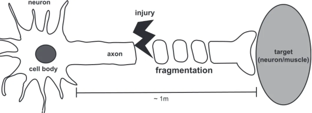
The nervous system is composed of numerous connections between neurons. Neurons connect with each other via their axons, which are often very long. Axon survival is key to maintain and control nervous system function, and axon degeneration is linked to nervous system diseases. Mechanical damages to axons induce a specific axon destruction process in the axonal segment distal to the injury site. The distal axonal segment becomes fragmented and loses its integrity within 48 hours after injury at least in mouse (Figure 1).1 This type of axon
degeneration, Wallerian degeneration, which was named after Waller who first described this phenomenon in 1850,2 is the most well-known model to study axon degeneration.
Figure 1. Wallerian degeneration. Neurons are connected to each other or their targets by their processes, especially axons. When axons get damaged, the destruction program called Wallerian degeneration undergoes in distal segment. The key feature of this process is fragmentation of distal axons.
cell body axon target (neuron/muscle) neuron ~ 1m injury fragmentation
It took more than 130 years after its initial description until the most important tool to study Wallerian degeneration was discovered. A spontaneous mouse strain with a spontaneous mutation which shows delayed Wallerian degeneration was discovered.3 , and a series of genetic studies revealed that this phenotype is caused
by a single gene in an autosomal dominant manner. This gene, named as Wallerian degeneration slow (wldS),4 delays Wallerian degeneration for up to 3
weeks after injury.5-7 The first clue as to how Wallerian degeneration is regulated molecularly came from the landmark paper in 2001 that described the structure of the wldS gene.8 WldS gene results from a gene fusion event and encodes a fusion protein of the N terminal 70 amino acids of ubiquitination factor E4B (ube4b) and the whole nicotinamide mononucleotide adenylyltransferase 1 (nmnat1). Although exact molecular mechanisms of WldS action remain unclear, the current model suggests that the enzymatic function of Nmnat1 is required for WldS
function. The N terminal of Ube4b promotes cytosolic localization of WldS,9 and ectopic cytosolic presence of Nmnat activity is likely to mediate axon protective effect of WldS.10 As such, cytosolic targeting of Nmnat proteins protects axons from Wallerian degeneration11 and knocking out the cytosolic nmnat gene,
nmnat2, leads to axon degeneration in mouse.12 Over a decade after discovery, the second mutation that prevents Wallerian degeneration was discovered in 2012. In contrast to wldS, which is a gain-of-function mutation, this mutation is loss-of-function in sarm1 gene, sterile alpha and HEAT/Armadillo motif containing 1, originally identify by its role in toll-like receptor 4 (TLR4) signaling. Intriguingly,
sarm1 loss-of-function protects axons from Wallerian degeneration both in fly
induced by loss of Nmnat2 function is rescued by inhibiting Sarm1.15 Therefore,
axon degeneration is a regulated, active self-destruction process that involves pro-survival (e.g. Nmnats) and pro-degeneration (e.g. Sarm1) factors and can be thought as highly localized programed death. Molecular dissection of this program may provide new insight into how axon survival and degeneration is regulated.
Figure 2. Functions of WldS and Sarm1 in Wallerian degeneration. WldS acts
as additional cytosolic Nmnats that originally degrade rapidly after axon damage. Sarm1 induces Wallerian degeneration at the downstream of Nmnat effects.
Sarm1 activation
injury Nmnat loss Wallerian degeneration
wld RNA (Nmnat gain-of-function)
sarm1 MO (sarm1 loss-of-function) S
WldS gain-of-function and sarm1 loss-of-function can delay Wallerian
degeneration for weeks. How can severed axons can survival this long without their cell bodies, where genetic information is localized? Recent studies showed that local protein synthesis in axons plays a key role in axon matineance16-17 , and
particularly revealing information was that nmnat2 and sarm1 mRNAs are locally translated in axons in vivo in axon branching and pruning stages, respectively18 .
These findings strongly suggest that local protein synthesis may regulate axon survival and degeneration.
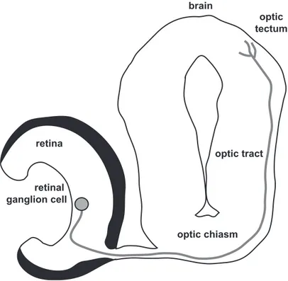
In this thesis, I hypothesized that Wallerian degeneration-resisting axons continue to locally synthesize proteins, a process essential for their long-term survival, and used the visual system of Xenopus tropicalis frog as an experimental model to test this hypothesis. Retinal ganglion cells are the projection neuron of the visual system, their cell bodies localizing to the retina of the eye and long axons terminating in the optic tectum of the contralateral midbrain hemisphere. This retinotectal pathway is homologous to the retinocollicular pathway, where retinal ganglion cell axons directly connect to the superior colliculus in mammals (Figure 3). Together with rich information on anatomy and development of the visual pathway, rapid development and availability of powerful embryological techniques make Xenopus retinotectal pathway an ideal model to study axon development, survival and degeneration. The results of this study show that
sarm1 loss-of-function and wldS gain-of-function prevent Wallerian degeneration
in X. tropicalis retinal ganglion cells in vivo in an mRNA translation-dependent manner.
Figure 3. Experimental model: Xenopus tropicalis retinotectal pathway. Axons from retinal ganglion cell grow to the brain, cross the optic chiasm, and reach to the optic tectum. It is easy to axotomize by in the way of eye removal.
retinal ganglion cell retina optic chiasm optic tectum optic tract brain
II. MATERIALS AND METHODS 1. Xenopus tropicalis embryos
Xenopus eggs were fertilized in vitro and grown in 0.1X modified Barth’s saline (MBS). The jelly coat was removed by 2% L-cysteine in 1X MBS for 5 minutes in room temperature, and washed in 0.1X MBS.
2. Microinjection
To control genetic expression in vivo, 1 ng of morpholino and 100 pg of RNA were injected in each dorso-animal blastomere at 8-cell-stage to target CNS with RFP RNA for tracer. Morpholino used in this research was non-targeting morpholino as a control, 5’-TATAAATTGTAACTGAGGTAAGAGG-3’ for targeting sequence, and sarm1 which targeting sequence is 5’- GAAGGATTCATGGTTCTCACTCTTC-3’, based on Xenopus sequence (Genetools, Philomath, Oregon, USA). 5’-capped mRNA used in this experiment was FLAG-wldS and synthesized in vitro using mMESSAGE mMACHINE kit
(Invitrogen, Carlsbad, California, USA). The schematic diagram is shown in Figure 4.
2. Electroporation
To label retinal ganglion cell axons, I used electroporation technique on optic vesicle of embryo at Nieuwkoop and Faber stage 2619 as reported in previous research.20 One 𝜇g/𝜇L of pEGFP-C1 was injected into the cavity of optic vesicle
4. Exposed brain preparation and chemical treatment
To inhibit local translation in axon, cycloheximide was treated in 0.1X MBS that embryos raised and final concentration was 5 𝜇M. The scalp of embryo was removed for easy penetration of chemical into the brain just after the axotomy. 5. Open book preparation of brain and imaging
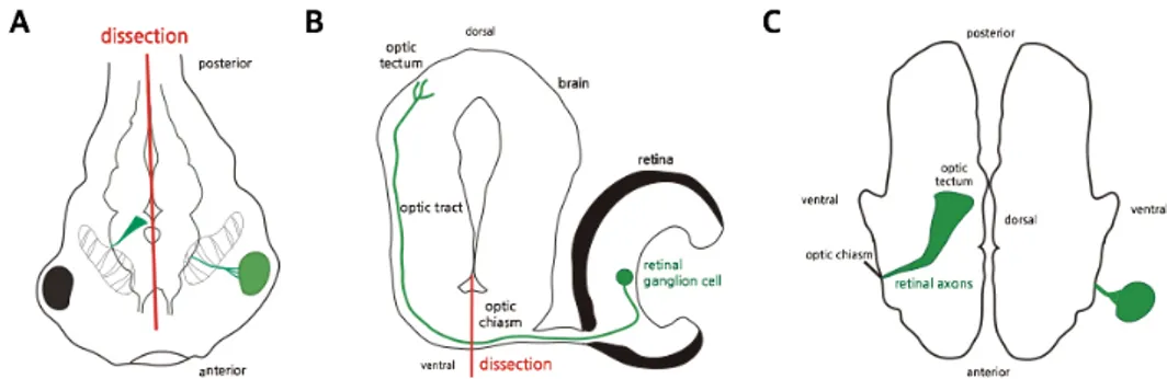
The tadpoles were fixed in 4% paraformaldehyde with from 0.2 to 0.8% glutaraldehyde in phosphate buffered saline (PBS) after appropriate time point, usually 48 hours in this thesis. The brains of tadpoles were separated from their body, and divided into two parts by cutting midline boundary of left and right brain (Figure 5). These flattened brains were mounted in the slide glass, named open book preparation. The patterns of retinal ganglion cell axons were observed using LSM700 confocal microscope (Carl Zeiss, Oberkochen, Baden-Württemberg, Germany). Stacked images were obtained as 400.11 𝜇m × 400.11 𝜇m size with 16-bit depth and 1,024×1,024 pixel numbers using EGFP wavelength, 488 nm.
6. Image analysis and statistical analysis
All images were analyzed by ImageJ (National Institute of Health, Maryland, Oregon, USA), and stacks were merged into one image in each sample. Axons with discontinuous forms were classified into the fragmented group, and embryos with continuous axons of one or more were intact group. The number of each group was counted and analyzed statistically using Fisher’s exact test of Prism5
fertilization
gene expression control labeling axotomy
1.5 hrs 29 hrs 4 days 48 hrs
fixation and imaging + cycloheximide
(optional)
Figure 4. Overall experimental scheme. To confirm the function of wlds and
sarm1, wlds RNA and sarm1 morpholino was injected in fertilized eggs, and
electroporated eyes were removed to induce Wallerian degeneration in vivo. To investigate the effect of translation inhibition in degeneration-resisting axons, exposed brain preparation and cycloheximide treatment were added just after the axotomy.
Figure 5. Open book preparation of Xenopus brain. (A) Tadpole at stage 45 are presented, and green line indicates EGFP-expressing neuron. Brain was dissected along the midline showed in diagram. (B) Dorsoventral section of the tadpole brain. Midline is presented in bold line. (C) The configuration of dissected brain is showed, optic axons in green reach to the optic tectum. This orientation is suitable for brain imaging.
III. RESULTS
1. Wallerian degeneration of retinal ganglion cell axons in Xenopus in vivo can be quantitatively measured
Before confirming neuroprotection effect by wldS and sarm1, I defined
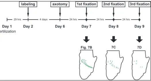
efficient time point to decide whether the Wallerian degeneration is induced by axotomy (Figure 6). At stage 26, a stage when retinal ganglion cell begins to appear, one eye in a wildtype embryo was electroporated with pEGFP-C1 to label retinal ganglion cell axons projecting to the contralateral optic tectum. At stage 45, when most retinal ganglion cell axons have reached the optic tectum, the electroporated eye was dissected out to induce axotomy. In this experiment, I fixed embryos at 24 hour intervals after axotomy and imaged EGFP-expressing axons, because Wallerian degeneration was reported that degradation occurs within 24 hours and completed within about 48 hours1. The entire contralateral
brain was imaged under a laser scanning confocal microscope, as shown in Figure 7. The embryo without any surgery maintains intact axonal morphology, which is evident in dense labeling of the entire retinotectal pathway by EGFP florescence (Figure 7A, white arrow). In contrast, desomatized axons showed clear signs of axon degeneration as early as 24 hours, with discontinuous EGFP puncta indicative of axon fragmentation (Figure 7B, red arrow). Wallerian degeneration showed a time-dependent progress over next additional 48 hours, showing gradual disappearance of EGFP puncta (Figure 7B and 7C, red arrow). After 72 hours, most axonal fragments were cleared (Figure 7D), making it difficult to judge whether axons had degenerated or had not been efficiently
fertilization
labeling axotomy
29 hrs 4 days 24 hrs
1st fixation
Day 1 Day 2 Day 6 Day 7
24 hrs Day 8 24 hrs Day 9 2nd fixation 3rd fixation Fig. 7B 7C 7D
Figure 6. Experimental scheme for Wallerian degeneration time course analysis. Eyes of embryos were electroporated to label retinal ganglion cell axons and removed to induce Wallerian degeneration in these axons. Embryos were fixed at intervals of 24 hours for 72 hours and their axons were analyzed.
Figure 7. Time course of Wallerian degeneration of retinal axons in vivo. (A) Retinal ganglion cell axons not severed show in white, normal and continuous structures. These axons grow to the optic tectum which is presented to left top part of the image. The scale bar is showed as while line at left bottom corner, 100 µm. (B) At 24 hours after axonal injury, the axons degrade but still intact in some strands. It is the evidence that Wallerian degeneration starts within 24 hours. (C) Damaged axons present dramatic differences with Figure 7A, granulation into small particles and are clearance at 48 hours. (D) Only a few fragments are remained after 72 hours (Telen, telencephalon; Dien, diencephalon; Tec, optic tectum).
2. WldS expression delays Wallerian degeneration in vivo
After establishing a good condition to image axon degeneration, I investigated whether gain-of-function of wldS delays Wallerian degeneration in Xenopus
tropicalis, as in other species such as mouse and fly. Using microinjection
technique, I injected in vitro transcribed wldS RNA into two dorsal animal
blastomeres of an 8-cell-stage embryo, which are fated to constitute around 90% of the central nervous system. I co-injected a trace amount of monomeric red fluorescence protein 1(mRFP1)-encoding mRNA to aid visual screening of successfully injected embryos. The same amount of control mRNA was injected into the control group, to ensure the same amount of total mRNA was injected in each group.
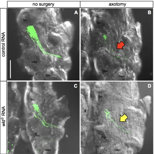
At stage 26, I screened embryos expressing mRFP1 in the central nervous system, and electroporated one eye with pEGFP-C1 to label the retinotectal pathway unilaterally. At stage 45, I screened embryos, whose eyes were successfully labeled by EGFP fluorescence, and removed the labeled eyes to induce axotomy. Forty eight hours after axotomy, embryos were fixed in 4% paraformaldehyde, 0.2% glutaraldehyde in 1X PBS and severed axons terminating in the contralateral optic tectum was imaged under a confocal microscopy (Figure 8). As expected, wldS-expressing axons appeared to be
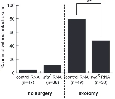
protected from Wallerian degeneration, when compared to the control (Figure 9). To quantify this result, I categorized brains into the following two categories: brains with at least one intact axon (lateral optic tectum was imaged under a
control group, and this difference was statistically significant (p value, 0.0028). The reasons why intact axons still exist in control group which eye removed is that some of the lesioned axons may do not undergo degeneration process enough until 48 hours or judgement error whether intact or fragmented. This result provides the first evidence that the wldS delays Wallerian degeneration in X. tropicalis, and suggest that molecular mechanism by which WldS exerts its axon
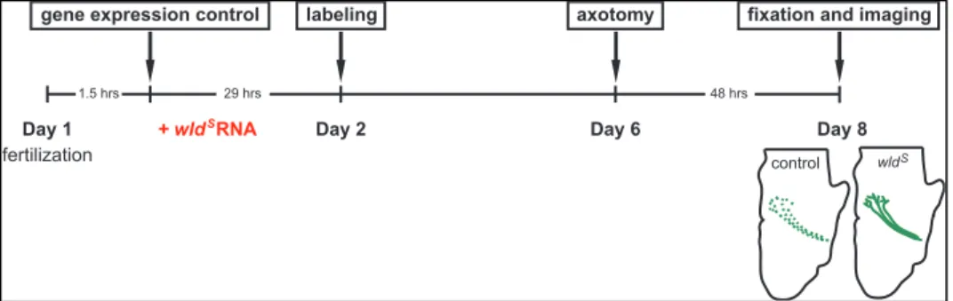
Figure 8. Experimental scheme for wldS experiments. WldS RNA injected embryos were damaged in their electroporated eyes at stage 45 and fixed in paraformaldehyde after 48 hours.
fertilization
gene expression control axotomy
1.5 hrs 29 hrs 48 hrs
fixation and imaging
Day 1 Day 2 Day 6
labeling
Day 8 control wld
+ wld RNAS
Figure 9. Effect of wldS in Xenopus in vivo. (A) Control group axons expressing EGFP show in green and are intact. These axons pass optic chiasm and branch in the optic tectum. The scale bar is in the left lower corner of this image, 100 µm. (B) Axons are destructed into fragments by Wallerian
Figure 10. Quantification of wldS effect. Non-surgery groups show continuous axons in both control and wlds group. Damaging axons, axons degenerated in 39 of 49 embryos in control but only 18 of 38 in wlds RNA-injected embryos. This
difference between the number of control and wlds group is statistically significant (p value, 0.0028). Statistics is analyzed by Fisher’s exact test in
0 20 40 60 80 100 fragmented
no surgery
axotomy
control RNA (n=47) control RNA (n=49) wld RNA (n=38)% animal without intact axons
**
S S
wld RNA (n=38)
3. Sarm1 knockdown delays Wallerian degeneration in vivo
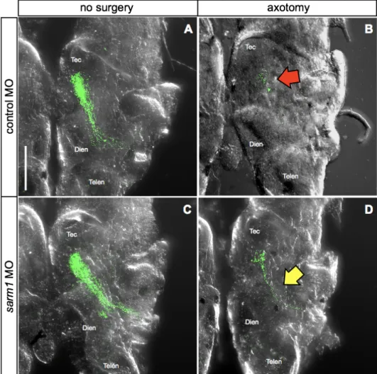
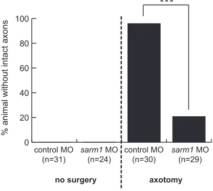
Next, I sought to investigate whether sarm1 loss-of-function delays Wallerian degeneration in Xenopus tropicalis. To this end, I injected sarm1 MO into central nervous system-fated blastomeres (i.e. two dorsal animal blastomeres at 8 cells stage). The same amount of control MO, which contains sequence that targets no known genes in Xenopus genome, was injected into control embryos. The tracer RNA (mRFP1) was co-injected as described in the previous section. Unilateral eye electroporation with pEGFP-C1 was performed to stage 26 embryos to label the retinotectal pathway. Successfully electroporated embryos were selected and desomatized at stage 45, and their brains were imaged as described in the previous section. In controls, desomatized axons showed clear Wallerian degeneration, which was evident in discontinuous EGFP signal along the retinotectal pathway (Figure 12A-B, red arrow). In contrast, many sarm1 MO-injected embryos still maintained intact retinotectal pathway even 48 hours after axotomy (Figure 12C-D, yellow arrow). Quantification of this result shows that majority of sarm1 knockdown embryos, 23 of 29, show axons protected from Wallerian degeneration, in contrast to control condition, where only 1 of 30 embryos showed protected axons. This difference was statistically significant (Figure 13). This result provides the first evidence that the sarm1 loss-of-function delays Wallerian degeneration in X. tropicalis, and together with the results of the previous section, suggest that molecular mechanism underlying axon survival and degeneration is evolutionarily conserved.
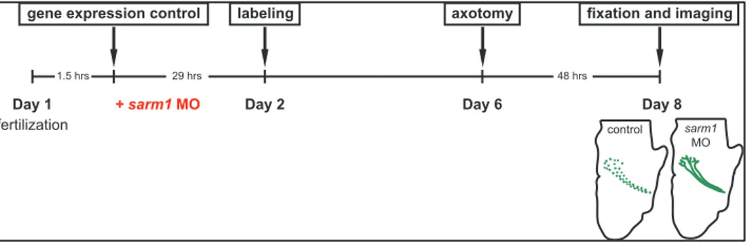
Figure 11. Experimental scheme for sarm1 knockdown experiments. Sarm1 morpholino (MO) was injected in embryos to inhibit translation of sarm1 and EGFP was electroporated in their eyes. These embryos were fixed in paraformaldehyde 48 hours after axotomy.
fertilization
gene expression control axotomy
1.5 hrs 29 hrs 48 hrs
fixation and imaging
Day 1 Day 2 Day 6
labeling
Day 8
control sarm1
MO
Figure 12. Effect of sarm1 on Wallerian degeneration of retinal axons in vivo . (A) Control axons showed continuous structure from optic chiasm to optic tectum. (B) Lesioned axons degraded and showed particles in control after 48
Figure 13. Quantification of sarm1 knockdown effect. All non-surgery xons in control and sarm1 knockdown are intact. When axotomized, 29 of 30 in control undergo Wallerian degeneration but only 6 of 29 in sarm1 knockdown, and this is significant (p value < 0.0001). Fisher’s exact test, *** p ≤ 0.001.
0 20 40 60 80 100 fragmented control MO (n=31) control MO (n=30) sarm1 MO (n=29) sarm1 MO (n=24) no surgery axotomy
***
4. Cycloheximide inhibits the ability of wldS expression and sarm1 knockdown
to delay Wallerian degeneration in vivo
Next, to investigate the roles of local protein synthesis in axon survival and degeneration, I designed experiments using cycloheximide, a translation elongation inhibitor, to pharmacologically inhibit protein synthesis in axons. Before treating cycloheximide in wldS or sarm1 MO injected embryos, I studied
maximum concentration which does not affect viability of embryos. Eyes of non-injected embryos were removed to induce Wallerian degeneration, and skin and meninges overlying the contralateral optic tectum was peeled off to expose the brain region where retinal axons terminate. Cycloheximide was treated right after axotomy and embryos in this ‘exposed brain’ preparation were raised in different concentrations of cycloheximide (200, 100, 50, 25, 10, 5, and 2.5 𝜇M) for 48 hours, as an earlier study used bath application of 10 𝜇g/mL (about 35.5 𝜇M) cycloheximde to Xenopus laevis.21 Some embryos were left in the same solution without any surgery. Same amount of vehicle (i.e. ethanol) was treated to control group. Fifty 𝜇M or higher concentration of cycloheximide was toxic, even to embryos in non-surgery groups (Figure 14). All embryos in 10 𝜇M and 25 𝜇M cycloheximide died (Figure 15), but only one among three embryos in each 2.5 𝜇M and 5 𝜇M died and all the non-operated tadpoles survived in the same concentration. Inhibition of protein synthesis, not the toxicity of ethanol that cycloheximide dissolves in, may lead to death of non-surgery tadpoles because none of them died in same concentration of ethanol. Therefore, I determined that 5 𝜇M is maximal non-lethal dose for cycloheximide and used this concentration for all the following experiments.
(A-Figure 15. Dose determination of cycloheximide in Xenopus in vivo (Ⅱ). (A) Treating 2.5 𝜇M cycloheximide (CHX), only one embryo through three was dead
After defining sub-lethal dose of cycloheximide, wldS RNA- or sarm1
MO-injected, desomatized embryos were treated with 5 𝜇M cycloheximide for 48 hours in exposed brain preparation (Figure 16). For control, same volume of ethanol as that of cycloheximide was added to 0.1X MBS. In all groups, the same amounts of RNA and MO were injected. Total amount of RNA which was injected in a blastomere was 200 pg including 100 pg of RFP RNA for tracing, and MO was 1 ng. Groups except cycloheximide-treated wldS-expressing or
sarm1 knockdown were added up to each experiment groups.
In accordance with the result of previous sections of this thesis, wldS expression
delayed Wallerian degeneration in 50% of embryos examined (24 of 48 embryos showed intact axons) (Figure 18). Cycloheximide treatment in same condition reduced this effect to 18% (2 of 11 embryos showed intact axons). Although the same trend of results was obtained in six biological replicates, this different was not statistically significant (p value, 0.0911). Treating cycloheximide to sarm1 MO expressing embryos led to a decrease in embryos with protected axons (83% to 38%. 30 of 36 embryos protected in control, vs. 6 of 16 embryos protected in cycloheximde group), and in this case the difference was statistically significant. (p value, 0.0024). Although the result in wldS experiments is not statistically
significant (p value, 0.0911) due to small sample (Table 1), sarm1 MO results shows definite decrease of protection ability under cycloheximide. As conclusion, cycloheximide treatment increases Wallerian degeneration in both wldS -expressing and sarm1 knockdown retinal ganglion cell axons in Xenopus in vivo.
Figure 16. Experimental scheme to test the effect of cycloheximide treatment on Wallerian degeneration-resisting axons. One ng/nL of non-targeting or
sarm1 morpholino and 100 pg/nL of control or wldS RNA were co-injected in
developing eggs, and these embryos were axotomized and raised in cycloheximide at stage 45 for 48 hours.
- CHX
fertilization
gene expression control axotomy
1.5 hrs 29 hrs 48 hrs
fixation and imaging + cycloheximide
Day 1 Day 2 Day 6 Day 8
labeling
+ sarm1 MO
+ wld RNAS
Figure 17. Effect of cycloheximide on Wallerian degeneration of wldS -expressing or sarm1 knockdown axons. (A-B) In control group, axons degenerated in the consequence of Wallerian degeneration independently to cycloheximide treatment. (C) WldS axons presented continuous structures as
described in previous result. (D) In cycloheximide, wldS-expressing axons degraded within 48 hours after injury. (E) Sarm1 knockdown showed delay of Wallerian degeneration. (F) Cycloheximide treatment in sarm1 morpholino-injected group led axons to fragmentation (scale bar, 100 µm).
Figure 18. Quantification of the effect of cycloheximide on Wallerian degeneration of wldS axons. 50% of wldS RNA-injected embryos presented
continuous axons, but only 18% with cycloheximide (CHX). Cycloheximide treatment in wldS axons facilitated Wallerian degeneration in vivo (statistically
0
20
40
60
80
100
intact
+
+
CHX_
+
% animal with intact axons
n.s.
wld RNAS
Figure 19. Quantification of the effect of cycloheximide on Wallerian degeneration of sarm1 knockdown axons. Injecting sarm1 morpholino, cycloheximide (CHX) treatment decrease the ratio of intact embryos (p value,
0
20
40
60
80
100
intact
+
+
**
sarm1 MO
CHX
_
+
% animal with intact axons
Table 1. Results of six independent experiments to test the effect of cycloheximide on Wallerian degeneration of wldS-expressing and sarm1 knockdown axons
During six experiments, all control embryos show discontinuous axons after axotomy in both cycloheximide non-treated and treated condition. Injection of
wlds RNA and sarm1 morpholino delays Wallerian degeneration, but less in cycloheximide. Null of number is presented as a hyphen.
IV. DISCUSSION
Wallerian degeneration is a process that dissipate injured axons in nervous system. Non-neuronal cells, especially astrocytes in central nervous system, engulf axonal segments and axons degrades gradually. The Wallerian degeneration is similar to apoptosis in regard to active process that eliminates cell compartments, but WldS do not affect to apoptosis,22 and actual pathway of the
Wallerian degeneration and delay process is not well-known yet. Because axon needs protein synthesis locally to maintain and control its function,16-17 it is rational thought that axons severed to the cell body will also synthesize proteins to survive when resisted to the Wallerian degeneration. Although the translation in the cell body comprises the large proportion to entire protein synthesis,23 it is
disputative that translation also occurs in axons.18 Inhibition of translation in axons will reduce delay of degeneration hypothetically.
In this thesis, I confirmed neuroprotection activity of wldS and sarm1 applied in retinal ganglion cell axons of Xenopus tropicalis and studied alteration of degeneration pattern when cycloheximide, a protein synthesis inhibitor, was treated at the first time. It is well known that wldS expression and sarm1 knockout suppress Wallerian degeneration in mouse and fly in vivo and in vitro, but not tried in X. tropicalis.
First, I showed that wldS expression and sarm1 knockdown also delay
Wallerian degeneration in optic system in vivo. These data present that wldS and
sarm1 are also involved in degeneration in X. tropicalis and that this pathway is
Protection of axons against the Wallerian degeneration is not the effect of other cells including any signaling molecule or contact because of evidences that neurons of wldS gain-of-function or sarm1 loss-of-function remained intact when Wallerian degeneration was induced13, 24in cell culture, without any other cell. In
X. tropicalis, neuroprotection ability will also be cell-autonomous effect in the
manner of performing retinal extract culture.
Caenorhabditis elegans, the simplest model organism that nervous system
exists in, also undergo Wallerian degeneration after injury. WldS and Nmnats do
not show delaying of degradation.25 The mutant form of Sarm1 homologues pertain in C. elegans, but have no protection activity. These results reveal the existence of another pathway of Wallerian degeneration which is independent of Nmnats and Sarm1 in C. elegans. Because these molecules have been reported as key survival and promotive factors in Drosophila and vertebrates, these proteins may acquire the linkage between Wallerian degeneration after branching to nematodes.
Second, I confirmed that axons of wldS expressing or sarm1 knockdown showed Wallerian degeneration until 48 hours after axotomy, when inhibiting translation. This strongly supports a hypothesis that axons need local translation to prolong Wallerian degeneration. To remain intact, a group of mRNAs are translated in Wallerian degeneration-resisting axons, and identification of these translating mRNAs using latest techniques will shed light on discovery of survival mechanisms during delayed degeneration (Figure 20). Cycloheximide did not affect survival of axons in control group because 85% of non-treated axons undergo Wallerian degeneration within 48 hours (68 of 80) compared to
wide area, not specific to retinal axons because of open brain preparation. The astrocytes, discarding fragmented axons, may also be influenced by cycloheximide, blocking translation. It may disrupt absorbance of axon debris by glial cells when Wallerian degeneration proceeds. To conclude the effect of local translation in surviving axons, more experiments are required using cycloheximide and other translation inhibitors as rapamycin.
Figure 20. The model for the role of local translation in axon maintenance. The axons remain intact after damage when existence of Wlds or loss of Sarm1,
and may need local translation to survive. Blocking translation may lead to axon degeneration in these separated axons.
axon terminal
Wallerian degeneration-resisting axon
ribosome unknown mRNAs proteins axon survival axon degeneration translation inhibitor blocked protein synthesis
Figure 21. Decreased clearance of axon fragments under cycloheximide condition. When cycloheximide was treated, more fragmented axon particles were observed than non-treating ones, indicating that less engulfment occurs in the consequences of translation inhibition during Wallerian degeneration. Scale bar, 100 µm, is presented in left lower side (Telen, telencephalon; Dien, diencephalon; Tec, optic tectum).
V. CONCLUSION
In this thesis, I show, for the first time, that ability of wldS gain-of-function and
sarm1 loss-of-function are conserved in Xenopus tropicalis in vivo. This is the
first research that local translation inhibition affects survival against Wallerian degeneration. The key result of this thesis is summarized to two parts.
First, wldS expression and sarm1 knockdown suppresses Wallerian
degeneration in axons of retinal ganglion cells in X. tropicalis. Second, blocking protein synthesis in these axons deprive survival ability of wldS gain-of-function or sarm1 loss-of-function against Wallerian degeneration. This study is strong evidence that local protein synthesis is required for axons to resist degeneration to remain intact, and will give an insight into mechanisms underlying axon survival and degeneration.
REFERENCES
1. Wang JT, Medress ZA, Barres BA. Axon degeneration: molecular mechanisms of a self-destruction pathway. J Cell Biol 2012;196(1):7-18. 2. Waller A. Experiments on the section of the glossopharyngeal and
hypoglossal nerves of the frog, and observations of the alterations produced thereby in the structure of their primitive fibers. Phil Trans R Soc Lond 1850;140:423-29.
3. Lunn ER, Perry VH, Brown MC, Rosen H, Gordon S. Absence of Wallerian degeneration does not hinder regeneration in peripheral nerve. Eur J Neurosci 1989;1(1):27–33.
4. Lyon MF, Ogunkolade BW, Brown MC, Atherton DJ, Perry VH. A gene affecting Wallerian nerve degeneration maps distally on mouse chromosome 4. Proc Natl Acad Sci USA 1993;90(20):9717-20.
5. Perry VH, Lunn ER, Brown MC, Cahusac S, Gordon S. Evidence that the rate of Wallerian degeneration is controlled by a single autosomal dominant gene. Eur J Neurosci 1990;2(5):408–13.
6. Perry VH, Brown MC, Lunn ER. Very slow retrograde and Wallerian degeneration in the CNS of C57BL/Ola mice. Eur J Neurosci 1991;3(1):102–5.
7. Beirowski B, Adalbert R, Wagner D, Grumme DS, Addicks K, Ribchester RR, et al. The progressive nature of Wallerian degeneration in wild-type and slow Wallerian degeneration (WldS) nerves. BMC Neurosci 2005;6:6.
degeneration. J Cell Biol 2009;184(4):501-13.
10. Milde S, Gilley J, Coleman MP. Subcellular localization determines the stability and axon protective capacity of axon survival factor Nmnat2. PLoS Biol 2013;11(4):e1001539.
11. Babetto E, Beirowski B, Janeckova L, Brown R, Gilley J, Thomson D, et al. Targeting NMNAT1 to axons and synapses transforms its neuroprotective potency in vivo. J Neurosci 2010;30(40):13291-304.
12. Gilley J, Coleman MP. Endogenous Nmnat2 is an essential survival factor for maintenance of healthy axons. PLoS Biol 2010;8(1):e1000300.
13. Osterloh JM, Yang J, Rooney TM, Nicole Fox A, Adalbert R, Powell EH, et al. dSarm/Sarm1 is required for activation of an injury-induced axon death pathway. Science 2012;337(6093):481-4.
14. Gerdts J, Brace EJ, Sasaki Y, DiAntonio A, Milbrandt J. SARM1 activation triggers axon degeneration locally via NAD+ destruction. Science 2015;348(6233):453-7.
15. Gilley J, Orsomando G, Nascimento-Ferreira I, Coleman MP. Absence of SARM1 rescues development and survival of NMNAT2-deficient axons. Cell Rep 2015;10(12):1974-1981.
16. Wu KY, Hengst U, Cox LJ, Macosko EZ, Jeromin A, Urquhart ER, et al. Local translation of RhoA regulates growth cone collapse. Nature 2005;436(7053):1020-4.
19. Nieuwkoop PD, Faber J. Normal table of Xenopus Laevis (Daudin): a systematical & chronological survey of the development from the fertilized egg till the end of the metamorphosis). 1st ed.Abingdon (VA):Taylor & Francis Inc;1994.
20. Falk J, Drinjakovic J, Leung KM, Dwivedy A, Regan AG, Piper M, et al. Electroporation of cDNA/Morpholinos to targeted areas of embryonic CNS in Xenopus. BMC Dev Biol 2007;7:107.
21. Zhang T, Guo X, Chen Y. Retinoic acid-activated Ndrg1a represses Wnt/b-catenin signaling to allow Xenopus pancreas, oesophagus, stomach, and duodenum Specification. PLoS One 2013;8(5):e65058.
22. Adalbert R, Nógrádi A, Szabó A, Coleman MP. The slow Wallerian degeneration gene in vivo protects motor axons but not their cell bodies after avulsion and neonatal axotomy. Eur J Neurosci 2006;24(8):2163-8.
23. Lee SK, Hollenbeck PJ. Organization and translation of mRNA in sympathetic axons. J Cell Sci 2003;116(21):4467-78.
24. Conforti L, Fang G, Beirowski B, Wang MS, Sorci L, Asress S, et al. NAD+
and axon degeneration revisited: Nmnat1 cannot substitute for WldS to delay Wallerian degeneration. Cell Death Differ 2007;14(1):116-27.
25. Nichols AL, Meelkop E, Linton C, Giordano-Santini R, Sullivan RK, Donato A, et al. The apoptotic engulfment machinery regulates axonal degeneration in C. elegans neurons. Cell Rep 2016;14(7):1673-83.
ABSTRACT (IN KOREAN)
wld
s와 sarm1에 의한 축삭 생존에서 국소적인 단백질 번역의 역할
<지도교수 정호성>
연세대학교 대학원 의과학과
장정임
신경계가 제대로 기능하기 위해서는 정상적인 구조를 유지하는 것이 중요하다. 신경세포는 축삭이라는 구조를 통해 서로 신호를 전달한다. 축삭 구조를 유지하는 것이 얼마나 중요한지 나타내는 단적인 예로, 여러 신경퇴행성질환을 들 수 있다. 이들 질병에서는 공통적으로 축삭 변성이 관찰되며, 그 결과로 신경계의 심각한 기능 상실이 발생한다. 따라서 축삭 변성 과정을 연구하는 것은 여러 신경계 질환을 이해하기 위한 밑바탕이다. 축삭에 손상이 주어졌을 때 세포체로부터 먼 쪽의 축삭이 단편으로 분해되는 현상을 월러 변성이라고 하며, 신경퇴행성질환이 진행되며 나타나는 축삭 변성과 유사하여 변성 과정을 연구하는 대표적인 모델로 사용되고 있다.위해 국소적인 단백질 합성이 필요하다고 알려졌기 때문에, 월러 변성이 지연된 축삭은 장기간 정상 구조를 유지하기 위해 국소 번역을 필요로 할 것이라는 가설을 세웠다. 본 연구에서는 Xenopus tropicalis라는 개구리 배아의 시각계를 모델로 하여 wldS와 sarm1의 기능이 양서류에서도 보존되어 있음을 확인하였으며, 단백질 합성 저해제인 시클로헥시미드를 처리 시 월러 변성을 억제 효과가 감소한다는 것을 밝혔다. 이 연구 결과는 월러 변성이 저해될 때 축삭 내부에서 번역 과정이 일어난다는 가설을 뒷받침하는 강력한 증거이며, 앞으로 축삭의 생존과 퇴화가 조절되는 원리를 밝히기 위한 중요한 이론적 근거가 될 것이다.