의학 석사학위 논문
True Anterior Hip Approach by
Anatomical Dissection
아 주 대 학 교 대 학 원
의 학 과
True Anterior Hip Approach by Anatomical
Dissection
by
Gu Young Chung
A Dissertation Submitted to The Graduate School of Ajou University in Partial Fulfillment of the Requirements for the Degree of
MASTER OF MEDICAL SCIENCES
Supervised by
Ye Yeon Won, M.D.
Department of Medical Sciences
The Graduate School, Ajou University
정구영의 의학 석사학위 논문을 인준함.
심사위원장 원 예 연 인
심 사 위 원 전 영 수 인
심 사 위 원 조 재 호 인
아 주 대 학 교 대 학 원
2005년 12월 22일
감사의 글 본 논문을 완성할 수 있도록 세심한 지도와 격려로 이끌어주시고 도움을 주신 원예연 교수님께 깊은 감사를 드립니다. 그리고 논문 체제와 내용을 바로 잡아주신 전영수 교수님, 부족한 저에게 처음부터 끝까지 논문완성을 위해 하나하나 짚어주신 조재호 교수님께 깊은 감사를 드립니다. 또한 틈틈이 조언을 아끼지 않으신 강신영 교수님, 민병현 교수님, 전창훈 교수님, 정민석 교수님께 감사를 드립니다. 논문을 위해 바쁜 시간 중에도 도와준 후배 동현, 선경, 지영이와 동고동락한 정형외과 의국식구들께 감사의 마음을 전합니다. 아울러 인생의 중요한 기간에 깊은 사랑을 주신 부모님께 감사드립니다. 마지막으로 언제나 곁에서 힘이 되어준 사랑하는 아내와 장인 장모님 그리고 앞으로 태어날 아이에게 깊은 사랑을 보내며, 하나님의 사랑이 언제나 가득하기를 기도합니다. 저 자 씀
- ABSTRACT -
True Anterior Hip Approach by Anatomical Dissection
Purpose: To evaluate safe triangle within femoral triangle for the potential space of
true anterior hip approach
Material and Methods: Cadaveric dissections were done on 51 femoral triangles of
26 cadevers. We measured length of the direct head of rectus femoris from anterior superior iliac spine (ASIS) to patella upper pole, ASIS to lateral border of femoral nerve, and entry point of femoral nerve and vessel branches to rectus. We analyzed the safe portion within the risky femoral triangle and applied it to two clinical situations, femoral head fracture and septic hip arthritis.
Result: Usually, there were three terminal branches to rectus femoris from the
femoral nerve. The entry point of the first branch was at the proximal 17.5 – 31.4% portion of the rectus femoris. The second and the third branch entered at the proximal 22.5 – 40.7% and 26.3 – 42.3%, respectively. The vessel entry was at 20.2 – 37.3%. The length from ASIS to femoral nerve was 3.5 – 8.5cm. Using this space, the operation was done much easier in the two situations mentioned above.
Conclusion: We suggest that the space within femoral triangle between proximal
17.5% of rectus femoris and iliacus could be used as one of the potential space for femoral head fracture fixation, septic hip irrigation in children and arthroscopy portal.
Key Words: Hip, Rectus femoris, Femoral nerve, Femoral head fracture, Hip
TABLE OF CONTENTS
ABSTRACT ··· ii
TABLE OF CONTENTS ··· iii
LIST OF FIGURES ··· iv
LIST OF TABLES ··· v
I. INTRODUCTION ··· 1
II. MATERIALS AND METHODS ··· 3
A. MATERIALS ··· 3
1. Cadavers ··· 3
B. METHODS ··· 4
1. Surgical dissections of femoral triangle ··· 4
2. Measuring ··· 4
III. RESULT ··· 6
A. RESULTS OF SURGICAL DISSECTIONS ··· 6
B. CLINICAL APPLICATIONS ··· 7
1. Femoral head fractures ··· 7
2. Pyogenic arthritis of hip ··· 9
IV. DISCUSSION ··· 10
V.CONCLUSION ··· 15
REFERENCES ··· 16
LIST OF FIGURES
Fig. 1. Identifying all of branches to rectus femoris from femoral nerve and measuring
the exact nerve entry point to the muscle belly. True anterior arthroscopic or irrigation portal. ··· 5 Fig. 2. Open reduction and internal fixation through safe triangle by Hervert screws
··· 8 Fig. 3. Acute osteomyelitis with septic hip was irrigated and was done bone drilling
through safe triangle. ··· 9 Fig. 4. This figure shows potential space of anterior hip approach, which consists of
LIST OF TABLES
I. INTRODUCTION
There are variable surgical approaches, which allow for accessibility to the anterior, the lateral, or the posterior aspect of the hip through variable incisions for total hip replacement arthroplasty, fracture dislocations of the hip, septic hip drainage and arthroscopy. However, there were no true direct anterior hip approaches. The usual approach to the anterior aspect, such as Smith Peterson method, has internervous plane between the tensor fascia lata innervated by superior gluteal nerve, and sartorius and rectus femoris muscle, which are innervated by the femoral nerve. In Judet and Letounel approach and its variants, the direct head of rectus femoris and sartorius muscle had to be detached from their muscle origin.
No single approach exists that would permit the treatment of all injuries in an ideal fashion. The best approach for an individual patient depends on the type of injury and patients’ variables, such as, age, preexisting disease, and concomitant injuries. The decision is further influenced by the timing of surgery, the kind of fracture stabilization, and by complications typically seen with certain approaches.
Up to these days, the femoral triangle has not been used for surgical approaches because there are many risky structures, such as the motor branch of rectus femoris, minor cutaneous branches and lateral femoral cutaneous nerve, superficial circumflex iliac artery (SCIA), deep circumflex iliac artery (DCIA) and ascending branch of later femoral circumflex artery (LCFA). Among these, the most significant and surgically vulnerable one is the motor branch of rectus femoris. Though dangerous, we wanted to check the direct approach between rectus femoris and
iliacus for its clinical usages.
The authors of this article have reported on the safe space in this area by dissecting the femoral triangles of 28 hips on 14 cadavers(Cho at all,2004). Afterwards, clinical approaches were done using this space. The purpose of this article is to get statistically accurate data by dissecting on additional 23 hips of 12 cadavers.
The initial question of this study is whether the true anterior hip approach through the femoral triangle is risky or not and which space could be used for rectus femoris medial approach. The purpose of this study is to find the landmarks of safe zone within the femoral triangle. Through this area, performing arthrogram will be possible using new arthroscopic portal. Further more, this area could provide a better approach for operation of septic arthritis on a child’s hip, or some kinds of femoral head fracture.
II. MATERIAL AND METHOD
A. MATERIALS1. Cadavers
Surgical dissections of femoral triangle were done on 51 sides of 26 cadavers. There were 19 males and 7 females, and the mean age was 58.8. There were no anatomic deformities of femoral triangle in all cadavers. Anatomic analysis of femoral nerve innervations to rectus femoris was done. The length between ASIS and femoral nerve, and the length between ASIS and several entry point of femoral nerve to rectus femoris were measured. We were also using this space with observing the first branch during the two operative cases. One was femoral head fracture and the other was septic hip case. The exact location of the motor nerve of sartorius was also measured. In the lateral area of femoral triangle, there were many vascular structures such as SCIA, DCIA and ascending branch of LFCA. We measured the length between ASIS and the midline of patellar upper pole. The usual surgical positions of anterior approach were supine position or semi-lateral position with hip and knee extended(Byrd,2001). The length between ASIS of ilium and patella upper pole was also measured in each position. There were no difference between true supine position and semi-lateral position. Though rectus femoris inserts to tibial tuberosity via patella, the length of patella and patella tendon usually did not have any change by the knee motion. Therefore, we measured the length between ASIS and upper pole center of patella in supine position.
B. METHODS
1. Surgical dissections of femoral triangle
With the dissection to the femoral triangle and Smith-Peterson anterior approach, we delicately dissected the femoral nerve and its branch to sartorius and rectus femoris.
2. Measuring
We identified all of the branches to the rectus femoris from femoral nerve, and measured the exact nerve entry point to the muscle belly (Fig. 1). The nerve diameter was also measured for determining the main bundle to the rectus femoris. The number of nerve branch and anatomic characteristics were also surveyed. The most proximal feeding artery from LFCA was also detected and the exact length from ASIS to its entry point to the rectus femoris was measured. We measured the shortest length between the ASIS to the lateral border of the most lateral bundle of the femoral nerve, parallel to the inguinal ligament. We evaluated safe triangle within risky femoral triangle.
Fig. 1. Identifying all of branches to rectus femoris from femoral nerve and measuring the exact nerve entry point to the muscle belly. true anterior arthroscopic or irrigation portal.
III. RESULT
A. RESULTS OF SURGICAL DISSECTIONSThe point at which the motor branch of the rectus femoris divided into smaller subbranches was identified. There were many nerve branches to rectus femoris and usually one main feeding artery. The motor branch of the rectus femoris was divided into 2-3 subbranches just before it reached the muscle. Finally, it was divided into 3-4 branches near the muscle belly. It nearly touched the medial margin of the rectus femoris at the proximal one-fourth to one-fifth point of entire rectus femoris length. The superior subbranch penetrated the muscle fascia at the posterior surface of the proximal one third or one forth portion of the muscle,
whereas the inferior subbranch penetrated the muscle fascia at the medial border of the muscle. There were some anatomic variations.
The distance between ASIS and patella upper pole ranged from 38.0 to 48.0 cm. The mean distance was 41.7cm. The distance between ASIS and the most lateral border of femoral nerve which paralleled to the inguinal ligament ranged from 3.5 to 8.5 cm. The mean distance was being 6.3 cm. The distance from ASIS to the first branch was 7.0-13.4 cm and the range of the nerve diameter was 0.2-1.5 mm. The distance to the second branch was 9.0-17.5 cm, and the diameter, 0.3-2.2 mm. The third branch was 10.0-18.2 cm, diameter, 0.5-2.0 mm. Among the cadavers, nine were absent of the third branch. In most of the cases, the main branch(ie., the the thickest branch) was usually the second or third branch.
cm from ASIS. All of these vessel entries were more distal than the first branch of femoral nerve entering the rectus femoris.
Each of the length of the first, the second and the third branches from ASIS were divided by the length of rectus femoris for standardization of individual variation. We converted the data to percentage.
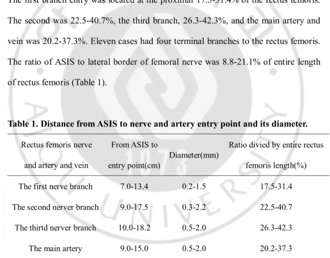
The ratio of the entry point and the entire length of rectus femoris were calculated. The first branch entry was located at the proximal 17.5-31.4% of the rectus femoris. The second was 22.5-40.7%, the third branch, 26.3-42.3%, and the main artery and vein was 20.2-37.3%. Eleven cases had four terminal branches to the rectus femoris. The ratio of ASIS to lateral border of femoral nerve was 8.8-21.1% of entire length of rectus femoris (Table 1).
Table 1. Distance from ASIS to nerve and artery entry point and its diameter.
Rectus femoris nerve and artery and vein
From ASIS to entry point(cm)
Diameter(mm)
Ratio divied by entire rectus femoris length(%) The first nerve branch
The second nerver branch The thirtd nerver branch
The main artery
7.0-13.4 9.0-17.5 10.0-18.2 9.0-15.0 0.2-1.5 0.3-2.2 0.5-2.0 0.5-2.0 17.5-31.4 22.5-40.7 26.3-42.3 20.2-37.3 B. CLINICAL APPLICATIONS 1. Femoral head fractures
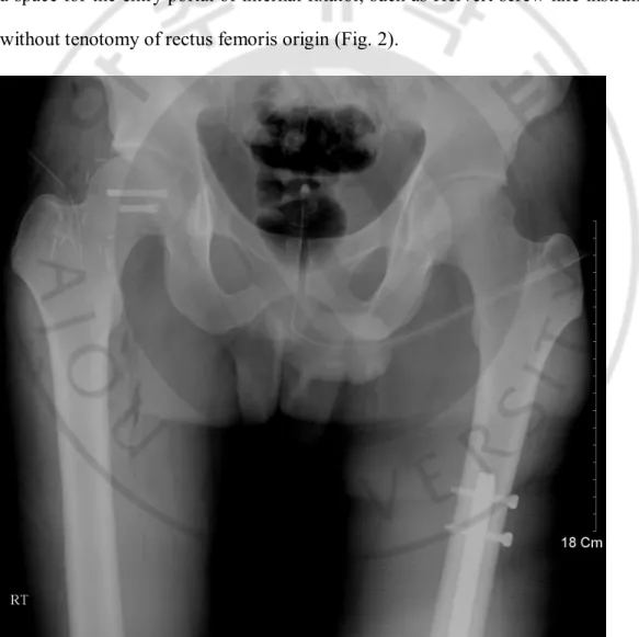
fragment with posterior dislocation. The reduction has been achieved by closed or open maneuver, but in some fracture types, the internal fixation has not been easy. Rectus femoris tenotomy from its origin site was sometimes needed for an adequate internal fixation. However, we were able to mobilize the rectus femoris to the medial and lateral directions. The proximal 17.5% of rectus femoris medial border could be a space for the entry portal of internal fixator, such as Hervert screw like instrument, without tenotomy of rectus femoris origin (Fig. 2).
Fig. 2. Open reduction and internal fixation through safe triangle by Hervert screws.
2. Pyogenic arthritis of hip
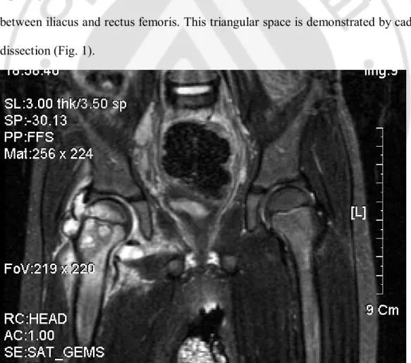
In another case of a 3 year-old male, who was admitted due to a right hip pain with fever, the Patrick test was positive, WBC count 15,000/dl, and neutrophil was 73.7%. The ESR was 74 mm/hour, CRP was 11.05 mm/dl. The plain radiogram showed diffuse radiolucency on proximal femoral metaphysis. In MRI, diffuse hip effusion was shown with high signal intensity on T-2 weighted image (Fig. 3). Under the impression of septic hip with acute osteomyelitis of proximal metaphysis of femur, hip irrigation was done at the medial border of rectus femoris through the space between iliacus and rectus femoris. This triangular space is demonstrated by cadaver dissection (Fig. 1).
Fig. 3. Acute osteomyelitis with septic hip was irrigated and was done bone drilling through safe triangle.
IV. DISCUSSION
The anterior approach of the hip is a relatively infrequent operative method for the orthopedic armamentarium. However, it could be used for arthroscopy, femoral head fracture and septic hip drainage. The usual anterior approach concept involves the lateral border of rectus femoris. In this study, however, we found there is a safe triangle at the medial border.
Surgically, the most important structure of safe triangle within dangerous femoral triangle is the femoral nerve, especially the motor nerve to the rectus femoris. The length between ASIS and entry point of the first branch to rectus femoris provides enough space for true anterior approach.
Even if there were some damages to the motor branch to rectus femoris, the functional loss is not a significant problem. Injury to the first branch does not have any significant meaning because there are several branches to rectus femoris. The main nerve to rectus femoris was the second or the third branch in our cadevaric study. Some authors have described the intramuscular neurovascular anatomy of the rectus femoris muscle and evaluated whether the muscle can be split into two functional units for this reason.
Anatomic study of vessels to the rectus femoris had been done for the free flap. The vascular supply of the rectus femoris muscle emanated mainly from the lateral femoral circumflex artery. In our cadevaric study, some types of variation were found in the origin of a nutrient artery of the muscle. The most common type was one in which the artery is derived from the descending branch of the lateral femoral
circumflex artery and the result is similar to the previous reports. However, this finding is not important for our safe area because the location is far from the zone in which we are interested. We did not consider cutaneous nerve location because the important lateral femoral cutaneous nerve was located on the lateral portion of this area, and the other minor cutaneous nerves usually did not have any surgical significance. The motor branch of sartorius muscle also did not have any significance in open surgical procedure because sartorius retraction to the medial side was not difficult for its anatomic characteristics and there was other nerve supply to sartorius muscle on more distal portion.
Hip arthroscopy is a technically difficult procedure to perform(Hawkins,1989). Some authors have reported that a limited anterior approach to the joint has made hip arthroscopy technically less difficult to treat a wide range of hip pathology(Yang and Morris,1999). All conventional portals are located at the lateral aspect of the femoral triangle(Byrd,2001;Glick,2001;Monllau at all,2003). This means that there is no true anterior portal in the hip area. The so-called anterior portal is a kind of anterolateral portal, which avoids the risky femoral triangle(Swiontkowski at all,1992). In this study, some triangular area could be used for anterior portal within the risky femoral triangle. This portal could be a potential space that makes the approach and viewing of medial joint space easier.
Typical complications of femoral head fractures are posttraumatic necrosis of the femoral head with its prevalence being 15-66%, and arthritis of the hip joint. It is yet uncertain whether the type of surgical approach can influence the rate of necrosis. Some authors have suggested principles such as posterior approach for posterior
displacement and anterior approach for anterior displacement. They have reported that by using these principles, the rate of necrosis of the femoral head clearly diminishes(Jessberger at all,2002). Some authors described that there were no cases of avascular necrosis of the femoral head associated with an anterior approach. Because of the greater ease of access to the fracture, the anterior approach is recommended when operative reduction of a displaced Pipkin type I or II is indicated(Bauer and Sarkar,1997). In these kinds of situations, we could use this potential safe space as an entry point of internal fixation with internal fixator such as Hervert screws without tenotomy of rectus femoris origin (Fig. 2).
Usually, tenotomy of rectus femoris is not harmful because there is another forceful hip flexor, iliopsoas, and another knee extensor. No cases of hip flexion contracture have been reported, however less traumatic technique will make postoperative rehabilitation easier by avoiding tenotomy of rectus femoris.
Sometimes forceful rectus femoris medial traction makes the operation difficult. By using this space, we could not only avoid tenotomy of rectus femoris but also make the operation easier.
The usual drainage method of children’s septic hip is through the anterior approach. There are many studies for septic hip drainage, including percutaneous drainage using pleurocath and open drainage by the anterior approach(Griffet and El Hayek,1996). This safe triangular potential space can be used for septic hip drainage. Some authors used this area for ultrasonography in the diagnostic approach of septic arthritis(Sekiya at all,2000).
arthroscopy, septic hip irrigation, and head fracture case with anterior fragment, a true anterior approach can be attained through femoral triangle.
The safety zone could be extended to more distally because the main nerve branch was usually the second or the third branch, and the feeding vessel was located more distal to the first branch in all cadavers in this study. In some clinical situations, the first branch will be sacrificed, because it will not cause any significant damage to the rectus femoris function.
Through this study, we have found margin of safe triangle within femoral triangle. Hopefully it could be useful in some clinical situations (Fig. 4).
Fig. 4. This figure shows potential space of anterior hip approach, which consists of mainly proximal 1/5 of rectus femoris between rectus femori and iliacus. Rectus femoris Safe triangle Within femoral triangle
V. CONCLUSION
In the clinical situations mentioned above, the space between proximal 17.5% of medial border of rectus femoris and iliacus can be used for true anterior hip approach, without damaging the femoral nerve.
REFERENCES
1. Bauer GJ, Sarkar MR: Injury classification and surgical approach in hip dislocations and fractures. Orthopade. 26(4):304-16, 1997
2. Byrd JW: Hip arthroscopy. The supine position. Clin Sports Med. 20(4):703-31, 2001
3. Cho JH, Chung GY, Chung MS, Won YY: Safe triangle within dangerous femoral triangle for the potential space of the anterior hip approach. J of Kor Ortho Asso. 39(5):476-81, 2004
4. Glick JM: Hip arthroscopy. The lateral approach. Clin Sports Med. 20(4):733-47, 2001
5. Griffet J, El Hayek T: Percutaneous drainage of septic hip arthritis in children Rev Chir Orthop Reparatrice Appar Mot, 82(3):251-54, 1996
6. Hawkins RB: Arthroscopy of the hip. Clin Orthop, (249):44-7,1989
7. Jessberger S, Blattert TR, Wagner R, Weckbach A: Reducing approach-associated morbidity in fracture dislocation of the femoral head-a longitudinal study (1982-2000) Zentralbl Chir,127(6):485-9, 2002
8. Monllau JC, Solano A, Leon A, Hinarejos P and Ballester J: Tomographic study of the arthroscopic approaches to the joint. Arthroscopy, 19(4):368-72, 2003 9. Sekiya JK, Wojtys EM, Loder RT, Hensinger RN: Hip arthroscopy using a
limited anterior exposure: an alternative approach for arthroscopic access. Arthroscopy,16(1):16-20, 2000
posterior approaches for Pipkin I and Pipkin II fractures. J Orthop Trauma. 6(4):437-42, 1992
11. Yang D, Morris SF: Neurovascular anatomy of the rectus femoris muscle related to functioning muscle transfer.Plast Reconstr Surg, 104(1):102-6, 1999
- 국문요약 –
진정한 고관절 전방도달에 사용할 수 있는
안전한 공간을 위한 해부학적 고찰
아주대학교 대학원의학과 정구영 (지도교수: 원예연) 목적: 위험한 대퇴 삼각 부위에서 안전한 부위를 알아내 진정한 고관절 전방 도달 공간을 알아보고자 한다. 대상 및 방법: 사체 해부 연구를 통해 51 부위의 대퇴 삼각 부위를 관찰 하였다. 전상장골극에서 슬개골 상부 중심까지의 거리로 대퇴 직근의 길 이를 측정하였으며, 전상장골극에서 서혜 인대와 평행하게 대퇴 신경까지 의 거리를 측정하였다. 또한 전상장골극에서 대퇴 직근으로 분포하는 대 퇴 신경 가지에 이르는 거리와 대퇴 직근으로 분포하는 혈관까지의 거리 를 측정하고 그 해부학적 변이를 관찰하였다. 해부를 통해 알아낸 안전한 공간을 대퇴 골두 골절과 급성 세균성 고관절염 환자에 적용하였다. 결과: 대퇴 직근으로 가는 대퇴 신경의 가지는 주로 3개 였다. 첫 번째 가 지는 전체 대퇴 직근 길이의 근위 17.5-31.4% 부분에서 근육에 들어갔 고, 두번째 가지는 근위 22.5-40.7%, 세번째 가지는 26.3-42.3%, 혈관 은 20.2-37.3% 부위로 근육에 분포 되었다. 전하 장골극에서 대퇴 신경의 외측까지의 길이는 3.5-8.5cm 였다. 이 공간을 이용해 두 예에서 기 존의 방법(Smith-Peterson)에 더하여 보다 용이하게 수술이 시행되었다. 결론: 대퇴 삼각 부위 가장 중요한 구조물인 대퇴 직근 운동 신경의 위치 로, 대퇴 직근 근위 17.5%영역과 전상장골극에서 대퇴 신경 외측까지의 적어도 3.5 cm의 삼각형 공간, 특히 대퇴 직근과 장요근 사이의 공간은 고관절의 진정한 전방도달 수술 즉 대퇴 골두 골절, 소아 세균성 고관절 염, 고관절 관절경의 수술 시 사용 가능한 공간으로 사료된다. 핵심어 : 고관절, 대퇴 직근, 대퇴 신경, 대퇴 골두 골절, 고관절 관절경, 전방도달법