The J。띠nal 01Medicineand Ufe Science
경동맥
초음파의 기초와
임상적
응용
주승재
쩨주대학교 의학전문대학원 내과학교실
(Received May 31. 2013: Revised June 7,2013: Accept.edJune 14,2013)
Abstract
Vol. 10,No,1(June),2013
Basics and clinical applications of the carotid ultrasound
Scung-Jae .100
Jeμ Nalional UniversitySCh
∞
1이MedicineCarotid ullrasound is performed according to the steps of the recommended guideline. but the purpose 01 the study should be considered. SCreen;ng of the carotid stenosis needs the careful examinalion 01 carotid Dop미er ullrasound. To evaluate the future cardiovascu넘r risk,the exacl measurement 이the carotid intima-media lhickness (C1Mn and careful screening for carotid plaques is req미red. CIMT is usually measured al the 1ar wall of the common
∞
rotid artery just befαe the branch;ng of Iheintemal and exlernal carotid arteries. Mean CIMT ~O. 7 mm or max CIMT of the internal carotid artery ~ 1.5 mm suggests the high risk group for the luture cardiovascular diseases. Measurement 01 CIMT in already diagnosed patients 에th coronary artery disease or slroke has no clinical impact. For the patients 01 a high risk group,common cardiovascular risk I~Clors,such as hyperlipidemia or hypertension. should be managed according 10 the guidelines,and if needed ,coronary angiography may be considered. Car이id Doppler fjndings s니ggesling the carotid artery stenosis }50% usual!y req미re lurther studies such as CT or MR angiography 이Ihe carotid artery 10 evaiuate Ihe severily. (J Med Ufe Sci 201 3; 1O(1):81-87)
Key Words : UIσ'asound,Carotid stenosis ,Carotid intima-media lhickness. Plaque,Risk
서
론
경동맥 협칙중은 허연성 뇌쏠중의 중요한 원인이며 증상이 없 는 심한 경동액 협착도 수솔적 풍백내박정제출 {endar1er깅ctomy) 이나 경동액 스텐트 삽입술의 적용중이 된다 경동맥 혐착종의 선별 검사로 B형 빛 도플러 경동백 초음파가 임상에서 흔히 사 용된다 그런데 정동액월 B형 초음따로 검사하연 협좌뿐만 아니 라 내박충악 두께 <inψna-me이a thickne않 IMT)를 측정하고 plaque룹 발견항 수 있다 동맥경화는 전신 혈관에서 동시에 발 생하므로 경동액의 IM'J'!.f plaque이 잠재성 (subclinicaI) 동백경 화의 표지자 역할을 하여 심근경색증, 뇌혈중,심혈관계 사망 둥 올 예측환 수 있는 인자로 사용훨 수 있다는 많은 잉상 연구와 때타 분석 풍이 있다 경동백 초응파는 방사선 노출 없이 현관의 동맥 경화흘 명가하기 위해서 간단히 시행원 수 있기는 하나 IMT의 측정 부위와 방법‘경동액 초응따 선별 검사가 펼요한 대 상 등에 대한 다양한 의견이 존재하며 아직 국내 성인의 lMT 쩡 잭임저지,주융채 690-767 제주특별자치도제주시아린 13킬 15 제주대학쿄영원심징내과E-mail; sejj
。
α:PjeμnU.ac.kr상치 자료는 부촉한 실정이다
본론
1,경동액 초음파 방법 경동맥 초음파흘 위해서 7빠-lz이상의 linear-ar얘y transducer 가 멸요하다 겁λt"O't
고자 하는 경동액의 반대편으로 환자의 고개 환 툴리고 초응파의 깊이를 4cm으로 맞추며 중{zoom) 기능은 사 용하지 않고 다음의 순서로 검사한다” Slep 1 횡혹 B형스캔 탑촉자의 notch월 환자의 오른연 방향으로 위치하고 총경동액 (common carotid artery: CCA)의 근위부에서 위쪽으로 내경동 맥 (inrem 외 carotid 밍'lerγ ICA)의 충간 부위까지 서서히 횡축단연 스캔올 하여 현관의 위치 혈관벽 두째,plaque의 존재,주 위구조붙풍을관짤한다
Slep 2 내경동액과 외경동맥 도플러
각 분지의 근위부 1cm에서 간혈파형 도플러를 기꽉한다 혈 류의 방향과 도흘러 빙 사이의 각도는 60도 이하가 되도록 조절
8eung-Jae Joo 한다 만약 협착이 있으연 협착 전후의 혈류 속도를 기록한다 내경동맥은 크고 대개 경동맥 팽대부(bulb
잉+
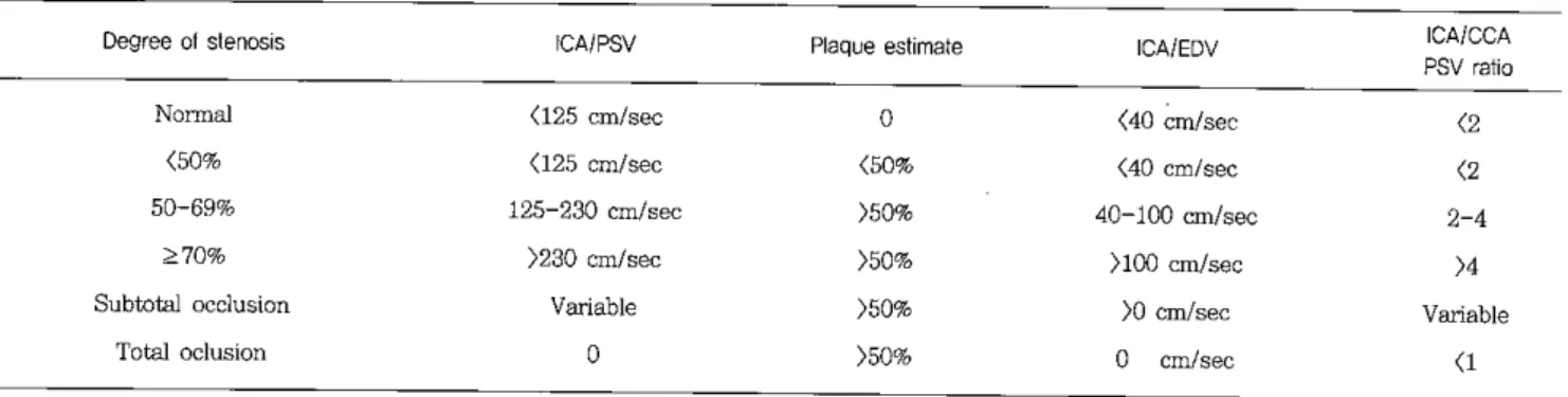
연결되어 있고 뒤 면,외측으로 주행하며 목에서 분지가 갈라지지 않고 처항이 낮 은 도플러 혈류 파형을 보인다 외경동백 (external carotid arærγ ECA)은 비교적 작고 대개 경동맥 팽대부와 연결되지 않 으며 앞편,내측으룩 주행하고 목에서 8개의 분지가 갈라지며 저 항이 큰 도플러 혈류 파형을 보인다.(Table 1,Fig. 1)2J3)경동맥 협착은 주로 내경동맥에 발생하며 B헝 초음파를 이용하여 협착 정도를 평가할 수 었으냐 부정확할 수 있기 때문에 도플러 파형 을 분석하여 협착 여부를 판단한다(Table 2)" .sup<:r[ '"t"'"'poro.l Mo.~;llo.,..y F<:ic;QJFigure 1. Anatomy of the carotid arterγ and its branchesS)
Table 1. Differentiation bet\veen internal and external carotid arteries2J
Inlernalcar이띠a↑ery Externalcarotid arterv Usually larger
Usual1y lateral and posteηor u
,
u밍ly incorporates carotid bulb No branches in the neck Iρ,w resistance spectral waveformUsually no oscil1ations in Doppler on temporal tap test
U잉ally smaller
U잉ally medial and anterior
Usually does not incorporate bulb Eight branches in the neck
High resistance spectral wavefoπn at rest Visible and 8udible os이llations on Doppler
sígnal wavefonn on temporal tap test
Table 2. Criteria for the classification of severity of the Întemal c
앙。
tid arteη 잉Degree이slenosis ICAIPSV Plaque esti미ate ICAfEDV ICAICCA
PSV ra↑10
Normal <125cm/sec o (40 cm/sec (2
(50% (125 cm/sec (50% (40 cm/sec (2
50-69% 125-230 cm/sec )50'< 40-100 cm/sec 2-4
::?70% )230 cm/sec )50% )100 αn/sec )4
S니btota1 occlusion Variable )50%
m
αn/sec VariableTota1 oclusion o )50% o cm/sec (1
Figure 2. Patient position and probe orientation for the carotid 앙ieIγ
’”
Step 3 장촉 plaque 선별 스캔 횡측 단면 위치에서 탑촉자의 notch가 환자의 머리를 향하게 90도 회전시킨다 귀월 중심a
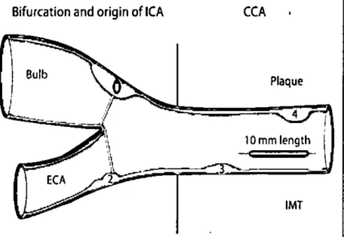
로 당축자흘 앞짝,옆쪽,뒤쪽으로 회전시켜서 총경동맥?맹대부,내정동액에 plaque가 있는지 관찰 한다{Fig. 2)". Plaque이 가장 크게 관찰되는 위치와 각도툴 기혹 하고 탑촉자륜 횡측으로 회전시켜서 확인한다 Plaque은 적어도 0.5 mm혹은 주위 tMT수치의 50%이상 동맥내강으로 돌출하 였거나 중막-외박 정계에서 내악-내강사이의 두께가 )1.5 mm 일 때 진단된다{Fig. 3)"Bifurcationand origin of ICA
「기
p
。앙erior
CCA
Basics and미inic.alapplicationsof the cnrotid ul뼈sound 경계가 이중 선으로 선영하게 보이는 각도툴 선택하여 내경동액 과 외경통액으로 분지되기 직전 1αn을 수동,혹은 반자동으로 측정히여 명균값과 최대값을 구한다 tMT측정 범위에 plaque이 포항되지 않도록 측갱 위치할 조절한다 혈관 외막은 밝은 초용 파 영상을 띄고 있어서near wall의 초음파 영상에서 혈관 외악 과 중막의 경계가 부정확하게 표시되어 측정된 αIT 값이 실제 값을 반영하지 않을 수 있기 때분에far wall에서반 IMT플 측정 한다S 미국 심초음따협회 지침에는 경동액 맹대부와 내경동맥 1MT측정이 부정확할 수 있고 재현성이 떨어지기 때문에 총경동 액 IMT측갱안을 권장하고 있으나 최근 연구에서 모든 부위 IMT를 측쩡하여 명균값을 사용하였을 때 심혈관께 사건 밥생융 더 갱확히 예측하였다는 결과도 있어서 영상이 선영하여 측정이 가능하다연 총경동액,맹대부,내경풍액 모두에서 IMT를 촉갱하 는 것이 권장되기도 한다찌 정동백 IMT값은 연령이 중가한수흑 중가하고 여성에 비해서 냥성에서 더 크며 인종간 차이가 있다 경동액 IMT값이 75 percenLile이상이연 중가되어 있는 것으로 판정하고 심혈관계 합병중 발생의 고위험군으로 간주한다 25-75 percentile은 영균 치이며 심혈관계 합병증 받생의 워혐이 증가하지 않는 군이다 25 percentile 이하의 값올 보이연 심혈관제 합영중 발생의 위험 이 낮은 군이다 여러 관찬적 코호트 연구에서 사용된tMT옥정 부위와 위치에 약간의 치이가 있으므로(Fig. 4)꺼 각각의 코호트 연구에서 체시된 IMT정상치틀 창고할 때는 측정 부위와 방법이 같。빠 시용 가능하다” 국내에서는ARlRANGE호트에서 정상 인의 경통액IMT값이 제시되었다{Table 3)" 한국인의경동백tMT 평균값이 0.7 mm이상이연 모든 연령 군에서 75 percentiIe 01 상에해당된다
Fi밍lre 3. V잉ious fonns of carotid plaques; 1; thickness
) 1.5 mm. 2: lumen encroaching )0.5 mm,3,4: surrounding intima-media Utickness )5σ%
‘
lStep 4 경동맥IMT측정
각 총경통맥의 이완기발 내경이 가장 직올 때 원위부의 far wall에서 1 cm을 측정한다 수축기에 측정하면 내경이 증가하여
IM'I가 신장되므로 실제보다 착께 측정될 수 있다 총겸통백의
near wall과 far wall모두에서 혈관내강-내박 경계와 중막-외막
Bulb
-i Plaque 4 10mmlength →一.
--:r::::
,-IMT-흩톨톨뚫뚫팩즐뚫
薦뚫뿔뚫쩔
αS' CII를CI u.æc 종!lIJ
EAS
1m
1NY3gc
l..•l3U앨펌걷”
햄
i
이@蠻鍵
Figure 4. Different segment definitions of c1inical studies.71
CCA; common carotid aroory. BIF: carolid bifurcation. ICA: intemal c앙-otid aπeη ,'CCA definition in the CHS: If the
beginning of the bulbus widening is detenninable,a 1 cm segment proxirpa1. else a segment extending from 8 to 18mm proximal to the tip of the f10w divider. #CCA definition in the KJHD:A 1
ω
1.5 cm segment proximal to the bulbar wideningSeung-J ae J 0
。
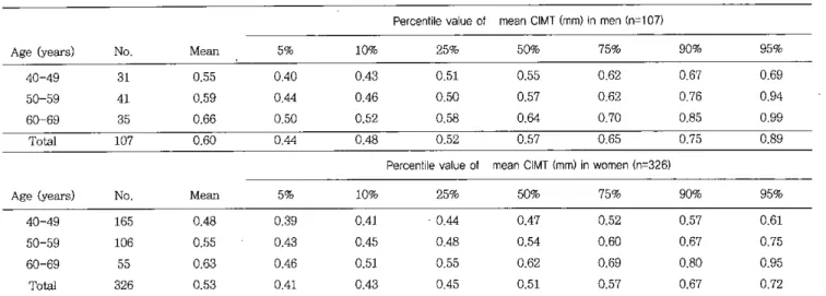
Table 3. Nonnal values of the carotid intima-media thickness in Korean populationB1
Percenlileva비e of mean CIMT(m떼In πen (n::0107)
Age (years) No Mean 5% 10,,* 25% 50"* 75% 90% 95%
40-49 31 0.55 0.40 0.43 0.51 0.55 0.62 067 0.69
50-59 41 0.59 0.44 0.46 050 0.57 0.62 076 0.94
60-69 35 0.66 0.5C 0.52 0.58 0.64 0.70 0.85 0.99
Total 107 0.60 0.44 0.48 0.52 0.57 0.65 0.75 0.89
Percentilevalue 01 mean CIMT(mm)in women(n::0326)
Age (years) N
。
Mean 5% 10% 25% 50% 75% 90,,* 95%40-49 165 0.48 0.39 0.41 0.44 0.47 0.52 0.57 0.61 50-59 106 0.55 0.43 0.45 0.48 0.54 0.60 0.67 0.75 60-69 55 0.63 0.46 0.51 0.55 0.62 0.69 0.80 0.95 Total 326 0.53 0.41 0.43 0.45 0.51 0.57 0.67 0.72 2. 경동맥 IMT와 plaque 의 임상적 의의 동백경화가 진행되면 내막에 동맥경화반 (atherosc1erotic plaque)이 발생하고 내박의 두께가 전반적으로 두꺼워진다 동맥 경화의 진행 정도를 펑가하려연 통맥의 내막 두께를 측정해야 하 나 경동맥 초음파에서 내막의 두께만 측정하는 것이 힘들기 때문 에 IMT를 측정하여 섬혈관계 사건 발생의 위험도를 평가한다 경동맥 1M맴}심혈관계 사건 사이의 연관성은 여러 관찰적 코 호트 연구와 메타 분석에서 증명되었다 8개 코호트 연구의 메타 분석 결과 총겸통백 IMT 값이 1- 표준면차와 0.1 rnm 증가할 때 마다 심근경색증은 각각 26%와 15% 증가하였고 뇌졸중은 각각 32%와 18% 증가하였다의 Framingham Offspring 연구 코호트 에서 총경통백 평균 IMT 가 1-표준연차 증가하면 심혈관계 질환 이 13% 증가하였메이 최근의 147B 코호트 연구의 베타 분석에 도 총경동맥 IMI와 심근경색증 뇌졸중 발생 사이에 양의 상관관 계가 있었는데 IMT 가 0.1 rnm 증가할 때마다 심큰정색증은 8%, 뇌졸중은 12% 증가하였다 (Fig. 5)111
12
=
12.30%;Q
lest lor helerogeneily,
P=
.24 Source ARIG,251994 GAPS,262006 Ghartottesvil1e,
272006 GHS.2B2007 FATE,B 2011 Hoorn Slu여,292003 KIHD,
30 1991 Malmo,31 2000 MESA,32 2007 Nijmegen Sludy ,33 2009 NOMAS ,34 2007。
SACA2 Sludy ,35 2007 Rotte띠am Sludy,
36 1997 η。
msø Sludy,37 2000 Conlribu1ion to Tolal USE-IMTPop 미ation , % ofTotal 31 8 1 7 3 1 2 10 13 3 2 1 8 g Hazard Rati。
(95% CI)a 1.11 (1.08-1.14) 1.10 (0.99-1.23) 0.88 (0.56-1.36) 1.11 (1.06-1.1 히 1.20(1.01-1.42) 1.07 (0.72-1.59) 1.05 (0.96-1.1 히 1.10(1.04-1.17) 0.98 (0.89-1.08) 1.34 (0.94-1.9 이 1.36 (0.99-1.8 히 1.09 (0.96-1.24) 1.13 (1.06-1.20) 1.04 (0‘,
8-1.1이 1.09 (1.07-1.12) 0.5•
←---를l←•
•
←←---←
----
--←--
•
-
----→--
<>
,---,
1.0Hazard Ralío (95% CI)"
2.0
Figure 4. Relation of common caro디d intima-media thickness (CIMT) with frrst-time myocardial infarction or stroke; a meta-ana1ysis of 14 population-based cohortsl1l ~Hazard ratios are per O.1-mm increase in co~on CIMT
정통맥 1M]는 연령,혈중 지질치,혈압 등과 갇은 기존의 심혈 관계 위험 인자와도 양의 상관관계가 있기 때문에 과연 경통맥 1M]가 심혈관계 발생 위힘을 예측히는 기존의 Fran뼈ham risk 8core모델 동에 비해서 우월한 위험도 정보를 제꽁하고 심혈관계 위험도를 재분류히는데 도움을 줄 수 있는가에 대해서는 논란이 있다 ARlC (Atheros 이erosis Risk in Comrnunities) 연구에서 Frarningham risk score모델 단독 사용시 보다 겸통맥 IMT이 포 함된 모델을 사용하였을 때 심혈관계 위험도 평가를 향상시킬 수 있었으며 겸통맥 1M]와 plaque 정보를 추가하였을 때 23%의 환
BasÎcs and clinicaJ applîcations of the carotid ultrasound 자의 위험도가 재분류 되었다l잉 그러나 메타분석에서는 총경동
맥 IMT 정보를 추가하였을 때 위험도 순 재분류 지표 (net reclassification index: NRI)가 매우 낮아서 일반인의 심혈관계 위험도 평가에 경통맥 1M]가 굳이 필요한가에 대한 의문이 제기 되고 있다 기존의 심혈관계 위험 인자에 총겸통맥의 펑균 IMI 를 추가하였을 때 Framingham Offspring 연구의 NRI는 0%였고 (Tahle 4)찌’다른 147H코호트 연구의 머타 분석에도 NRI는 0.8%
였으며 중간 정도의 위험군에서 조차 NRI는 3.6%에 지나지 않았 대Table 5)이
Table 4. Rec1assification into new Framingham risk score (FRS) cat.egories as a fùTIction of original FRS categories for common carotid intima-media thickness; 상18 Framîngham Offspring study cohort띠
Degree이slenosis Non-evenæ (n=2707) Low risk ((6%) Moderaæ ri하,(6-20%) High risk C>20%) 'Ev8nts (n=258) Iρw risk ((6%) Moderate risk (6-20%) High risk ()20%)
Reclassillcatîon for non-events: -0.4% [(36+25~45-26)/2707] Reclassification for events: -0.4% (3+4-8/258)
Net Reclassification Index: -0,4% - (-0.4%) = 0%
New lowrisk 1151 45 0 33 O O
New moderate risk New high risk
36 0 1185 25 26 239 3 0 120 4 8 90
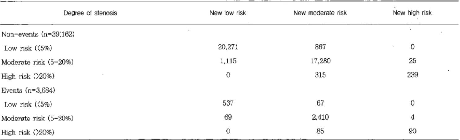
Table 5. Reclass 퍼cation with carotid intima-media thickness added to Framingham risk score categories; a meta-analysis
。
f 14 population-based cohortslllDegree 01stenosís Newlow risk New moderale risk New high risk Non-events (n=39;162) Low risk ((5%) 20,271 867 o Moderate risk (5-20%) 1,115 17,280 25 Hi링1 risk 020%) 0 315 239 Events (n=3,684) Low risk (<5%) 537 67 0 Moderate risk (5-20%) 69 2,410 4 High risk ()20%) 0 85 90
Reclassification for non-events: -0.5% ((867+362-1,115-315)/39,162] Reclassification for events: 0.4% [(67+102-69-85)/3 ,684]
Net Reclassification Index: 0,4% - (-0.5%) = 0.9%
Net Reclass버cation Irnprovement (NRI): 0.8% (95% CI; 0.1-1.6%) NRI in inteπnediate risk group: 3.6% (95% CI; 2.7-4.6%)
Seung-Jae Joo 정동액 IMT는 내악뿐만 아니라 충막 두께도 갇이 측정하는데 현관명환근 세포로 구성되어 있는 중악은 동맥경화 보다는 노화 나 혈압의 영향쏠 더많아 받기 때문에 전신 동액정화 진행을 명 가하는 지표로서 샤용되는데 째한점이 있다 이에 비해서 경동액 plaque는 주로 동맥경화 진행과정에서 발생하기 때문에 경동액 IMT보다 동맥경화의 더예민한 지표로 작용할 수 있다 경동백 plaque과 심혈관계 사건 사이의 연관성올 관쌀한 전향적 관찰 연구 결과에서 경풍백 1M1에 비해서 상대 위험도가 유사하거나 약간 더높았다” 대규모 인구 집단이 포함된 ARTC연구에서 경 풍백 plaque이 있울 때 관동액 짙환 받생 상대위험도는 acoustic shadov띠얘이 있융 때 2.96. 없을 때 2.02였고m뇌혈충 딸생의 상대위혐도는 여지쩨서 각각 1.92. 4.01였고 남자에서 각각 1.99 2.23였다… 또한 경동백 IMT 뿐만 아니라 plaque 정보틀
Frami뼈ham risk score모헨에 모두 추가하였월 때 환자의 위현
도 재분류플 향상 시원 수 있었다12).Fγamm앙13m Offspring 연 구 표호트에서는 싼amin앙1am risk score모띤에 총경동액 Wπ
월 포항시켰을 때 환자의 위험도 채분류 지수는 0%였으나 정동 액 plaque한 포항시켰을 때는 7.3%여서 총정동액 αIT 보다는 경풍백 plaque이 싱혈관계 짚환 예측에 더도응을 출 수 있음융 보여 주었다@
3
심혈관계 위험도 평가톨 위해서 경동맥 초응따 선볕검사 가띨요한인구집딘 2010 ACCF/AHA 진료 지칩에서 경동액 1MI는 섬혈관계 위험 도가 충간 정도인 부증상 성안에서 심혈관계 위험을 명가하는데 필요한 검사로 권유하고 있다{권고 수준 TIa증거 수준 B)I51미 국심초음파협회 진료 지침에도 관동백 첼환이냐 말초혈관질환, 대동맥류 퉁이 없는 성인의 향후 10년심근경색증이냐 심혈관계 사망 받생 위험원이 6-20%인 경우 경동액 IMT와 plaque는 심 현관계 위협도 명가를 더 정확히 개선하는 검사 방법으로 권고되 고 있다 이외에도 기존의 Framin양am 위험도 모헨에서 정확 히 명가될 수 없는 (1)조기 심혈관 질환의 가측력 (2)예외적으 로 심한 하나의 위험인자만 갖고 있는 60셰 이만의 성인(예‘가 축성 고할레스태찬현중 환자) (3) 적어도 2개 이상의 심현관계 위험인자플 갖고 있는 %세 미안의 여성 둥에서도 권고된다” 그 러나 이미 심혈관계 진황을 진단받았거나 검샤 결과가 향후 치료 방정에 영항을 주지 않올 것으로 예상되는 경우에는 시행하연 안 된다 반복 검사블 하여 명가한 정동백 IMT의 진행 정도가 심혈 관계 짚환 발생음 예측한 수 없기 때문에” 입상 연구흘 위한 경 우 이외의 반복 검시는 권고되지 않는다 결론
경동액 초음따 검시는 권고안에 있는 순서대로 시행하되 검사 목적이 무엇인지 우선적으로 고려한다, 경동백 협약 유무틀 진단 하고자 할 때는 경동액의 도플러 소견을 주의 김게 관싼하고 심 현관계 집황의 위험도플 명가하고자 한 때는 정통액 IMT블 정확 히 욕정하고 탑촉지원 여러 각도로 돌려서 plaque 유무관 검사 한다 경풍백 1MI‘는내경통액과 외경동백으로 분지되기 직전 1 cm의 far waTI에서 명균치 측정하여 0.7 mm이상이거나 내경통 액의 최대 1MT7f 1.5 mm 이상이연 심혈관 사건 발생의 고위험 군으로 분휴된다 이미 관동맥질환, 뇌줍중 등의 심혈관 진환이 진단된 환자의 TMT측갱은 입상적 유용성이 없다 경동백 초음 따에서 심현판계 질환의 고위험군으로 판명되었윤 때는 우선 고 지혈중, 고혈앙 풍의 위험 인지관 적극적으로 관리하고 연요 시 관동맥 조영울을 시행한다 경동액 초음파에서 50%이상의 협학 이 의심될 때는 경동액 CT나 MRI틀 촬영하여 협착의 심한 정도 릎판정한다참고문헌
1. Stein JH‘KOI'CmτCE. Hurst RT'. Lonn E. Kendall CB
Mohler ER. et al: Arnerican Society of Echocardiography Carolid Intima-Media Thickness Task Force. Use of carotid UT빼sound 10 identify subclinical vascular disease
and evaluate cardiovascular disease risk: a consensus
statement from the American Society o(
Echocardiography C와。tid ]nÜ1na-Media Thickness Task Force. Endorsed by the Society for Vascular Medicine. J Am soc Echo
∞
rdi。양 2008:21:93-1112. Gerhard-Herman M. Gardin JM. Jafr M. Mohler E.
Roman M. Naqvi TZ: American Society of
Echocardiography: Society of Vascular Medicine and Biol。양 Guidelines for noninvasive vasc미양 laboratory
testing: a report from the American Society of Echocardiography and the Society of Vasc띠강 Medicine
and Biology. Arn soc Echocardiogr 2006:19:955-72 3. Anderson JE. Grant' s atlas of anatomy. 7th edition
Baltimore: W~liams& WI1kins:1978. p.9-10
4. Touboul PJ,Hennerici MG. Meairs S. Adams H,
Amarenco P. Bornstein N. et al. Mannheim carotid intima-media thickness consensus (2004-2006). An update on behaTf of the Ad찌sory Bo밍'C!of the 3rd and 4th WaLching the Risk Symposium. 13th and 15th European Slroke Conferences. Mannheim‘Germany
2004‘and Brussels. Belgium. 2006. Cerebrovasc Dis
2007:23:75-80
5. Wù<strand J. MethodologicaTconsiderations of 띠trasoW1d measurement of c않。tid artery intima-media thickness and lumen diameter. CJin Physiol Funct Tmaging 2007:27:341-5
6. Bots ML. Sut!<m-TyrrellK Lessons from the past and promises for the future for carotid intima-media thickness. J Arn Coll C와'C!ioI2012:60:1599-604
H,Tuomainen TP,et al; PROG- IMT Study Group Carotid intima-media thickness progression to predict cardiovascular events in the general population (the PROG→IMT collaborative project): a meta-analysis of
indi띠dua! participant data. Lancet 2012;379;2053-62 8. Youn YJ. Lee NS. Kim JY,Lee JW ,Sung JK ,Ahn SG,et
al Normative values and correlates of mean common carotid intima-media thickness in 야1e Korean rural middle-aged population: the Atherosclerosis RIsk of Rural Areas 이 Korea General Population (ARIRANG) study. J Korean Med Sci 2011;26:365-71
9 1ρrenz MW,Markus HS,Bots :ML,Rosvall M,Sitzer M Prediction of clinic외 cardiovasc버ar events with carotid intima-media thickness: a systematic review and meta-analysis. Circulation 2007;115;459-67
10. PoJak JF,Pencina MJ,Pencina KM:,O'Donnell CJ ,Wolf PA,D'Agostino RB Sr. Carotid-wal1 intima-media thickness and cardiovascular events. N Engl J Med 2011:365;213-21
11. Den Ru:ijter 1:묘'v1,Peters SA,Anderson TJ,-Britton AR, Dekker JM,Eijkemans MJ,et a1. Common carotid intima-media thickness mea잉llTements in cardiovascular risk prediction; a meta-ana!ysis. JAMA 2012;308;796-803
Basics and clinical applications of the carotid ultrasonnd 12. Nambi V,Chambless L,Folsom AR,He M,Hu γMosley
T,et a!. C않ntid intima-media 상llckness and presence or absence of plaque improves prediction of coronarγ heart disease risk: the ARIC (Atherosclerosis Risk In Communities) study. J Am CoJ]'C암diol 2010;55;1600-7
13. Hunt KJ,Sharrett AR,Charnbless IE ,Folsom AR,Evans GW,Heiss G.. Acoustic shadowing on B-mode 버trasound
。
f the carotid artery predicts CI:ID. illtrasound Med Biol 2001;27:357-6514. Hunt KJ,Evans GW,Folsom AR,Sharrett AR,Chambless IE,Tegeler CH,et 외 Acoustic shadowing on B-mode ultrasound of the carotid arterγ predicts ischemic stroke the Atherosc1erosis Risk in Communities (ARIC) study Stroke 2001:32;1120-6
15.0Greenland P,Alpert JS ,Be이er GA,Benjamin EJ ,Budoff MJ,Fayad ZA,et al; American College of Cardiology Foundationl American Heart Association Task Force on Practice Guidelines. 2010 ACCF/AHA guideline for assessment of cardiovasc띠ar risk in asymptomatic adults a report of the American College of Cardiology Foundationl Ameπcan Heart Association Task Force on Practice Guidelines. Circulation 2010;122;e584-636