의 학
의 학
의 학
의 학 석사학위
석 사 학 위
석 사 학 위
석 사 학 위 논문
논 문
논 문
논 문
Novel mechanism of gastric proton
pump inhibitor (PPI) : Suppression of
Helicobacter-pylori induced
angiogenesis and selective induction of
apoptosis in gastric cancer cells.
아
아
아
아 주
주
주 대
주
대
대
대 학
학
학 교
학
교
교
교
대
대
대
대 학
학
학 원
학
원
원
원
의
의
의
의 학
학
학
학 과
과
과
과
김
김
김
김
동
동
동
동
규
규
규
규
Novel mechanism of gastric proton pump inhibitor
(PPI) : Suppression of Helicobacter-pylori induced
angiogenesis and selective induction of apoptosis in
gastric cancer cells.
by
Dong Kyu Kim
A Dissertation Submitted to The Graduate School of Ajou University
in Partial Fulfillment of the Requirements for the Degree of
MASTER OF MEDICAL SCIENCE
Supervised by
Sung Won Cho, M.D., Ph.D.
Department of Medical Sciences
The Graduate School, Ajou University
김동규의
김동규의
김동규의
김동규의
의학
의학
의학
의학
석사학위를
석사학위를
석사학위를
석사학위를
인준함
인준함
인준함
인준함.
심 사 위 원 장
심 사 위 원 장
심 사 위 원 장
심 사 위 원 장
조
조
조
조
성
성
성
성
원
원
원
원
인
인
인
인
심 사 위 원
심 사 위 원
심 사 위 원
심 사 위 원
이
이
이
이 광
광 재
광
광
재
재
재
인
인
인
인
심 사 위 원
심 사 위 원
심 사 위 원
심 사 위 원
정
정
정
정 재
재 연
재
재
연
연
연
인
인
인
인
아
아
아
아 주
주
주 대
주
대
대
대 학
학
학 교
학
교
교
교
대
대
대
대 학
학
학 원
학
원
원
원
2008
2008
2008
2008
년
년 6
년
년
6
6
6
월 23
월
월
월
23
23
23
일
일
일
일
ABSTRACT
Novel Mechanism of Gastric Proton Pump (PPI) Inhibitor :
Suppression of Helicobacter pylori Induced Angiogenesis and
Selective Induction of Apoptosis in Gastric Cancer Cells.
To survival in an ischemic microenvironment with a lower extracellular pH, ability to up-regulate proton extrusion is critical for cancer cell survival. Gastric H+/K+-ATPase exchanges luminal K+ for cytoplasmic H+ and is the enzyme primarily responsible for gastric acidification. On the basis of the fact that blocking the clearance of acidic metabolites are known to induce the cell death, we hypothesized that pantoprazole (PPZ), one of gastric H+/K+-ATPase inhibitors used frequently to treat acid-related diseases, could inhibit growth of tumor cells. And, although activation of mitogen activated protein kinases (MAPKs) by
Helicobacter pylori infection is associated with induction of host angiogenesis, which may
contribute to H.pylori associated gastric carcinogenesis, the strategy for its prevention has not been identified. As we previously reported a strong inhibitory action of gastric proton pump inhibitors (PPIs) on MAPK extracellular signal regulated kinase (ERK) 1/2 phosphorylation, we investigated whether PPIs could suppress the H.pylori induced angiogenesis via inhibition of MAPK ERK1/2.
To detect PPZ-induced apoptosis, we performed that Genomic DNA fragmentation, terminal deoxynucleotidyl transferase (Tdt)-mediated nick end labeling assay, and annexin V
staining. And mitogen-activated protein kinase activation and heat shock proteins expression were determined by immunoblot with specific antibodies. The antitumor effect of PPZ was evaluated in vivo by a xenograft model of nude mice. And, to address the relationship between H.pylori infection and angiogenesis, comparative analysis of density of CD34+ blood vessel was performed in tissues obtained from 20 H.pylori positive gastritis and 18
H.pylori negative gastritis patients. Expression of hypoxia inducible factor 1 (HIF-1α) and
vascular endothelial growth factor (VEGF) was tested by reverse transcription-polymerase chain reaction and secretion of interleukin 8, and VEGF was measured by ELISA. To evaluate the direct effect of H.pylori infection on the tubular formation of human umbilical vein endothelial cells (HUVEC), an in vitro angiogenesis assay was employed. Activation of MAPK and nuclear factor κB was detected by immunoblotting.
After PPZ treatment, apoptotic cell death was seen selectively in cancer cells and was accompanied with extracellular signal-regulated kinase deactivation. By contrast, normal gastric mucosal cells showed the resistance to PPZ-induced apoptosis through the overexpression of anti-apoptotic regulators including HSP70 and HSP27. In a xenograft model of nude mice, administration of PPZ significantly inhibited tumorigenesis and induced large-scale apoptosis of tumor cells. And, H.pylori positive gastritis patients showed a higher density of CD34+ blood vessels (mean 40.9(SEM 4.4)) than H.pylori negative gastritis patients (7.2±0.8), which was well correlated with expression of HIF-1α. Conditioned media from H.pylori infected gastric epithelial cells directly induced tubular formation of HUVEC and the increase of in vitro angiogenesis was suppressed by PPI treatment. Infection of
and expression of proangiogenic factors was mediated by MAPK activation and partially responsible for NFκB activation. PPIs effectively inhibited the phosphorylation of MAPK ERK1/2 that is a principal signal for H.pylori induced angiogenesis.
In conclusion, PPZ selectively induced in vivo and in vitro apoptotic cell death in gastric cancer, suggesting that proton pump inhibitors could be used for selectively anticancer effects. And, the fact that PPZ could downregulate H.pylori induced angiogenesis indicates that antiangiogenic treatment using a PPZ could be a promising protective therapeutic approach for H.pylori associated carcinogenesis.
Key words : Proton pump inhibitor, Apoptosis, Gastric cancer cell, H+/K+-ATPase, anticancer effect, Helicobacter pylori, Angiogenesis, VEGF, HIF-1α, Carcinogenesis
TABLE OF CONTENTS
ABSTRACT ... 1
TABLE OF CONTENTS ... vii
LIST OF FIGURES ... ix
I. INTRODUCTION ... 1
II. MATERIALS AND METHODS ... 6
A. Cell culture, bacteria strain, and reagents ... 6
B. Tissue samples ... 7
C. Measurements of Intracellular pH ... 8
D. Detection of Apoptosis . ... 8
E. Western Blot Analysis ... 9
F. Reverse transcription-polymerase chain reaction (RT-PCR) analysis ... 10
G. Enzyme linked immunosorbent assay (ELISA) ... 11
H. Immunohistochemistry for counting vessles in the gastric mucosa . ... 11
I. Immunofluorescence Staining ... 12
J. In vitro angiogenesis assay ... 12
K. Tumor Xenograft . ... 13
L. Statistics . ... 13
III. RESULTS ... 14
IV. DISSCUTION ... 43
국문요약 국문요약 국문요약
LIST OF FIGURES
Fig. 1. Effects of pH of culture medium on cell viability and cellular expression of
H+/K+-ATPase...15
Fig. 2. Inhibition of cancer cell viability by PPZ treatment...18
Fig. 3. Effect of proton pump inhibitors on gastric cancer cell viability. ...19
Fig. 4. Selective induction of apoptosis with PPZ. ...21
Fig. 5. Distinct MAPK signaling in cancer cells (A) and noncancer cells (B) by PPZ. ...23
Fig. 6. Effect of PPZ on expression of HSP70 and HSP27 in gastric cancer and noncancer cells. ...25
Fig. 7. Antitumorigenic potency of PPZ in xenograft model...27
Fig. 8. Immunohistochemical staining of the CD34 endothelial cell marker. ...30
Fig. 9. Expression of hypoxia inducible factor 1 (HIF-1α) mRNA in human gastric mucosa... 31
Fig. 10. In vitro angiogenesis assay. ...33
Fig. 11. Release and expression of vascular endothelial growth factor (VEGF) and interleukin 8 (IL-8) from Helicobacter pylori infected AGS cells. ...36
Fig. 12. Effects of proton pump inhibitor (PPI) on expression of angiogenic growth factors interleukin 8 (IL-8), hypoxia inducible factor 1 (HIF-1α), and vascular endothelial growth factor (VEGF)...37
Fig. 13. Involvement of mitogen activated protein kinase and nuclear factor κB (NFκB) in Helicobacter pylori induced mRNA expression of hypoxia inducible factor 1 (HIF-1α) and vascular endothelial growth factor (VEGF). ...39
Fig. 14. Deactivation of mitogen activated protein kinase (MAPK) extracellular signal regulated kinase (ERK)1/2 signaling with proton pump inhibitor (PPI). ...41
I. INTRODUCTION
The H+/K+-ATPase of gastric parietal cell exchanges luminal K+ for cytoplasmic H+ and is the enzyme primarily responsible for gastric acidification (Marie et al, 2004; Scott et al, 1993; Besancon et al, 1993; Pouyet et al, 1992). The enzyme consists of two subunits, a 114 kDa α-subunit and a 35 kDa β-subunit. The α-subunit containing ATP and cation binding sites carries out the catalytic and transfporting function of the proton pump. The heavily glycosylated β-subunit is required for endocytic retrieval of the H+/K+-ATPase from the canalicular membranes and is also essential for protecting proton pump from the environment of acid milieu. Because abnormally controlled gastric acids secreated by H+/K+-ATPase caused several gastrointestinal acid-related diseases including gastroesophageal reflux disease, gastric ulcer, duodenal ulcer, and Barrett’s esophagus, gastric proton pump inhibitors have been developed as the treatment for these acid-related diseases (Horn et al, 2000; Sachs et al, 1997; Sachs et al,1995; Fitton et al, 1996). Pantoprazole (PPZ) of these proton pump inhibitors, a substituted 2- pyridylmethyl/sulfinyl benzimidazole derivative, is a prodrug requiring protonation for functional activation at acidic conditions, accumulating selectively in acidic gastric luminal space and ultimately inhibiting acid secretion by the covalent binding with cystein residues in α-subunit of H+/K+-ATPase.
Besides of gastic H+/K+-ATPase, several kinds of vacuolar-type H+-ATPase are ubiquitously found on the membrane of various intracellular compartments of eukaryotic cells such as lysosomes, endosomes, the Golgi complex, and secretary granules (Nelson et al,
1995). Essential regulateion of pH in cytoplasmic, intraorganellar, and local extracellular spaces through vacuolar-type H+-ATPase has been suggested to play an important role in the mechanism of cell survival. These facts can be credited by several studies showing that vacuolar-type H+-ATPase inhibitor, bafilomycin A or concanamycin A, strongly induced apoptotic cell death (Nishihara et al, 1995; Ishisaki et al, 1999; Ohta et al, 1998; Aiko et al, 2002).
Cell survival and cell death are tightly controlled by numerous signal enzymes and regulators such as mitogen-activated protein kinases (MAPKs) and heat shock proteins (HSPs). MAPK signal enzymes are divided into three major groups, extracellular signal-regulated kinases (ERKs), c-Jun NH2-terminal kinases (JNKs)/stress-activated kinases, and
p38 (Franklin et al, 2000; Johnson et al, 2002). The ERKs appear to play a crucial role in the process of extracellular signals to the nucleus leading to induction of cellular growth, proliferation, and differentiation (Ballif et al, 2001; Dent et al, 1998). Heat shock proteins, which function mainly as molecular chaperones, allow cells to adapt to their environmental changes and to survive in otherwise lethal conditions (Garrido et al, 2001; Mehlen et al, 1996; Garrido et al, 1999; Bruey et al, 2000; Mosser et al, 1997; Buzzard et al, 1998; Beere et al, 2000). Because HSPs include pro- and antiapoptotic proteins that interact with a variety of cellular proteins, the type of HSP induced and its level of expression can determine the fate of a cell in response to a death stimulus. Generally, HSP60, HCS70, and HSP90 are constitutively expressed in mammalian cells, whereas HSP70 and HSP27 are strongly induced by different stresses, such as heat, oxidative stress, or anticancer drugs. It is well known that HSP27 and HSP70 play a cytoprotective role in gastrointestinal damage
including apoptosis (Tsukiml et al, 2001; Rokutan et al, 2000; Ethridge et al, 1998; Shichijo et al, 2003).
Maintenance of intra- or extracellular pH is very much important for cell function, and cancer cells in vivo often exist in an ischemic microenvironment with a lower extracellular pH than surrounding normal cells (Stubbs et al, 1999; Helmlinger et al, 1997; Stubbs et al, 1992; Suubbs et al, 1994; Frenzel et al, 1994; Gillies et al, 1994). The acidity in tumors is due to the increased production of acidic metabolites from rapid and large amounts of glycolysis and is provoked by the limited ability of the tumor vasculature to remove these acidic products. To overcome this hypoxic microenvironment and to prevent the accumulation of the increased acidic metabolites, the ability to dispose of intracellular protons is critical for cancer cell survival (Mccoy et al, 1995; Tannock et al, 1989; Maritinez-et al, 1993; Lee Maritinez-et al, 1998). These findings support the rationale of the present study that the inhibition of proton extrusion might be more susceptible or vulnerable to cell death of cancer cells than normal cells.
In this study, we have demonstrated for the first time that PPZ, the H+/K+-ATPase inhibitor, induced apoptosis selectively in gastric cancer cells and significantly inhibited tumorigenesis in a tumor xenograft model. We also documented the mechanism of the selectivity of this proton pump inhibitor on apoptosis of cancer cells. Our novel finding suggests that proton pump inhibitors could be considered as the selective anticancer agents in gastric cancer.
And another PPZ study, we investigated that PPZs could exert angiogenesis actions through MAPK inhibitions in H.pylori induced angiogenesis. That chronic persistent gastric
inflammation associated with Helicobacer pylori may play a crucial role in either the development or progression of gastric cancers has been generally agreed (Peek et al, 2002; Stolte et al, 2002; Scheiman et al, 1999) but the exact molecular mechanisms of how longstranding H.pylori infection can induce and make the procancer microenvironment favourable for the survival of tumor cells have not yet been clearly identified. The mechanisms fostering the neoplastic process of H.pylori infection have been revealed to include : (1) induction of neoplastic mutation by a considerable burden of oxidative stress (Touati et al, 2003; Bagchi et al, 1996); (2) imbalance between cell proliferation and apoptosis (Maeda et al, 2002; Gupta et al, 2001); (3) production of proteases and growth factors providing the environment for cell migration (Betten et al, 2001; Allen et al, 2000; Crawford et al, 2003); and (4) induction of host angiogenesis.
Among the diverse host cellular response related to H.pylori associated inflammation or carcinogenesis, some investigators reported that angiogenic growth factors induced by
H.pylori might be primarily important (Kitadai et al, 2003; Cox et al, 2001; Strowski et al,
2004; Innocenti et al, 2002; Franceschi et al, 2002). Kitadai and colleagues and Cox and colleagues (Cox et al, 2001) found that H.pylori infection induced several angiogenic factors and proteases, such as interleukin 8 (IL-8), vascular endothelial growth factor (VEGF), angiogenin, urokinase-type plasminogen activator, and metalloprotease 9 using high
throughput technology of cDNA microarray analysis. Strowski and colleagues (Strowski et al, 2004) also reported that H.pylori stimulated host VEGF gene expression via
a mitogen activated protein kinase (MAPK) pathway. These data imply that H.pylori are capable of inducing host angiogenesis, which may play a critical role in the development and
progression of gastric cancer. However, trials documenting the precise mechanism and preventive therapeutic approaches have not been performed.
The last decade has seen stanardisation of the treatment regimens for H.pylori eradication, with the use of triple therpy consisting of a PPI and two antibiotics, mainly clarithromycin and amoxicillin. Blockage of gastric acid secretion by PPIs contributes towards eradication of H.pylori via the rising pH of the gastric lumen. Appropriately high pH values increase antimicrobial susceptibility of H.pylori because the minimum inhibitory concentration of most antibiotics against H.pylori is very dependent on the pH of the environment (Iwahi et al, 1991; Nakao et al, 1998; Hirai et al, 1995; Tsuchiya et al, 1995; McGowan et al, 1994; Mauch et al, 1993).
Previously, we found that PPIs could exercise selective induction of apoptosis in gastric cancer cells, which was due to a significant inhibitory action of PPIs on MAPK activation (Yeo et al, 2004). As Stowski and colleagues (Hirai et al, 1995) reported that H.pylori stimulated host VEGF gene expression via the MAPK pathway, we hypothesized that PPIs could exert antiangiogenesis actions through MAPK inhibition in H.pylori induced angiogenesis. Here, we have found that infection with H.pylori significantly upregulated angiogenesis of the gastric mucosa by strong induction of proangiogenic factors, including IL-8, hypoxia inducible factor 1 (HIF-1α), and VEGF and, remarkably, angiogenesis induced by H.pylori was attenuated by PPI treatment.
II. MATERIALS AND METHODS
A. Cell culture, bacteria strain, and reagents
Human gastric cancer cell lines (AGS, Kato III, SNU-1, SNU-601, MKN-28, and MKN-45) were cultured in RPMI 1640 (Life Technologies, Inc., Grand Island, NY) containing 10% fetal bovine and 100 units/ml penicillin in a humidified 5% CO2 atmosphere.
As counter cells of these cancer cells, we cultured normal rat gastric mucosal RGM-1 cells and normal rat intestinal epithelial IEC-6 cells, which were maintained with DMEM-F12 and DMEM, high glucose (Life Technologies, Inc.) supplemented with 10% bovine insulin, respectively, and COS-1 cell, normal human fibroblast cells with RPMI 1640. Human umbilical vein endothelial cells (HUVEC) were obtained by collagenase treatment of umbilical cord veins, as previously described (Jappe EA et al, 1973). Cells were cultured on gelatin coated dishes and propagated in RPMI 1640 medium supplemented with 20% bovine calf serum, 90 ug/ml heparin (Sigma Chemical Co, St Louis, Missouri, USA), and 50 ug/ml endothelial cell growth factor.
A cagA+ and vacA+ standard strain of H.pylori (ATCC 43504) was obtained from the American Type Culture Collection (ATCC, Manassas, Virginia, USA). H.pylori were recovered from frozen stock by seeding on a blood agar plate including 7% sheep blood at 37℃ for five days under microaerophilic conditions (5% O2, 10% CO2) generated with
campy pouch (Becton Dickinson Microbiology Systems, Sparks, Maryland, USA). For inoculation of the bacteria, H-pylori were resuspended in phosphate buffered saline (PBS) to an A450 of 1.2 units, which corresponds to a bacterial concentration of 5 x 10
8
units (CFU)/ml, and cocultured with AGS cells at a concentration of 5 x 10⁷ CFU/ml. Solutions of pantoprazole (PPZ) and omeprazole were obtained from Altana Pharma AG (Konstanz, Germany) and AstraZeneca, respectively, and lansoprazole (Takeda, Japan) was solved in PBS (adjusted pH 2.0) with HCl overnight at room temperature. PD98059 (50µM, extracellular signal regulated kinase (ERK) 1/2 inhibitor; Cell Signaling Technology, Beverly, Massachusetts, USA), SB203580 (10µM, p38 inhibitor; Cell Signaling Technology), 1-pyrrolidinecarbodithioic acid (PDTC 100µM, ammonium salt, nuclear factor κB (NF-κB) inhibitor; Calbiochem, La Jolla, Califonia, USA), and BAYII-7082 (5µM, NF-κB inhibitor; Calbiochem) were used in the cell culture experiments. Briefly, to evaluate the effect of these inhibitors or PPIs, they were preincubated with AGS cells for eight hours, washed with PBS, and inoculated with H.pylori.
B. Tissue samples
Biopsied samples were obtained from five patients (mean age 48 years) with functional dyspepsia without H.pylori infection, 20 patients (means age 55 years) with chronic active
H.pylori positive gastritis, and 18 patients (means age 54 years) with H.pylori negative
gastritis induced mostly by non-steroidal anti-inflammatory drugs or other cause during gastroscopy. The presence of H.pylori was determined using the following tests : haematoxylin-eosin staining and Giemsa staining of biopsied tissues, rapid urease test, and urea breath test. When all of the above were negative, the case was defined as H.pylori negative and if more than two of these tests were positive, the case were defined as H.pylori positive. Gastritis was evaluated histologically and scored according to a modified Sydney
classification (Dixon MF et al, 1996); two different pathologists scored the degree of gastritis independently. Informed written consent was obtained from patients and the study was approved by Institutional Review Board.
C. Measurements of Intracellular pH.
Intracellular pH was measured in the monolayers using the pH-sensitive fluorescent probe 2',7'-bis-(2-carboxyethyl)-5-carboxyfluorescein (BCECF). Cells were loaded with BCECF for 10 minutes at room temperature in solution A containing 2.5µM/L BCECF-AM and mounted in the miniature Ussing chamber described for [Ca2+]ί measurements. BCFCF fluorescence was recorded and calibrated using a protocol described previously (Namkung W et al, 2003). Briefly, the fluorescence at excitation wavelengths of 490 and 440nm was recorded using a recording setup (Delta Ram; PTI Inc., St Louis, MO), and the 490;440 ratios were calibrated intracellularly by perfusing the cells with solutions containing 145mmol/L KCl, 10mmol/L HEPES, and 5µmol/L nigericin with the pH adjusted to 6.2 to 7.6.
D. Detection of Apoptosis.
Induction of apoptosis was determined by assaying a genomic DNA fragmentation. Briefly, cells were lysed for 15minutes in 10 mmol/L Tris.Cl (pH 7.4), 5 mmol/L EDTA, and 1% Triton X-100 and centrifuged at 12,000 rpm for 15 minutes. The supernatant was incubated with 0.1mg/ml proteinase K at 37℃ for 1 hour and extracted with an equal volume of phenol-chloroform, and the cellular DNA was precipitated with 1:10 volumes of
0.3 mol/L sodium acetate and 2 volumes of absolute EtOH overnight at -70℃. The precipitate was dissolved in 20 µL TE buffer containing 200 µg/ml RNase and incubated for 1 hour at 37℃. The extracted DNA was resolved on 1.8% agarose gel and stained with ethidium bromide.
Caspase-3 and poly(ADP-ribose) polymerase (PARP) proteolysis were assessed by immunoblotting with specific antibodies (Cell Signaling Technology, Beverly, MA). Terminal deoxynucleotidyl transferase (Tdt)-mediated nick end labeling (TUNEL) and annexin V/propidium iodide staining were detected by Fluorescent FragEL DNA fragment detection kit (Oncogene, Boston, MA) and annexin V-FITC apoptosis detection kit (BD Bioscience, San Diego, CA), according to the manufacture’s instructions, respectively.
E. Western Blot Analysis.
Human gastric cancer cell line, MKN-45 and normal gastric mucosal cell line, RGM-1 cells were incubated with PPZ(0.5 mmol/L PPZ for 0.5, 2, 4 or 8 hours and 0.3 or 0.6 mmol/L PPZ for 24 hours). And Human gastric cancer cell line, AGS cells were incubated with 0, 100, 200, 400 µmmol/L PPZ for eight hours, washed with PBS three times, and then inoculated with H.pylori (5 x 10⁷ CFU/ml) for 15 minutes (western blotting for ERK1/2) or two hours (western blotting for NFκB). Cells were resuspended in lysis buffer (20mM Tris, pH 7.5, 150mM NaCl, 1% Triton X-100, 1 mM EDTA, 1 mM EGTA, and protease inhibitor cocktail; Roche, Mannheim, Germany). The suspension was sonicated for approximately 30 seconds and centrifuged at 15,000g for 30 minutes. For documenting NFκB activation, nuclear/cytosolic fractionation was performed using NE-PER Nuclear and Cytoplasmic
Extraction Kit (Pierce, Rockford, Illinois, USA) following the manufacturer’s protocol. Isolated protein was subjected to western blotting. Proteins were extracted from the cells, electrophoresed on 12% sodium dodecyl sulphate-polyacrlyamide gels, and transferred to PVDF membranes using a semidry transfer system (Hoeffer Phamacia Biotech, San Francisco, California, USA). Membranes were blocked in 5% non-dry milk and probed with 1:1000 dilution of specific antibodies corresponding to phospho-p38 (Cell Signaling Technology, Beverly, MA), phosphor-ERK, phosphor-JNK, HSP70, HSP60, total ERK, NFκB p65, HSP27 or α-tubulin; all antibodies were purchased from Santa Cruz Biotechnology (Santa Cruz, California, USA).
F. Reverse transcription-polymerase chain reaction (RT-PCR) analysis
Total RNA was isolated from cells with appropriate treatment using TRIzol reagent (Life Technologies, Milan, Italy), and 2 µg total RNA were reverse transcribed according to the manufacturer’s instructions (M-MLV reverse transcriptase; Promega, Madison, Wisconsin, USA). PCR was performed using the Premix Ex Tag kit (Takara, Chiba, Japan) with specific primers as follows : 5'-CTC AAA GTC GGA CAG CCT CA-3' and 5'-CCC CGC AGT TTT CTG CT-3' for HIF-1α; TCG GGC CTC CGA AAC CAT G-3' and 5'-GGT TCC CGA AAC CCT GAG G-3' for VEGF; 5'-TTG TTG CCA TCA ATG ACC CC-3' and 5'-TGA CAA AGT GGT CGT TGA GG-3' for GAPDH. The PCR reaction was carried out for 28 thermal cycles of 94℃ for one minute at 55℃ (for HIF-1α and GAPDH) or 60℃ (for VEGF) for one minute, and 72℃ for one minute. The product was resolved on 1% agarose gel and stained with ethidium bromide.
G. Enzyme linked immunosorbent assay (ELISA)
Immunoreactive human IL-8 and VEGF were measured in culture supernatants of AGS cells using enzyme linked immunosorbent assay (ELISA) kits according to the manufacture’s instructions (HyCult Biotechnology, Uden, the Netherlands). AGS cells were grown in six well cell culture dishes, incubated in the presence or absence of PPZ for eight hours, and after washing with PBS, cocultrued with H.pylori for various times. Culture supernatant (200µl) was used for analysis of IL-8 and VEGF production.
H. Immunohistochemistry for counting vessles in the gastric mucosa.
Immunohistochemistry staining of CD34+ endothelial cells was performed to analyse the degree of angiogenesis in the gastric mucosa of gastric patients. For immunohistochemical detection, 10% buffered formalin fixed paraffin embedded sections were deparaffinised, rehydrated, and then boiled in 100 mM Tris buffered saline (pH 7.6) with 5% urea in an 850 W microwave oven for five minutes, followed by two more treatments of five minutes each. Then sections were stained with Histostain-Plus kit (Zymed Laboratories Inc., San Francisco, California, USA) according to the manufacture’s instructions. Primary antibodies against the CD34 endothelial cell marker were purchased from Novocastra Laboratories (clone QBEnd/10; UK). Sections were counterstained with haematoxylin. CD34 positive blood vessles were counted on three separate sites (x100 magnified field) and presented as mean (SEM) of 20 H.pylori positive and 18 H.pylori negative cases.
I. Immunofluorescence Staining.
Dispersed single cells (2 x 10⁵ cells per well) were grown on 22 x 22 x 1 ㎣ glass coverslips in 6-well cultrure plates. After 24-hour culture, cells were fixed in ice-cold methanol for 5 minutes in a -20℃ freezer and permeabilized with 1% Triton X-100/PBS for 10 minutes at room temperature. The cells were blocked with 5% bovine serum albumin for 30 minutes and probed with anti-α subunit of H+/K+-ATPase antibodies (1:100 diluted in 5% bovine serum albumin, Santa Cruz Biotechnology) for 2 hours. Cy3-conjugated secondary antibodies were used to visualize under a confocal microscope (BX50F, Olympus, Japan).
J. In vitro angiogenesis assay
In vitro angiogenesis assay was slightly modified from Kitadal and colleagues. Briefly, AGS cells (1 x 10⁷ cells/100 ㎟ culture dish) were incubated with 0,100,200, or 400
µM PPZ for eight hours, washed with PBS three times, and then inoculated with H.pylori (5
x 107 CFU/ml) for 24 hours. The cell culture supernatant was harvested and centrifuged at 5000 g for 30 minutes. Conditioned media was prepared by 1:1 dilution of the culture supernatant with HUVEC endothelial cell medium. The conditioned media were filtered through 0.4µM pore filters (Millipore, Boston, Massachusetts, USA) to remove H.pylori and then the media were added to HUVEC culture and changed every three days. After nine days, the HUVEC were observed for tubular formation under microscopy and confirmed expression of the endothelial cell marker, CD31, by immunocytofluorescence staining. HUVEC were fixed in ice cold methanol for five minutes, frozen at -20℃, and treated with
1% Triton X-100/PBS for 10 minutes at room temperature. Cells were blocked with 5% bovine serum albumin for 30 minutes and probed with anti-CD31 antibodies (1:100 dilution in 5% bovine serum albumin; Santa Cruz Biotechnology) for two hours. Cy3-conjugated secondary antibodies were used to visualize under a inverted fluorescence microscope (Olympus, IX71, Tokyo, Japan).
K. Tumor Xenograft.
Subconfluent MKN-45 cells were dissociated with 0.25% trypsin and 1 mmol/L EDTA (Life Technology, Inc.) and suspended in PBS at density of 5 x 10⁷ cells/ml. Each mouse was s.c. inoculated with MKN-45 cells (5 x 10⁶ per site) on the left and right side of the back on day 0. The animals were randomly divided into two groups (9 per group). Group A received an intratumoral injection of PBS daily from day 14; Group B daily received an intratumoral injection of 0.4 ㎎/㎏ PPZ daily from day 14. The shortest and longest intervals, and the volume of each tumor (㎣) was calculated. Mice were sacrified at day 22, and isolated tumor tissues were analyzed for microscopic gross finding and TUNEL stain. These studies were approved by the Institutional Animal Care and Use Committee and complied with the highest international criteria for human use of animals in research.
L. Statistics.
All values are expressed as mean (SEM) and the Mann-Whitney U test and Friedman ANOVA test were used for statistical calculations.
III. RESULTS
Human gastric cancer cells were more tolerant of acidity in the culture media than normal cells.
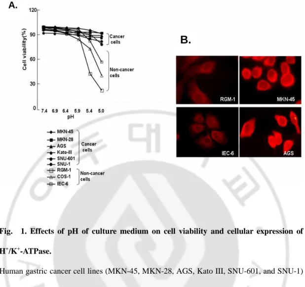
To determine whether cancer cells tolerate acidic conditions better than noncancer cells, we cultured several gastric cancer cell lines (AGS, KATO III, MKN-28, MKN-45, SNU-1, and SNU-601) and noncancer cell lines (RGM-1, IEC-6, and COS-1) in media maintained at different pHs, including 7.4, 6.9, 6.4, 5.9, and 5.4. The cancer cells adapted well to lower pH, whereas the normal cells were much less tolerant of acidity in the culture media (Fig. 1A). At pH 5.4, most of cancer cell lines tested showed >80% viability, but normal cell lines RGM-1 significantly decreased viability by 2RGM-1.6%.
To investigate whether this difference in cell survival at lower pH was due to ability to dispose or disperse H+, we immunocytochemically stained two cancer cells (AGS and MKN-45) and two noncancer cells (RGM-1 and IEC-6) with antibodies against the gastric proton pump, α subunit of H+/K+-ATPase. We found that there was a much larger amount of proton pump in cancer than noncancer cells (Fig. 1B). Immunofluorescence images showed a predominant expression of the α subunit of H+/K+-ATPase in the membrane and cytoplasm of cancer cells. These results indicated the enhanced ability to dispose of H+ due to overexpression of H+/K+-ATPase, which might contribute to cancer cell survival in an acidic microenvironment.
Fig. 1. Effects of pH of culture medium on cell viability and cellular expression of H+/K+-ATPase.
Human gastric cancer cell lines (MKN-45, MKN-28, AGS, Kato III, SNU-601, and SNU-1) and noncancer cell lines (RGM-1, Cos-1, and IEC-6) were cultured in media at different pH for 24 hours, and cell viability was measured by the 3-(4,5-dimethylthiazol-2-yl)-2,5-diphenyltetrazolium bromide assay (A). Expression of H+/K+-ATPase, α-subunit was detected with specific antibodies and visualized with Cy3-conjugated secondary antibodies. Note that the α-subunit of H+/K+-ATPase was more highly expressed in the plasma membrane and cytoplasm of cancer than noncancer cells (B).
A.
Treatment of PPZ significantly inhibits cancer cell viability in a pH-dependent manner.
Because PPZ is a protonatable weak base, which can convert to active form in an acidic environment with a low pH, we tested the effects of PPZ on cell growth in culture media maintained at various pHs (Fig. 2). Nevertheless, cancer cells showed tolerance of acidity in culture media as shown in Fig. 1A, and administration of the proton pump inhibitor PPZ led to significant reduction of cancer cell survival (Fig. 2A). At pH 5.0, PPZ treatment reduced cell viability by 6.9% on average in MKN-45 cancer cells (Fig. 2A). On the contrary, normal cell line RGM-1 did not showed a significant difference in cell viability between the PPZ-treated and nontreated groups (data not shown). Despite resistance of cancer cells to the acidic environment, cancer cells were much more susceptible to growth inhibition of PPZ at a lower pH.
We evaluated whether this attenuation of cancer cell viability is induced by a decrease of intracellular pH due to the blocking of disposal of intracellular H+ by specific protonation of this drug. As shown in Fig. 2B, PPZ treatment significantly increased acidity of intracellular pH in AGS, MKN-28, and MKN-45, but SNU-601 did not show a remarkable change (Fig. 2B). Of note, AGS decreased intracellular pH from 7.6 to 7.2. These findings showed that PPZ-induced cell death was caused by the specific protonation of this proton pump inhibitor.
We also investigated whether other proton pump inhibitors such as omeprazole and lansoprazole has a similar effect to pantoprazole on cancer cell viability. Human gastric MKN-45 cells were cultured with each proton pump inhibitor in pH 6.0 of media for 24 hours, and cell viability was determined by
3-(4,5-dimethylthiazol-2-yl)-2,5-diphenyltetrazolium bromide assay. The results showed that whereas the PPZ or omeprazole-treated cells remarkably reduced cell viability in a dose-dependent manner, lansoprazole did not show any changes in cancer cell viability (Fig. 3). This difference suggested that the decreased cell viability comes from the specific effect of proton pump inhibition. Omeprazole and PPZ are available for parenteral injection, but lansoprazole is only available for oral administration, suggesting that lansoprazole lost its proton pump inhibiting ability in aqueous form.
Fig. 2. Inhibition of cancer cell viability by PPZ treatment.
In different pH media (7.4, 6.9, 6.4, 5.9, 5.0), gastric MKN-45 cancer cells were incubated in the presence or absence of 0.5 mmol/L PPZ, and cell viability was measured by 3-(4,5-dimethylthiazol-2-yl)-2,5-diphenyltetrazolium bromide assay (A). Although MKN-45 showed resistance to acidity of culture media, PPZ significantly decreased this cancer cell viability in a pH-dependent manner. To evaluate effect of PPZ on intracellular pH (pHi) of cancer cell, gastric cancer cells were plated in a 6-well culture dish, treated with 0.5 mmol/L PPZ for 16 hours, and measured according to Materials and Methods as described previously (B).
Fig. 3. Effect of proton pump inhibitors on gastric cancer cell viability.
To compare effect of other proton pump inhibitors (omeprazole and lansoprazole) with PPZ on cancer cell viability, MKN-45 cells were cultured in the presence of each proton pump inhibitor indicated at Ph 6.0 for 24 hours and were measured cell viability by 3-(4,5-dimethylthiazol-2-yl)-2,5-diphenyltetrazolium bromide assay. Cell viability (%) was calculated by formula as follows ; Cell viability (%) = (sample absorbance - total absorbance)/(spontaneous absorbance - total absorbance) × 100. Total absorbance was obtained from absorbance of 0.1% Triton X-100-treated cells as negative control, and spontaneous absorbance indicated absorbance of PPZ-untreated control cells.
PPZ selectively induced apoptosis cell death in human gastric cancer cells.
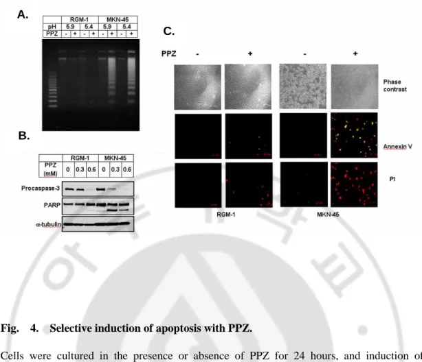
To determine whether PPZ preferentially induces apoptosis in cancer cells, we administered this agent to two cell lines, a normal gastric mucosal cell line, RGM-1, and a gastric cancer cell line, MKN-45 (Fig. 4). Although normal gastric mucosal RGM-1 cells significantly reduced cell viability in a low pH media as shown in Fig. 1A, an apoptotic genomic DNA fragmentation was detected by neither pH change nor PPZ treatment (Fig.4A). However, human gastric cancer cell MKN-45 showed a significant apoptotic genomic DNA fragmentation, caspase-3 and its substrate, PARP, were cleaved by PPZ treatment in a dose-dependent manner in gastric cancer cells but not detected in normal gastric mucosal cell (Fig. 4B). PPZ also induced alteration of phosphatidylserine distribution and permeability of plasma membrane only in cancer that which were detected by annexin V/propidium iodide staining (Fig. 4C). These results indicated much higher vulnerability and selective sensitivity to apoptosis in gastric cancer MKN-45 cells than normal mucosal RGM-1 cells.
Fig. 4. Selective induction of apoptosis with PPZ.
Cells were cultured in the presence or absence of PPZ for 24 hours, and induction of apoptosis was detected by genomic DNA fragmentation (A), caspase-3 activation and PARP cleavage (B), and localization of annexin V and propidium iodide (C). Annexin V-positive cells were stained green, and red represented propidium iodide-positive cells.
A.
B.
PPZ suppressed ERK phosphorylation in human gastric cancer cells.
To assay the involvement of MAPKs in PPZ-induced apoptosis, phosphorylation of ERKs, JNKs/stress-activated kinases, and p38 after PPZ treatment was detected (Fig. 5A and
B). In MKN-45 cells, PPZ significantly and selectively inhibited the phosphorylation of ERK
and increased the phosphorylation of p38 but had no apparent effects on the phosphorylation of JNK. After 8-hours treatment of 0.5 mmolL PPZ, ERK phosphorylation was completely inhibited in MKN-45 cells despite an equal amount of total proteins controlled by α-tubulin (Fig. 5A). In adition, inhibition of p38 by SB203580 blocked PPZ-induced apoptosis in MKN-45 cells (data not shown). As shown is Fig. 5B, RGM-1 cells did not change the degree of phosphorylation of ERK and p38, but phosphorylation of JNK was significantly decreased in a time-dependent manner. Taken together, these findings suggest that inhibition of ERK phosphorlyation may be responsible for the attenuation of cancer cell survival, whereas activation of p38 contributes to proton pump inhibitor-induced apoptosis.
Fig. 5. Distinct MAPK signaling in cancer cells (A) and noncancer cells (B) by PPZ.
Cells were treated in the presence or absence of 0.5 mmol/L PPZ for various times, and total proteins were extracted from the cells, electrophoresed on 12% SDS-PAGE gels, and transferred to polyvinylidene difluoride membranes. The membranes were probed with specific antibodies for p-ERK, p-P38, p-JNK, and α-tubulin, respectively.
Overexpression of HSP27 and HSP70 played a cytoprotective role in PPZ-induced apoptosis of normal gastric mucosal cells.
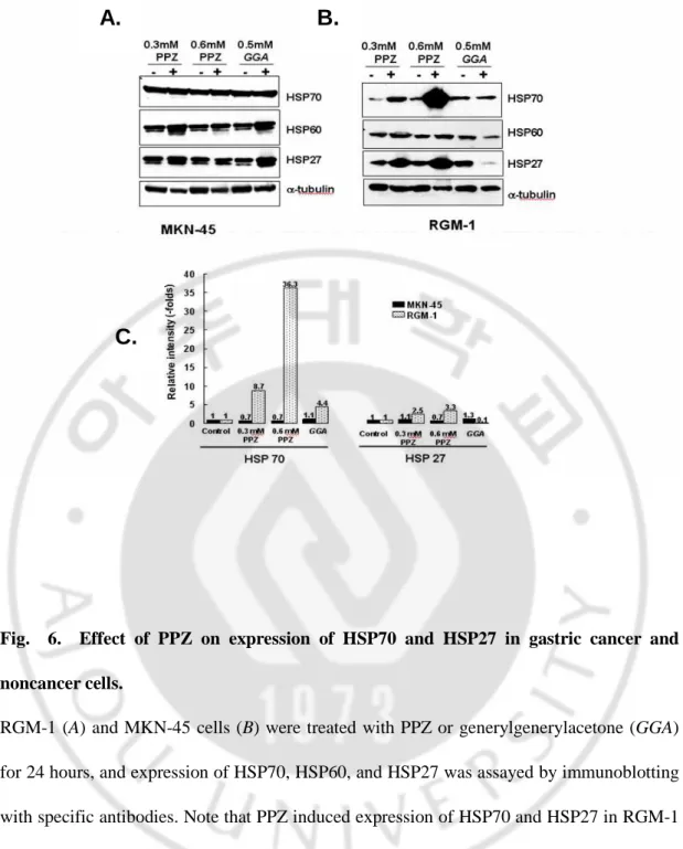
Interestingly, we found that HSP70 and HSP27 were overexpression in RGM-1 after PPZ treatment but not in MKN-45 cells (Fig. 6A and B). Notably, treatment of 0.6 mmol/L PPZ in RGM-1 cells showed a 3.3- and 36.3-fold increased of HSP27 and HSP70 compared with that of nontreated cells, respectively (Fig. 6C). However, HSP27 and HSP70, as well as HSP60, were not grossly changed in PPZ-treated gastric cancer MKN-45 cells (Fig. 6A). To compare the HSP induction, we also treated geranylgeranylacetone (GGA), which is well known as a HSP inducer in gastric mucosal cells. GGA slightly increased HSP60 and HSP27 in MKN-45 cell, but deceased HSP60 and HSP27 in RGM-1. In comparisom with GGA, the ablilty of PPZ to induce HSP70 is much higher at 5.6-fold than GGA. These data suggest that overexpression of HSP27 and HSP70 might play a cytoprotective role in PPZ-induced apoptosis of normal gastric mucosal cells.
Fig. 6. Effect of PPZ on expression of HSP70 and HSP27 in gastric cancer and noncancer cells.
RGM-1 (A) and MKN-45 cells (B) were treated with PPZ or generylgenerylacetone (GGA) for 24 hours, and expression of HSP70, HSP60, and HSP27 was assayed by immunoblotting with specific antibodies. Note that PPZ induced expression of HSP70 and HSP27 in RGM-1 normal gastric mucosal cells but not in MKN-45 gastric cancer cells. Relative intensities of HSP70 and HSP27 compared with nontreated control cells were represented in C.
A.
B.
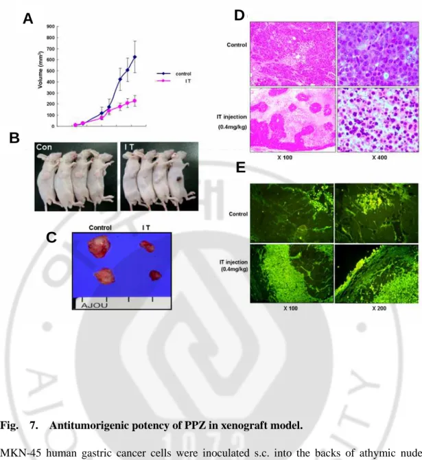
In a xenograft model of nude mice, administration of PPZ significantly inhibited tumorigenesis and induced large-scale apoptosis of tumor cells.
We also evaluated the antitumorigenic effects of PPZ in a gastric cancer xenograft model (Fig. 7). The animals were randomly divided into two groups: an intratumoral injection of PBS and an intratumoral injection of 0.4 ㎎/㎏ PPZ. Intratumor administration of PPZ significantly suppressed tumor growth in athymic nude mice, with decreased in tumor volume at day 22 of 44.69% compared with mice injected with PBS (Fig. 7A and B). The isolated tumor from mice with intratumor administration of PPZ was remarkably smaller than that of mice intramorally injected with PBS (Fig. 7C). Histopathological examination revealed that PPZ treatment provoked considerable apoptosis. Only a few tumor cells survived, and most tumor cells were replaced by apoptosis cells (Fig. 7D). TUNEL staining also showed considerably higher apoptotic cell death in the PPZ-treated group compared with the control group (Fig. 7E).
Fig. 7. Antitumorigenic potency of PPZ in xenograft model.
MKN-45 human gastric cancer cells were inoculated s.c. into the backs of athymic nude mice. PPZ (40mg/kg) was injected intrautmorally (IT) 14 days after xenograft. Mice were sacrificed 22 days after tumor inoculation. Tumor volume was measured and represented (A
and B). The isolated tumors (C) were sectioned and processed for H&E staining (D, ×100
and ×400 magnification) and TUNEL assay (E, ×100 and ×200 magnification); bars, ±SD.
A
B
C
D
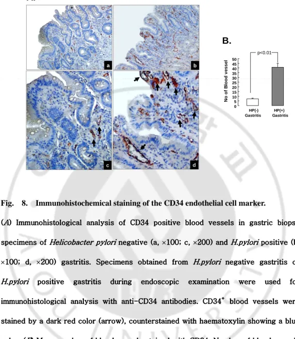
Distinct expression of CD34+ blood vessels between H.pylori positive and negative gastric patients
Stomach tissue samples were obtained from gastritis patients during endoscopy examination and were evaluated histologically by haematoxylin-eosin staining. Finally, we choose 20 H.pylori positive gastritis and 18 H.pylori negative gastritis cases with a similar degree of gastritis, scored according to the modified Sydney classification, as the degree of gastric inflammations itself can affect angiogenesis. Immunohistochemical staining using antibodies against the CD34 endothelial cell marker was performed to evaluate the difference in angiogenesis according to H.pylori infection. The results showed that patients with
H.pylori positive gastritis (Fig. 8A (b, d)) showed significantly higher expression of CD34
positive blood vessels in the gastric mucosa layer than that of patients with H.pylori negative gastritis (Fig. 8A (a, c)). The number of blood vessels was counted in three sites, for each specimen, with equal dimensions, and mean levels are shown in Fig. 8B. While H.pylori negative gastritis samples had a mean of 7.2 (SEM 0.8) blood vessels per x100 magnified field, H.pylori positive gastritis samples had a significant increased number (mean 40.9 (SEM 4.4)) of blood vessels (p<0.01). Moreover, blood vessels observed in cases with
H.pylori positive gastritis (Fig. 8A (b, d), arrow) were thicker and larger than those of H.pylori negative cases (Fig. 8A (a, c), arrow). Interestingly, blood vessels were found more
abundantly in the mesenchymal stromal layer below the mocosa layer but the number of blood vessels in the stromal layer was not different between H.pylori positive gastritis and
H.pylori negative gastritis cases, suggesting that H.pylori infection may be associated with
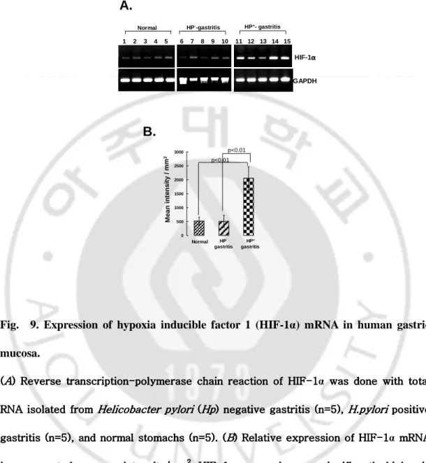
We then evaluated if there were any differences in expression of HIF-1α, the potent angiogenic transcriptional factor (Fig. 9). As a control, we used gastric biopsies obtained from five cases diagnosed with functional dyspepsia with no significant abnormal gastroscopic findings and no H.pylori infection. Compared with HIF-1α mRNA from five normal stomachs, expression was not altered in five H.pylori negative gastritis but was significantly increased in H.pylori positive gastritis (p<0.01), suggesting that HIF-1α is responsible for H.pylori induced angiogenesis (Fig. 9B).
0 5 10 15 20 25 30 35 40 45 50 HP(-) HP(+) Gastritis Gastritis N o o f B lo o d v e s s e l a b
A.
B.
p<0.01 c dFig. 8. Immunohistochemical staining of the CD34 endothelial cell marker.
((((AAAA) Immunohistological analysis of CD34 positive blood vessels in gastric biopsy ) Immunohistological analysis of CD34 positive blood vessels in gastric biopsy ) Immunohistological analysis of CD34 positive blood vessels in gastric biopsy ) Immunohistological analysis of CD34 positive blood vessels in gastric biopsy specimens of
specimens of specimens of
specimens of Helicobacter Helicobacter Helicobacter Helicobacter pyloripyloripylori negative (a, pylori negative (a, negative (a, ×100; c, negative (a, 100; c, ×200) and 100; c, 100; c, 200) and 200) and 200) and H.pyloriH.pyloriH.pyloriH.pylori positive (b, positive (b, positive (b, positive (b,
×100; d, 100; d, 100; d, 100; d, ×200) gastritis. Specimens obtained from 200) gastritis. Specimens obtained from 200) gastritis. Specimens obtained from 200) gastritis. Specimens obtained from H.pyloriH.pyloriH.pyloriH.pylori negative gastritis or negative gastritis or negative gastritis or negative gastritis or H.pylori
H.pyloriH.pylori
H.pylori positive gastritis during endoscopic examination were positive gastritis during endoscopic examination were used for positive gastritis during endoscopic examination were used for positive gastritis during endoscopic examination were used for used for immunohistological analysis with anti
immunohistological analysis with antiimmunohistological analysis with anti
immunohistological analysis with anti----CD34CD34CD34 antibodies. CD34CD34 antibodies. CD34 antibodies. CD34 antibodies. CD34++++ blood vessels were blood vessels were blood vessels were blood vessels were
stained by a dark red color (arrow), counterstained with haematoxylin showing a blue stained by a dark red color (arrow), counterstained with haematoxylin showing a blue stained by a dark red color (arrow), counterstained with haematoxylin showing a blue stained by a dark red color (arrow), counterstained with haematoxylin showing a blue color. (
color. (color. (
color. (BBB) Mean number of blood vessels stained with CD34. Number of blood vessels B) Mean number of blood vessels stained with CD34. Number of blood vessels ) Mean number of blood vessels stained with CD34. Number of blood vessels ) Mean number of blood vessels stained with CD34. Number of blood vessels with equal dimensions were counted in triplicate f
with equal dimensions were counted in triplicate fwith equal dimensions were counted in triplicate f
with equal dimensions were counted in triplicate for each sample and are represented or each sample and are represented or each sample and are represented or each sample and are represented as means (SD). There was a statistically significant difference between the two groups as means (SD). There was a statistically significant difference between the two groups as means (SD). There was a statistically significant difference between the two groups as means (SD). There was a statistically significant difference between the two groups
(p<0.01). (p<0.01). (p<0.01).
(p<0.01). Hp, Helicobacter pyloriHp, Helicobacter pyloriHp, Helicobacter pylori.Hp, Helicobacter pylori...
Normal HP--gastritis HP+- gastritis
HIF-1αααα GAPDH 1 2 3 4 5 6 7 8 9 10 11 12 13 14 15
A.
0 500 1000 1500 2000 2500 3000 Normal HP¯ gastritis HP+ gastritis M e a n i n te n s it y / m m 2B.
p<0.01 p<0.01Fig. 9. Expression of hypoxia inducible factor 1 (HIF-1α) mRNA in human gastric mucosa.
((((AAAA) Reverse transcription) Reverse transcription) Reverse transcription) Reverse transcription---polymerase chain reaction of HIF-polymerase chain reaction of HIF-polymerase chain reaction of HIFpolymerase chain reaction of HIF---1111α was done with total was done with total was done with total was done with total RNA isolated from
RNA isolated from RNA isolated from
RNA isolated from Helicobacter pyloriHelicobacter pyloriHelicobacter pyloriHelicobacter pylori ( ( ( (HpHp) negative gastritis (n=5), HpHp) negative gastritis (n=5), ) negative gastritis (n=5), H.pylori) negative gastritis (n=5), H.pyloriH.pyloriH.pylori positive positive positive positive gastritis (n=5), and normal stomachs
gastritis (n=5), and normal stomachs gastritis (n=5), and normal stomachs
gastritis (n=5), and normal stomachs (n=5). ((n=5). ((n=5). ((n=5). (BBB) Relative expression of HIFB) Relative expression of HIF-) Relative expression of HIF) Relative expression of HIF---1111α mRNA mRNA mRNA mRNA is represented as mean intensity/mm
is represented as mean intensity/mmis represented as mean intensity/mm
is represented as mean intensity/mm2222. HIF. HIF. HIF. HIF--1--11α expression was significantly higher in 1 expression was significantly higher in expression was significantly higher in expression was significantly higher in
gastric mucosa of gastric mucosa of gastric mucosa of
gastric mucosa of H. pyloriH. pyloriH. pyloriH. pylori positive gastritis than in positive gastritis than in positive gastritis than in H.pylori positive gastritis than in H.pyloriH.pylori negative gastritis in spite H.pylori negative gastritis in spite negative gastritis in spite negative gastritis in spite of similar expression of GAPDH (p<
of similar expression of GAPDH (p<of similar expression of GAPDH (p< of similar expression of GAPDH (p<0.01). 0.01). 0.01). 0.01).
Suppression of H.pylori induced in vitro angiogenesis by gastric PPZ
To prove a direct effect of H.pylori infection on angiogenesis, we performed an in vitro angiogenesis assay. Conditioned media obtained from H.pylori infected AGS cells were added to HUVEC culture flasks and morphological changes in the endothelial cells were observed. After nine days, HUVEC became long in shape and formed a tubular structure (Fig. 10B) compared with conditioned media of non-H.pylori infected AGS (Fig. 10A). CD31 immunofluorescence staining showed a dense intensity of CD31 molecules in HUVEC cells incubated with the culture supernatants of H.pylori infected AGS (Fig. 10C) while control media obtained from non-H.pylori infected AGS stimulated neither tubular formation of HUVEC nor expression of the CD31 endothelial cell marker (Fig. 10A, and F).
Results of in vitro angiogenesis assay strongly suggested that H.pylori infection stimulated infected gastric epithelial cells to secrete proangiogenic factors which induce growth and differentiation of endothelial HUVEC. Interestingly, pretreatment with PPZ (200µM or 400µM, for eight hours) on AGS cells prior to H.pylori inoculation significantly inhibited tubular formation (Fig. 10D, and E) and CD31 expression of endothelial HUVEC (Fig. 10I, and J). However, no significant changes were noted in HUVEC incubated with 400µM PPZ alone (Fig. 10C, and H), suggesting that PPZ itself did not influence tubular formation of HUVEC. The data clearly indicate that PPZ suppressed H.pylori induced in vitro angiogenesis, suggesting that antiangiogenic treatment with PPZ could be a promising therapeutic approach for H.pylori associated carcinogenesis.
Control H. pylori (24 h) H. pylori (24 h) +PPI (200 µµµµM, 8 h) H. pylori (24 h) +PPI (400 µµµµM, 8 h) P h a s e -c o n tr a s t αααα -C D 3 1 A b A B C D E F G H I J PPI (400 µµµµM, 8 h)
Fig. 10. In vitro angiogenesis assay.
AGS cells (1×10⁷ cells/100mm2 culture dish) were incubated with 0, 200, or 400 µM proton pump inhibitor (PPZ) for eight hours, washed with phosphate buffered saline three time, and inoculated with Helicobacter pylori (5×10⁷ CFU/ml) for 24 hours. Conditioned media were prepared from 1:1 dilution of the cell culture supernatant and the human umbilical vein endothelial cell (HUVEC) medium. Conditioned media were filtered through a 0.4 µM pore filter to remove H.pylori and then added to the HUVEC culture which was change every three days. After nine days, HUVEC were observed in a tubular formation under microscopy (A-E) and expression of the endothelial cell marker, CD31, was confirmed by immunocytofluorescence staining (F-J). (A, F) Control HUVEC cells; (B, G) HUVEC cells
incubated with conditioned media of H.pylori infected AGS; (C, H) HUVEC cells incubated with conditioned media of 400 µM PPZ treated AGS; (D, I) HUVEC cells incubated with conditioned media of 200 µM PPZ/H.pylori infected AGS; (E, J) HUVEC cells incubated with conditioned media of 400 µM PPZ/H.pylori infected AGS.
Production of proangiogenic factors from H.pylori infected gastric epithelium and its inhibition by PPZ
Following H.pylori infection, AGS cells significantly secreted VEGF and IL-8, well characterized as proangiogenic factors, in a time dependent manner (Fig. 11A). Maximal induction of IL-8 (mean 1019 (SEM 278) pg/ml) and VEGF (1597 (94) pg/ml) was observed after 24 hours of inoculation (Fig. 11A). We also examined mRNA expression of these angiogenic factors using RT-PCR analysis (Fig. 11B). Expression of VEGF mRNA, one of the HIF-1α target genes, was induced after 16 hours of H.pylori infection, showing the correlation with HIF-1α expression (Fig. 11B). IL-8 mRNA was also significantly induced after H.pylori infection (Fig. 11B). All of these results suggest that synthesis of angiogenic epithelial cells, which could induce proliferation and differentiation of endothelial cells.
Because PPZ showed a strong antiangiogenic action in the in vitro angiogenesis assay (Fig. 10), we measured the effect of PPZ on expression of these angiogenic factors (Fig. 12). Secretion of IL-8 in the supernatants of H.pylori infected AGS cells was found to be remarkably suppressed after PPZ treatment in a dose dependent manner (Fig. 12A). Following eight hours of infection with H.pylori, IL-8 production increased up to 870 pg/ml but this increment in IL-8 production was significantly attenuated by PPZ pretreatment. Pretreatment with PPZ showed a considerable regulatory effect on H.pylori mediated VEGF synthesis (Fig. 12A). Suppression of thses angiogenic factors by PPZ was evidenced by transcriptional inhibition of the genes (Fig. 12B). At 400 µM of PPZ, expression of VEGF and HIF-1α seemed to decline relevant to that of control AGS cells.
A.
0 200 400 600 800 1000 1200 1400 1600 1800 2000 IL -8 ( p g /m l)Times after H. pylori
0 0.5 1 2 4 8 16 24 h V E G F ( p g /m l) 0 200 400 600 800 1000 1200
Times after H. pylori
HIF-1αααα VEGF IL-8 GAPDH 0 1 2 4 8 16 24 h
B.
0.5 1 1.5 0.5 1 1.5 0.5 1 1.5 2 2.5 3Times after H. pylori (h) 0 1 2 4 8 16 24
HIF-1αααα
Times after H. pylori (h) 0 1 2 4 8 16 24
VEGF
Times after H. pylori (h) 0 1 2 4 8 16 24 IL-8 R e la ti v e in te n s it y ( -f o ld s ) R e la ti v e in te n s it y ( -f o ld s ) R e la ti v e in te n s it y ( -f o ld s )
Times after H. pylori
0 0.25 0.5 1 2 4 8 24h
C.
Fig. 11. Release and expression of vascular endothelial growth factor (VEGF) and interleukin 8 (IL-8) from Helicobacter pylori infected AGS cells.
(A) Production of VEGF (top) and IL-8 (bottom) was measured by ELISA with the culture supernatant of AGS cells infected with H.pylori for the indicated times. (B) Induction of hypoxia inducible factor 1 (HIF-1α), VEGF, and IL-8 mRNA by H.pylori infection was tested by reverse transcription-polymerase chain reaction analysis in AGS cells incubated with the bacterium for the indicated times. (C) Relative band intensity is presented as fold ratio.
IL -8 ( p g /m l) 00 100 200 300 400 500 600 700 800 900 1000 GAPDH HIF-1αααα VEGF H. pylori (16 h) - + + + + PPI (µM, 8h) - - 100 200 400
B.
A.
0 100 200 300 400 500 600 700 800 900 1000 V E G F ( p g /m l) - + + + + + H. pylori (16 h) - - 50 100 200 400 PPI (µM, 8 h) - + + + + + H. pylori (16 h) - - 50 100 200 400 PPI (µM, 8 h)Fig. 12. Effects of proton pump inhibitor (PPI) on expression of angiogenic growth factors interleukin 8 (IL-8), hypoxia inducible factor 1 (HIF-1α), and vascular endothelial growth factor (VEGF).
(A) To examine the inhibitory effect of PPI on (A) To examine the inhibitory effect of PPI on (A) To examine the inhibitory effect of PPI on
(A) To examine the inhibitory effect of PPI on Helicobacter pyloriHelicobacter pyloriHelicobacter pylori induced angiogenic Helicobacter pylori induced angiogenic induced angiogenic induced angiogenic growth factor expression, AGS cells (1
growth factor expression, AGS cells (1growth factor expression, AGS cells (1 growth factor expression, AGS cells (1×101010107777
cells/100 mm cells/100 mm cells/100 mm cells/100 mm2222
culture dish) were culture dish) were culture dish) were culture dish) were incubated with 0, 50, 100, 200, or 400
incubated with 0, 50, 100, 200, or 400 incubated with 0, 50, 100, 200, or 400
incubated with 0, 50, 100, 200, or 400 µM PPI for eight hours, washed with phosphateM PPI for eight hours, washed with phosphateM PPI for eight hours, washed with phosphateM PPI for eight hours, washed with phosphate buffered saline three times, and inoculated with
buffered saline three times, and inoculated with buffered saline three times, and inoculated with
buffered saline three times, and inoculated with H.pyloriH.pyloriH.pyloriH.pylori (5 (5 (5 (5×101010107 7 7 7
CFU/ml) for 16 hours. CFU/ml) for 16 hours. CFU/ml) for 16 hours. CFU/ml) for 16 hours. Production of IL
Production of ILProduction of IL
Production of IL---8 (top) and VEGF (bottom) was measured in culture supernatant of -8 (top) and VEGF (bottom) was measured in culture supernatant of 8 (top) and VEGF (bottom) was measured in culture supernatant of 8 (top) and VEGF (bottom) was measured in culture supernatant of the cells by ELISA. (B) Total RNA extracte
the cells by ELISA. (B) Total RNA extractethe cells by ELISA. (B) Total RNA extracte
the cells by ELISA. (B) Total RNA extracted from cells was used in the reverse d from cells was used in the reverse d from cells was used in the reverse d from cells was used in the reverse transcription
transcriptiontranscription
Expression of H.pylori induced angiogenic factors is mediated by activation of ERK1/2
As VEGF and IL-8 expression was found to be regulated by MAPK and NKκB on
H.pylori induced angiogenesis using specific inhibitors (Fig. 13). PD098059 (50µM), one of
the ERK inhibitors, strongly inhibited H.pylori induced HIF-1α and VEGF expression, and SB203580 (10µM), a p38 inhibitor, was also able to inhibit expression of these angiogenic factors. PDTC (ammonium salt, 100µM) a NFκB inhibitor, potently suppressed HIF-1α and VEGF expression induced by H.pylori. BAY11-7082 (5µM) reversibly increased expression of the genes. These data suggest that H.pylori induced VEGF induction was mediated via the MAPK pathway, and partially by the NFκB pathway.
Fig. 13. Involvement of mitogen activated protein kinase and nuclear factor κB (NFκB) in Helicobacter pylori induced mRNA expression of hypoxia inducible factor 1 (HIF-1α) and vascular endothelial growth factor (VEGF).
Prior to inoculation with H.pylori, AGS cells were treated with each inhibitor (50 µM PD08059, 10 µM SB203580, 100 µM PDTC, or 5 µM BAYII-7082 (BAY)) for eight hours, and their effects on H.pylori induced HIF-1α and VEGF expression were evaluated by reverse transcription-polymerase chain reaction. PD, PD098059, extracellular signal regulated kinase (ERK)1/2 inhibitor; SB, SB203580, p38 inhibitor; PDTC, 1-pyrrolidinecarbodithioic acid, ammonium salt, NFκB inhibitor; BAY, BAYII-7082, NFκB inhibitor.