ORIGINAL ARTICLE
YKL-40 in Induced Sputum After Allergen
Bronchial Provocation in Atopic Asthma
JH Lee, KH Park, JW Park, CS Hong
Division of Allergy and Immunology, Department of Internal Medicine, Institute of Allergy, Yonsei University
College of Medicine, Seoul, Republic of Korea
■ Abstract
Background: Serum chitinase-like proteins such as YKL-40 in asthmatic patients are known to positively correlate with disease severity
but controversy remains regarding their role. The allergen bronchial provocation test (ABPT) can induce allergic airway infl ammation in individuals with atopic asthma.
Objective: To evaluate the induction and kinetics of YKL-40 during allergen-induced airway infl ammation in atopic asthmatics.
Methods: Thirteen patients were enrolled from May to November 2008. They all underwent ABPT with Dermatophagoides farinae crude
extract. Induced sputums (IS) and serum were collected 3 times: 7 days before ABPT (baseline), 7 hours after ABPT, and 24 hours after ABPT. We examined the cytology of induced sputum (IS) and measured levels of YKL-40, interleukin (IL) 4, IL-5, IL-13, IL-33, tumor necrosis factor (TNF) _, and eosinophilic cationic protein (ECP) in IS and/or serum.
Results: Following ABPT, total infl ammatory cells, eosinophils, and neutrophils increased in a time-dependent manner in IS. YKL-40 levels
were increased in IS but not in serum at 7 or 24 hours after ABPT (P=.011 and P=.006, respectively). Similarly to YKL-40, IL-5 and ECP levels were also increased in IS at 7 and 24 hours after ABPT (P=.011 for IL-5 and P=.006 for ECP). Overall, YKL-40 levels were well correlated with ECP levels in IS (l=0.576, P<.001).
Conclusions: YKL-40 levels increased immediately in IS but not in the serum of atopic asthmatics. The correlation between YKL-40 levels
and ECP in IS suggests that YKL-40 may play a pathophysiologic role in human atopic asthma.
Key words: Chitinase-like protein. Allergen bronchial provocation test. Asthma. Allergic infl ammation. Induced sputum.
■ Resumen
Antecedentes y objetivo: Las proteinas séricas "chitinase-like" como YKL-40 podrían correlacionarse en pacienes asmáticos con la severidad
de la enfermedad. El objetivo de este estudio es evaluar la producción de YKL-40 y su cinética tras la provocación bronquial con alérgeno (PBA), una prueba que induce una potente respuesta infl amatoria en sujetos afectos de asma bronquial alérgica.
Metodos: Se incluyeron en el estudio un total de 13 pacientes asmáticos en un periodo comprendido entre mayo y noviembre del 2008.
a todos ellos se les realizá la PBA utilizando un extracto de Dermatophagoides farinae. Se obtuvieron muestras de suero y de esputo inducido (EI) en tres ocasiones — 7 días antes de la PBA (situación basal), y 7 y 24 horas tras la PBA. Se realizó un análisis citológico de las muestras de EI, y en el sobrenadante se cuantifi caron los niveles de YKL-40, interleuquina 4 (IL) 4, IL-5, IL-13, IL-33, factor de necrosis tumoral (TNF) _, y de la proteina catiónica del eosinófi lo (ECP). Estas determinaciones también se realizaron en las muestras de suero.
Resultados: tras la PBA se observó un incremento marcado en función del tiempo en el total de células infl amatorias, eosinófi los y
neutrófi los en las muestras de EI. Los niveles de YKL-40 también se incrementaron en el EI a las 7 y 24 horas de la PBA (p=0,011 y
p=0.006, respectivamente), mientras que no se modifi caron los niveles séricos. Los niveles de IL-5 y ECP en EI también se incrementaron
signifi cativamente (p=0,011 para la IL-5 y p=0,006 para la ECP) tanto a las 7 como a las 24 horas de la PBA. Se constató una correlación entre los niveles YKL-40 y ECP en EI (l=0,576, p<0,001).
Conclusiones: Los niveles de YKL-40 se incrementas de forma inmediata tras la provocación en EI de pacientes asmáticos alérgicos, y no así
en el suero. La correlación presente entre los niveles de ECP e YKL-40 en esputo, sugeriría un posible papel deYKL-40 en la fi siopatológía del asma alérgica.
Introduction
YKL-40, also called human cartilage glycoprotein 39 (HcGP-39), is a chitinase-like protein that was À rst discovered in synovial Á uid; its murine homolog is known as BRP-39 [1,2]. YKL-40 is known to be associated with a number of human inÁ ammatory diseases, such as rheumatoid arthritis [3], osteoarthritis [4], sarcoidosis [5], inflammatory bowel disease [6], hepatic À brosis [7], and several malignant tumors [8]. YKL-40 levels were recently shown to be related to endocrine diseases including obesity [9], diabetes mellitus, and atherosclerosis [10]. In addition, elevated YKL-40 levels have been reported in the serum, sputum [11], and bronchoalveolar lavage (BAL) Á uid [12] of patients with chronic obstructive pulmonary disease (COPD). The relationship between YKL-40 and various human diseases suggests that this protein could be a useful diagnostic and prognostic biomarker.
The activation of type 2 helper (TH2) lymphocytes and
their subsequent secretion of interleukin (IL) 13 are well-established key responses in the pathogenesis of allergic airway inÁ ammation [13]. Induction of IL-13 secretion results in the alternative activation of alveolar macrophages, and these activated macrophages have been reported to play a role in the propagation of inÁ ammation [14]. Respiratory epithelial cells, alveolar macrophages, and neutrophils are known to secrete YKL-40 in human airways, and hence, YKL-40 has been attributed a role in airway inÁ ammation [15]. Furthermore, an animal model study has shown that YKL-40 may play a critical role in various steps of asthma pathogenesis by increasing allergic sensitization, propagating TH2 inÁ ammation, and
regulating inÁ ammatory cell apoptosis [16].
In this study, we evaluated dynamic changes in YKL-40 levels in induced sputum (IS) following an allergen bronchial provocation test (ABPT) with house dust mite extract (Dermatophagoides
farinae [DF]) in the hope that our À ndings would shed light on
the mechanisms underlying atopic asthma [17].
Methods
Patients
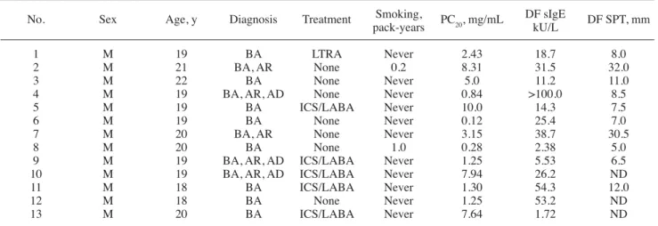
Thirteen patients who visited Severance Hospital to have their allergic asthma presumptively diagnosed by a physician were enrolled between May and November 2008. The Institutional Review Board of Yonsei University approved the study (IRB no. 4-2012-0002) and informed consent was obtained from all patients. According to international guidelines [18], the diagnosis of allergic asthma was based on clinical history, symptoms and signs, the methacholine bronchial provocation test, eosinophil count in IS, and speciÀ c immunoglobulin (Ig) E measurements by skin prick testing (Allergopharma) and/or the ImmunoCAP test (Pharmacia). DF-speciÀ c IgE was documented in all patients. All participants underwent ABPT with DF extract. IS and serum were collected 3 times: 7 days before ABPT (baseline) and at 7 and 24 hours after the À rst dose of allergen administration (Table 1).
Allergen Bronchial Provocation Test
DF whole body extract (1:100 w/v, containing 3.4 +g/mL of
Der f 1) cultured at the Institute of Allergy, Yonsei University
was used for ABPT. To prepare the inhalation material, a 1:100 extract of DF in phosphate buffered saline (PBS) was À ltered with an antiseptic À lter (Millipore) and diluted 10-, 25-, and 100-fold with isotonic saline. A baseline pulmonary function test was performed using a pneumotachometer system with a Lilly head (MasterScreen system, Erich Jaeger Co.), which measures maximum expiratory Á ow volume. The spirometric Á ow-volume curve was obtained according to international criteria [19].
ABPT was performed using the method described by Spector et al [20], with some modiÀ cations. First, DF solution (1:10 000 w/v) containing 34 ng/mL of Der f 1 was administered using a handheld nebulizer (Devilbiss
Table 1. Demographic and Laboratory Data of Patients Included in the Study
No. Sex Age, y Diagnosis Treatment Smoking, PC
20, mg/mL DF sIgE DF SPT, mm
pack-years kU/L
1 M 19 BA LTRA Never 2.43 18.7 8.0
2 M 21 BA, AR None 0.2 8.31 31.5 32.0
3 M 22 BA None Never 5.0 11.2 11.0
4 M 19 BA, AR, AD None Never 0.84 >100.0 8.5
5 M 19 BA ICS/LABA Never 10.0 14.3 7.5
6 M 19 BA None Never 0.12 25.4 7.0
7 M 20 BA, AR None Never 3.15 38.7 30.5
8 M 20 BA None 1.0 0.28 2.38 5.0
9 M 19 BA, AR, AD ICS/LABA Never 1.25 5.53 6.5
10 M 19 BA, AR, AD ICS/LABA Never 7.94 26.2 ND
11 M 18 BA ICS/LABA Never 1.30 54.3 12.0
12 M 18 BA None Never 1.25 53.2 ND
13 M 20 BA ICS/LABA Never 7.64 1.72 ND
Abbreviations: AD, atopic dermatitis; AR, allergic rhinitis; BA, bronchial asthma; DF, Dermatophagoides farinae; ICS, inhaled corticosteroid; LABA, long-acting ß2-agonist; LTRA, leukotriene receptor antagonist; ND, not done; PC20, methacholine provocation concentration for 20% decrease in forced
646, Devilbiss Healthcare Inc.) with tidal breathing for 2 minutes. After administration, spirometric data including forced expiratory volume in the À rst second (FEV1) were
obtained. A positive test was deÀ ned as a 20% decrease in FEV1 from baseline. If a negative result was observed, the next
concentration was administered up to 1:1000 diluents. After the À nal dose was administered for 2 minutes, serial spirometry was performed at 10, 20, and 30 minutes and at 1, 2, 4, 5, 6, and 7 hours. A signiÀ cant decrease in FEV1 60 minutes after
the À nal DF inhalation was deÀ ned as an early response, while a signiÀ cant decrease after 4 hours was deÀ ned an isolated late response. If both early and late responses were observed, a dual response was recorded. Patients who did not show any signiÀ cant spirometric changes after 7 hours of spirometric monitoring were considered nonresponders.
Induced Sputum Analysis
We used a previously reported method for sputum induction [21] with some modiÀ cations for processing [22]. Briefly, we checked each patient’s peak flow rate (PFR) with a mini-Wright peak Á ow meter. After administration of bronchodilator (salbutamol 200 +g, 2 puffs), sputum induction was performed with 3% hypertonic saline inhalation for 10 minutes. PFR was checked again if the patient complained of dyspnea. Sputum induction was stopped when a 20% decrease in PFR from baseline was observed. To prevent contamination with squamous epithelial cells, sputum expectoration and collection were performed after cleaning of the oral and nasal cavities.
Induced sputum samples were processed immediately as follows. Mucus components were collected separately from saliva, and the weight and volume of selected samples
Eosinophil cationic protein (ECP) was also measured in IS with the commercially available ImmunoCAP assay (Phadia). The levels of other inÁ ammatory cytokines, including interleukin (IL) 4 (detection limit, 10 pg/mL), IL-5 (detection limit, 3.0 pg/mL), IL-13 (detection limit,32 pg/mL), IL-33 (detection limit, 28 pg/mL), and tumor necrosis factor (TNF) _ (detection limit, 0.5 pg/mL) were also checked in IS and serum by ELISA (R&D Systems).
Statistical Analysis
All data were analyzed using SPSS statistical software (version 12.0). The Wilcoxon test was used for comparing medians, and the Spearman rank test was used for correlation analysis. Data are presented using means (SD).
Results
Cytological Analysis of Induced Sputum
Total cell count increased in a time-dependent manner after ABPT (baseline, 384.7 [407.3] ×103 cells/mL; 7 hours, 611.9 [593.1] ×103 cells/mL; 24 hours, 1160.5 [1232.8] ×103
cells/mL). SigniÀ cant increases were also detected after this test for eosinophil count (baseline, 29.1 [49.4] ×103 cells/mL;
7 hours, 157.9 [223.7] ×103 cells/mL; 24 hours, 546.0 [685.7]
×103 cells/mL) and neutrophil count (baseline, 84.0 [81.9] ×103
cells/mL; 7 hours, 247.2 [269.8] ×103 cells/mL; 24 hours, 381.4
[658.5] ×103 cells/mL) (Figure 1). 1600 1200 800 400 0
Total Macrophages Neutrophils Eosinophils Lymphopcytes
Cell Numbers in Induced Sputum,
ⴒ
10
3/mL
7 days before ABPT 7 hours after ABPT 24 hours after ABPT
Figure 1. Infl ammatory cell counts in induced sputum. Total cell, neutrophil, and eosinophil counts increased signifi cantly in a time-dependent manner after the allergen bronchial provocation test (ABPT). All data are represented by mean (SEM). *: P<.05 compared to baseline, #: P<.05 compared to 7 hours after ABPT.
* *# *# * * * measured. Then, an equal amount of 0.1%
dithiothreitol (Sigma) was added to each mucus sample, vortexed for 15 seconds, and incubated in a warm water bath (37°C) with shaking for 20 minutes. The samples were centrifuged and the supernatant was collected for subsequent measurements and stored at ï70°C. Remnant cell components were resuspended in PBS and À ltered through a nylon mesh with 60-+m pores to eliminate cell clots. A 10-+L aliquot of resuspension solution was mixed with 10 +L of 0.4% trypan blue solution to count total cell numbers and measure cell viability using a Neubauer counting chamber. Samples were then diluted in PBS to a concentration of 1×10⁶ cells/mL,
and cytospin was performed at 450 rpm for 6 minutes to prepare cytology slides. After staining the slides with Wright’s stain, over 400 inÁ ammatory cells were counted differentially under a light microscope.
Measurement of YKL-40, Eosinophil Cationic Protein, and Cytokines
YKL-40 levels were measured in IS and serum by enzyme-linked immunosorbent assay (ELISA) (Quidel) (detection limit, 20 ng/mL) [15].
100 80 60 40 20 0 300 250 200 150 100 0 50 140 120 100 80 60 40 20 0 700 600 500 400 300 0 200 100 120 100 80 60 40 0 20 50 40 30 20 10 0 2500 2000 1500 1000 500 0 Induced Sputum
Induced Sputum Serum Induced Sputum Serum Induced Sputum Serum
YKL-40, ng/mL IL-13, ng/mL IL-5, ng/mL IL-33, ng/mL IL-4, ng/mL TNF-_ , ng/mL ECP , ng/mL
A
E
B
F
C
G
D
Figure 2. Enzyme-linked immunosorbent assay results of YKL-40, infl ammatory cytokines, and eosinophil cationic protein (ECP) by immunoCAP in induced sputum (IS) and serum. YKL-40 (A), IL-5 (B), and ECP (D) levels increased signifi cantly in a time-dependent manner in IS but not in serum after the allergen bronchial provocation test (ABPT). Interleukin (IL) 4 (C), IL-13 (E), IL-33 (F), and tumor necrosis factor (TNF) _ (G) levels were not changed in IS or serum. All data are represented as means (SEM).
*P<.05 compared to baseline; #P<.05 compared to 7 hours after ABPT. *# * *# *# 1000 100 10 1 1000 100 10 1 1 10 100 1000 10 000 1 10 100 1000 10 000 l=0.576 P<.001 l=0.431 P<.006
ECP in Induced Sputum, +g/L Eosinophil Count in Induced Sputum, ⴒ103/mL
YKL-40 in Induced Sputum,
ng/mL
Figure 3. Scatter plots of YKL-40 vs eosinophil cationic protein (ECP) (A) and eosinophil count (B) in induced sputum for all paired samples. Each plot shows a substantial positive correlation. Closed squares represent baseline, closed triangles represent 7 hours after the allergen bronchial provocation test (ABPT), and closed circles represent 24 hours after ABPT.
Changes in YKL-40 Levels in IS and Serum After ABPT
YKL-40 levels in IS were increased signiÀ cantly at 7 and 24 hours after ABPT (baseline, 18.7 [11.8] ng/mL; 7 hours, 37.8 [29.7] ng/mL; 24 hours, 65.0 [52.9] ng/mL). However, there were no signiÀ cant differences in serum levels (baseline, 53.0 [23.7] ng/mL; 7 hours, 53.1 [34.4] ng/mL; 24 hours, 55.9 [30.6 ] ng/mL) (Figure 2A).
Changes in Cytokine and ECP Levels in IS and Serum After ABPT
Only IL-5 levels were signiÀ cantly increased in IS at 24 hours after ABPT (baseline, 6.6 [9.6] pg/mL; 7 hours, 12.1 [13.1] pg/mL; 24 hours, 29.3 [24.7] pg/mL) (Figure 2B). IL-4 was not detected in the samples and there were no signiÀ cant differences in IL-13, IL-33, or TNF-_ levels in either IS or serum (Figure 2C, E, F, G). ECP levels were signiÀ cantly increased in IS at 7 hours and 24 hours after ABPT (baseline, 96.4 [85.5] +g/mL; 7 hours, 380.9 [634.5] +g/mL; 24 hours, 1520.8 [2126.5] +g/mL) (Figure 2D).
Correlation Analysis of YKL-40 Levels and Other Variables in Induced Sputum
Eosinophil count and ECP levels were positively correlated with YKL-40 levels in IS (l=0.431, P=.006 for eosinophil count; l=0.576, P<.001 for ECP levels) (Figure 3). IL-5 levels and other inÁ ammatory cell counts showed no signiÀ cant correlation (Table 2).
Discussion
Chitin, which consists of N-acetylglucosamine polymers, is the second most abundant polysaccharide in nature. It is an important component of the cell wall of fungi, the exoskeleton of shellÀ sh and insects, and the sheath of nematodes [23]. Thus, human beings must have chitinase to protect their bodies from many kinds of pathogens containing chitin. Chitinase is a highly conserved endogenous enzyme found in insects, mammals, and many other organisms. The 2 active chitinases in humans, acidic mammalian chitinase and chitotriosidase, both have enzymatic activity for the degradation of chitin [23].
YKL-40 is known as a chitinase-like protein because it can bind to chitin but is unable to degrade it. Associations between YKL-40 and allergic diseases have been reported. Chupp et al [15] described a positive correlation between elevated serum YKL-40 levels and asthma severity, and the
same group also reported that a genetic variation of the CHI3L1 gene encoding YKL-40 was signiÀ cantly associated with serum YKL-40 levels and asthma susceptibility [24]. Furthermore, a single nucleotide polymorphism in the promoter region of the
CHI3L1 gene has been associated with the atopic phenotype
in children [25].
In this study, we observed an increment in YKL-40 levels in IS but not in serum at 24 hours after ABPT. The main cell sources of YKL-40 are bronchial epithelial cells, macrophages [26], and neutrophils [27]. ABPT is used to evaluate allergen-induced inÁ ammation in the laboratory. Like other insects and spiders, house dust mites contain chitin in their exoskeleton. However, YKL-40 elevation in IS might be an independent response to chitin, as chitin-free allergens also promote YKL-40 elevation in BAL Á uid after administration of the segmental bronchial challenge test [28].
Our results strongly suggest that allergen exposure may rapidly induce YKL-40 secretion from epithelial cells, macrophages, and other cells. The same pattern of increase observed for YKL-40 was also observed for IL-5 and ECP in IS, suggesting that YKL-40 may increase allergic eosinophilic inÁ ammation by inducing the accumulation and activation of local dendritic cells and reducing apoptosis of TH2 cells [16].
In this study, we used dithiothreitol for mucolysis of IS and therefore cannot exclude the possibility that this may have affected the measurement of cytokines by ELISA. IL-13 plays a critical role in relation to YKL-40 as it can induce YKL-40 expression from epithelial cells and macrophages, and IL-13-induced À brosis may be mediated by YKL-40 [29]. Further studies are needed to better understand the relationship between YKL-40 and allergic inÁ ammation (exacerbated by allergens, viruses, smoking, or pollution) and tissue remodeling.
Our results are not consistent with those of Kuepper et al [28], who reported that YKL-40 levels in BAL Á uid were markedly increased 24 hours but not 10 minutes after segmental allergen challenge. In the present study, we also measured YKL-40 in serum but did not À nd any changes in YKL-40 levels after ABPT, thus, diminishing the possibility of using serum YKL-40 as a biomarker for uncontrolled asthma. In contrast to our results, Kuepper et al reported that YKL-40 in serum was deÀ nitely increased 24 hours after allergen challenge, and other investigators have also reported that serum YKL-40 levels are higher in exacerbated asthma [30] and severe asthma [15]. However, contradictory results have also been reported [31].
The present study has limitations. First, we did not check YKL-40 levels in serum or IS after more than 24 hours following ABPT. Second, long-lasting uncontrolled inÁ ammation can induce the persistent release of YKL-40 Table 2. Correlation Analysis of YKL-40 and Other Variables in Induced Sputum
ECP IL-5 Macrophages Neutrophils Eosinophils
Correlation coefÀ cient (rho) 0.576 0.304 -0.031 0.245 0.431
P value <.001 .060 .850 .132 .006
from bronchial epithelial cells, which could affect serum levels of YKL-40. ABPT is a good model for studying allergen-induced allergic inÁ ammation. However, viral infection and pollution, in addition to allergens, are also equally important aggravating and/or etiologic factors in real-life asthma. Thus, ABPT does not sufÀ ciently reÁ ect real clinical situations of asthma exacerbation. Some investigators have studied sputum YKL-40 levels in asthma and COPD patients and shown that YKL-40 and chitinase are increased in COPD patients but not in asthmatics compared to healthy controls [11,32]. Therefore, whether or not YKL-40 is clinically related to the pathogenesis of asthma remains to be elucidated.
In conclusion, YKL-40 levels in IS increased during the early phase of allergic inÁ ammation induced by allergen exposure. This À nding suggests that YKL-40 may play an important role in the initiation of allergic inÁ ammation and that it may be useful as an acute phase biomarker in allergen-induced asthma exacerbations.
Acknowledgments
This study was supported by a faculty research grant of Yonsei University College of Medicine for 6-2008-0161.
Previous Presentation: The abstract of the pilot study
of this article was presented as a poster at the World Allergy Congress (WAC) 2009 held in Buenos Aires, Argentina on December 6-10, 2009 (abstract number: 142).
References
1. Hakala BE, White C, Recklies AD. Human cartilage gp-39, a major secretory product of articular chondrocytes and synovial cells, is a mammalian member of a chitinase protein family. J Biol Chem. 1993;268(34):25803-10.
2. Rejman JJ, Hurley WL. Isolation and characterization of a novel 39 kilodalton whey protein from bovine mammary secretions collected during the nonlactating period. Biochem Biophys Res Commun. 1988;150(1):329-34.
3. Johansen JS, Stoltenberg M, Hansen M, Florescu A, Hrslev-Petersen K, Lorenzen I, Price PA. Serum YKL-40 concentrations in patients with rheumatoid arthritis: relation to disease activity. Rheumatology. 1999;38(7):618-26.
4. Volck B, Johansen JS, Stoltenberg M, Garbarsch C, Price PA, Ostergaard M, Ostergaard K, Lvgreen-Nielsen P, Sonne-Holm S, Lorenzen I. Studies on YKL-40 in knee joints of patients with rheumatoid arthritis and osteoarthritis. Involvement of YKL-40 in the joint pathology. Osteoarthritis Cartilage. 2001;9(3):203-14.
5. Johansen JS, Milman N, Hansen M, Garbarsch C, Price PA, Graudal N. Increased serum YKL-40 in patients with pulmonary sarcoidosis--a potential marker of disease activity? Respir Med. 2005;99(4):396-402.
6. Koutroubakis IE, Petinaki E, Dimoulios P, Vardas E, Roussomoustakaki M, Maniatis AN, Kouroumalis EA. Increased serum levels of YKL-40 in patients with infl ammatory bowel disease. Int J Colorectal Dis. 2003;18(3):254-9.
7. Johansen JS, Mller S, Price PA, Bendtsen F, Junge J, Garbarsch C, Henriksen JH. Plasma YKL-40: a new potential marker of fi brosis in patients with alcoholic cirrhosis? Scand J Gastroenterol. 1997;32(6):582-90.
8. Cintin C, Johansen JS, Christensen IJ, Price PA, Srensen S, Nielsen HJ. Serum YKL-40 and colorectal cancer. Br J Cancer. 1999;79(9-10):1494-9.
9. Hempen M, Kopp H, Elhenicky M, Hbaus C, Brix J, Koppensteiner R, Schernthaner G. YKL-40 is elevated in morbidly obese patients and declines after weight loss. Obes Surg. 2009;19(11):1557-63. 10. Rathcke CN, Vestergaard H. YKL-40, a new infl ammatory marker
with relation to insulin resistance and with a role in endothelial dysfunction and atherosclerosis. Infl amm Res. 2006;55(6):221-7. 11. Otsuka K, Matsumoto H, Niimi A, Muro S, Ito I, Takeda T, Terada
K, Yamaguchi M, Matuoka H, Jinnai M, Oguma T, Nakaji H, Inoue H, Tajiri T, Iwata T, Chin K, Mishima M. Sputum YKL-40 levels and pathophysiology of asthma and chronic obstructive pulmonary disease. Respiration. 2011. 2012;83(6):507-19. Epub 2011 Sep 27.
12. Ltuv S, Kozhich A, Arouche N, Grandsaigne M, Reed J, Dombret M, Kiener PA, Aubier M, Coyle AJ, Pretolani M. YKL-40 is elevated in patients with chronic obstructive pulmonary disease and activates alveolar macrophages. J Immunol. 2008;181(7):5167-73.
13. Huang SK, Xiao HQ, Kleine-Tebbe J, Paciotti G, Marsh DG, Lichtenstein LM, Liu MC. IL-13 expression at the sites of allergen challenge in patients with asthma. J Immunol. 1995;155(5):2688-94.
14. Martinez FO, Helming L, Gordon S. Alternative activation of macrophages: an immunologic functional perspective. Annu Rev Immunol. 2009;27:451-83.
15. Chupp GL, Lee CG, Jarjour N, Shim YM, Holm CT, He S, Dziura JD, Reed J, Coyle AJ, Kiener P, Cullen M, Grandsaigne M, Dombret MC, Aubier M, Pretolani M, Elias JA. A chitinase-like protein in the lung and circulation of patients with severe asthma. N Engl J Med. 2007;357(20):2016-27.
16. Lee CG, Hartl D, Lee GR, Koller B, Matsuura H, Da Silva CA, Sohn MH, Cohn L, Homer RJ, Kozhich A, Humbles A, Kearley J, Coyle A, Chupp G, Reed J, Flavell RA, Elias JA. Role of breast regression protein 39 (BRP-39)/chitinase 3-like-1 in Th2 and IL-13-induced tissue responses and apoptosis. J Exp Med. 2009;206(5):1149-66.
17. Melillo G, Cocco G, D'Amato G. Bronchial provocation tests in etiologic diagnosis of asthma. Bronchopneumologie. 1979;29(4):329-36.
18. Global Initiative for asthma. Global strategy for asthma management and prevention. 2002. p. 02-3659.
19. American Thoracic Society. Standardization of Spirometry, 1994 Update. American Thoracic Society. Am J Respir Crit Care Med. 1995;152(3):1107-36.
20. Spector S, Farr R. Bronchial inhalation challenge with antigens. J Allergy Clin Immunol. 1979;64(6 pt 2):580-6.
21. Kang SM, Kim CW, Park JW, Hong CS. Induced sputum study via inhalation of hyperosmolar saline to investigate airway infl ammation in asthma: method of induced sputum study and examination of infl ammatory cells. Korean J Med. 1997;52(6):797-804.
22. Kim CW, Kim HJ, Park JW, Hong CS. Matrix metalloproteinase-9/ tissue inhibitor of matrix metalloproteinase-1 in induced
Manuscript received April 30, 2012; accepted for publication, June 4, 2013..
Chein-Soo Hong
Division of Allergy and Immunology Department of Internal Medicine Yonsei University College of Medicine 50 Yonsei-ro, Seodaemun-gu
120-752 Seoul, Republic of Korea E-mail: [email protected]
sputum of bronchial asthmatics. J Asthma Allergy Clin Immunol. 2000;20(6):916-26.
23. Lee CG. Chitin, chitinases and chitinase-like proteins in allergic infl ammation and tissue remodeling. Yonsei Med J. 2009;50(1):22-30.
24. Ober C, Tan Z, Sun Y, Possick JD, Pan L, Nicolae R, Radford S, Parry RR, Heinzmann A, Deichmann KA, Lester LA, Gern JE, Lemanske RF, Jr., Nicolae DL, Elias JA, Chupp GL. Effect of variation in CHI3L1 on serum YKL-40 level, risk of asthma, and lung function. N Engl J Med. 2008;358(16):1682-91.
25. Sohn MH, Lee JH, Kim KW, Kim SW, Lee SH, Kim KE, Kim KH, Lee CG, Elias JA, Lee MG. Genetic variation in the promoter region of chitinase 3-like 1 is associated with atopy. Am J Respir Crit Care Med. 2009;179(6):449-56.
26. Renkema GH, Boot RG, Au FL, Donker-Koopman WE, StijlandA, Muijsers AO, Hrebicek M, Aerts JM. Chitotriosidase, a chitinase, and the 39-kDa human cartilage glycoprotein, a chitin-binding lectin, are homologues of family 18 glycosyl hydrolase secreted by human macrophages Eur J Biochem. 1998;251:504-9. 27. Volck B, Price PA, Johansen JS, Sorensen O, Benfi eld TL, Nielsen
HJ, Calafat J, Borregaard N. YKL-40, a mammalian member of the chitinase family, is a matrix protein of specifi c granules in human neutophils. Proc Assoc Am Physicians. 1998;110:351-60. 28. Kuepper M, Bratke K, Virchow JC. Chitinase-like protein and
asthma. N Engl J Med. 2008;358(10):1073-5.
29. Lee CG, Da Silva CA, Dela Cruz CS, Ahangari F, Ma B, Kang MJ, He CH, Takyar S, Elias JA. Role of chitin and
chitinase/chitinase-like proteins in infl ammation, tissue remodeling, and injury. Annu Rev Physiol. 2011;73:479-501.
30. Tang H, Fang Z, Sun Y, Li B, Shi Z, Chen J, Zhang T, Xiu Q. YKL-40 in asthmatic patients, and its correlations with exacerbation, eosinophils and immunoglobulin E. Eur Respir J. 2010;35(4):757-60.
31. Specjalski K, Jassem E. YKL-40 protein is a marker of asthma. J Asthma. 2011;48(8):767-72.
32. Seibold MA, Donnelly S, Solon M, Innes A, Woodruff PG, Boot RG, Burchard EG, Fahy JV. Chitotriosidase is the primary active chitinase in the human lung and is modulated by genotype and disease. J Allergy Clin Immunol. 2008;122(5):944-50.