저작자표시-비영리-변경금지 2.0 대한민국 이용자는 아래의 조건을 따르는 경우에 한하여 자유롭게 l 이 저작물을 복제, 배포, 전송, 전시, 공연 및 방송할 수 있습니다. 다음과 같은 조건을 따라야 합니다: l 귀하는, 이 저작물의 재이용이나 배포의 경우, 이 저작물에 적용된 이용허락조건 을 명확하게 나타내어야 합니다. l 저작권자로부터 별도의 허가를 받으면 이러한 조건들은 적용되지 않습니다. 저작권법에 따른 이용자의 권리는 위의 내용에 의하여 영향을 받지 않습니다. 이것은 이용허락규약(Legal Code)을 이해하기 쉽게 요약한 것입니다. Disclaimer 저작자표시. 귀하는 원저작자를 표시하여야 합니다. 비영리. 귀하는 이 저작물을 영리 목적으로 이용할 수 없습니다. 변경금지. 귀하는 이 저작물을 개작, 변형 또는 가공할 수 없습니다.
A Dissertation
for the Degree of Doctor of Philosophy
Studies on the development of
biomimetic microenvironment for
stem cell culture
줄기세포 배양에 적합한 생체모사
배양미세환경 개발 연구
June, 2018
By
Myungook Lee
Department of Agricultural Biotechnology
Graduate School, Seoul National University
Studies on the development of
biomimetic microenvironment for
stem cell culture
줄기세포 배양에 적합한 생체모사
배양미세환경 개발 연구
지도교수 임 정 묵
이 논문을 농학박사 학위논문으로 제출함
2018년 6월
서울대학교 대학원
농생명공학부 바이오모듈레이션 전공
이 명 욱
이명욱의 박사학위논문을 인준함
2018년 7월
위 원 장 (인)
부위원장 (인)
위 원 (인)
위 원 (인)
위 원 (인)
SUMMARY
This research describes the development of a three-dimensional (3D) system for in vitro culture of mouse embryonic stem cells (mESCs) and human adipose-derived stromal cells (hADSCs) in a biomimetic, non-cellular microenvironment.
Because it was assumed that each type of stem cell requires its own customized microenvironment in a biomimetic culture system, a 3D microenvironment was developed for mESCs and hADSCs, and the results were compared.
First, a method to isolate and analyze high quality oocytes from young and aged mice was developed. Second, integrin expression on the surface of inbred R1 and hybrid B6D2F1 mouse ESCs were screened to analyze cell-to-cell extracellular matrix (ECM) interactions before constructing a biomimetic microenvironment. After functional analysis of integrin heterodimers, we confirmed that the integrin heterodimers α6β1 and
αvβ1 actively functioned on the surface of undifferentiated mESCs of
both strains. For the next step, to construct a biomimetic microenvironment customized for self-renewal and proliferation of mESCs, we evaluated whether different types of mESCs needed different types of 3D synthetic scaffolds for cell self-renewal. In
this step, the mechanical strength of microenvironments were varied, illustrating that different mESCs required different cellular interactions in the microenvironment, which indicated that each cell line required customization of the mechanical properties of the microenvironment.
For hADSCs, similar but slightly different processes were performed. The hADSCs were isolated from adipose tissue and stabilized through culture processes and magnetic-activated cell sorting. Transcriptional and translational analyses showed that hADSCs expressed α5, αv, and β1 subunitswhenunderconventional
culture methods. This result indicated that different combinations of integrin heterodimers were needed to create different 3D culture conditions for hADSCs when compared with those needed for mESCs. As a result, the suitable mechanical strength of microenvironments for hADSCs were different from those needed for the mESC cell lines.
Every cell line therefore needed a different biomimetic microenvironment for optimal culture. Overall, the establishment of a stabilized biomimetic microenvironmental culture system for stem cells will enable the development of in vitro biomimetics, which will help facilitate the development of clinical stem cell studies to develop novel therapies.
Key Words: Embryonic stem cells, Adipose-derived stromal cells, Integrin, adhesion peptide, three-dimensional biomimetic microenvironment, polyethylene glycol-based hydrogel
CONTENTS
SUMMARY
... iLIST OF FIGURES
... viiiLIST OF TABLES
... xLIST OF ABBREVIATIONS
... xi
CHAPTER 1 :General Introduction
... 1CHAPTER 2
: Literature Review
... 81. Embryonic stem cells ... 9
2. Adult Stem Cells………13
3. Stem cell niche ... 14
4. Three dimensional culture system ... 17
CHAPTER 3 : Developmental competence of embryo from superovulated mice of different ages by injection of different types of human chorionic gonadotrophin (hCG)……….………..………20
1. Introduction………..………...21
2. Materials and methods………..….……23
3. Results……….………..27
4. Discussion……….………35
CHAPTER 4
: Screeninig of integrin heterodimers fuctionally expressed on the surface of
undifferentiated embryonic stem cells in mice
... 361. Introduction ... 37
3. Results ... 47
4. Discussion ... 59
CHAPTER 5
: Difference in suitable mechanical properties of three-dimensional,
synthetic scaffolds for self-renewing mouse embryonic stem cells of
different genetic backgrounds
... 631. Introduction ... 64
2. Materials and methods ... 65
3. Results ... 74
4. Discussion ... 87
CHAPTER 6 : Development of biomimetic microenvrionment for human adipose derived strmal cell culture………..……….………89
1. Introduction………...……….90
2. Materials and methods………...…93
3. Results………..103
4. Discussion……….……….…..117
CHAPTER 7 : General Discussion & Conclusion ... 119
REFERENCES ... 126
LIST OF FIGURES
Figure Legend Page
1 Superovulation of ICR mice with different gonadotrophins 28
2
Transcriptional expression of integrin α and β subunit genes in undifferentiated ESCs derived from inbred R1 and hybrid F1 mice
48
3
Translational expression of integrin α and β subunit genes in undifferentiated ESCs derived from inbred R1 and hybrid F1 mice
50
4
Identification of integrin heterodimers interacting with fibronectin and laminin on the surfaces of undifferentiated ESCs derived from inbred R1 and hybrid F1 mice
54
5
Functional blocking of integrin heterodimers interacting with adhesive proteins in undifferentiated ESCs derived from inbred R1 and hybrid F1 mice
56
6
Effects of a three-dimensional synthetic hydrogel scaffold customized for E14 embryonic stem cells on maintaining ESC pluripotency of other cell line
75
7
Colony formation of mouse embryonic stem cells cultured inside three-dimensional polyelene glycol-based hydrogel with different mechanical properties
78
8
Proliferation activity of inbred R1 and hybrid B6D2F1 embryonic stem cells cultured inside three-dimensional polyethylene glycol-based hydrogel scaffolds of different mechanical properties
80
9
Transcriptional regulation of self-renewal-related genes in inbred R1 and hybrid B6D2F1 embryonic stem cells cultured in two-dimensional with or without mouse embryonic fibroblasts and inside three-dimensional polyethylene glycol-based hydrogel of optimal properties in this study
83
10
Analysis of self-renewal-related, alkaline phosphatase, Oct4 and SSEA-1 protein expression in inbred R1 and hybrid B6D2F1 (b) embryonic stem cells (ESCs)
85
11
Transcriptional expression of integrin subunits in CD45 -CD31- human adipose derived stromal cells (ADSCs) before primary culture
104
12
Translational expression of integrin subunits in CD45 -CD31- human adipose derived stromal cells (ADSCs) before primary culture.
105
13
Transcriptional expression of integrin subunits in CD45 -CD31- human adipose derived stromal cells (ADSCs) after cell culture.
106
14
Translational expression of integrin subunits in CD45 -CD31- human adipose derived stromal cells (ADSCs) after cell culture
15
Effect of fibronectin originated RGD motif in encapsulation of human adipose derived stromal cells (hADSCs) into polyethylene-glycol (PEG) hydrogel
109
16
Effect of mechanical properties of polyethylene-glycol (PEG) gel on BrdU uptake competence of human adipose derived stromal cells (hADSCs)
111
17
Effect of mechanical properties of polyethylene-glycol (PEG) gel on Cell Count Kit-8 (CCK-8) uptake competence of human adipose derived stromal cell (ADSCs) of 3arm PEG gel
114
18
Proliferation of adipose derived stromal cells (ADSCs) in polyethylene glycol (PEG) hydrogel. ADSCs were cultured in different concentration of RGD containing PEG hydrogel
LIST OF TABLES
Tables Legend Page
1
Maturational status of oocytes retrieved from the mice superovulated with different gonadotrophins. 30
2
Activation of mature oocytes retrieved from 6 to 8 weeks old or 45 to 65 weeks old mice ovulated naturally or superovulated with different gonadrophins.
32
3
Activation of mature oocytes retrieved after different human chorionic gonadotrophin (hCG) injections into aged (over 45 weeks old) mice.
34
4 Oligonucleotide primers and PCR cycling conditions of mouse integrin subunits. 45
5 Primary and Secondary Antibodies of mouse
surface integrin 46
6 Oligonucleotide primers and PCR cycling conditions of mouse stemness-related genes 73
7 Primary and Secondary Antibodies of mouse
stemness-related marker 73
8 Oligonucleotide primers and PCR cycling
conditions of human integrin subunits. 101 9 Primary and Secondary Antibodies of human
LIST OF ABBREVIATIONS
2D : two-dimensional 3D : three-dimensionalADSC : adipose-derived stromal cells ANOVA : analysis of variance
DMEM : Dulbecco’s modified eagle’s medium FBS : fetal bovine serum
ECM : extracellular matrix ESC : embryonic stem cells ICM : inner cell mass
LIF : leukemia inhibitory factor MEF : mouse embryonic fibroblast MMP : matrix metalloproteinases PEG : polyethylene glycol
CHAPTER 1
Stem cells are undifferentiated cells that are able to differentiate into specialized cell types. The main characteristics of stem cells are their self-renewal capabilities and their ability to differentiate. Stem cells are a source of specialized cells that make up living tissues and organs of animals and plants. Because of these characteristics, stem cells have the potential to be used in the development of therapies for cell replacement, organ regeneration, and a variety of disorders and injuries, such as Parkinson’s disease, heart disease, and other serious disorders. (Parekkadan and Milwid, 2010, Park et al., 2008)
There are two major types of stem cells: ESCs and adult stem cells (ASCs). ESCs are stem cells that are derived from the inner cell mass of blastocysts. Pluripotency is a unique characteristic of ESCs. They can differentiate into a variety of cell types that include all three embryonic germ layers. (Thomson et al., 1998). ASCs, which are also called somatic stem cells, are generally isolated from several types of adult tissues or organs, such as the umbilical cord, bone marrow, adipose tissue, and other tissues. ASCs exist throughout the body after embryonic development, and maintain and repair the tissue in which they are located. (Wagner et al., 2005, Kern et al., 2006)
from embryos or adult tissues and placed in a culture dish. After isolation, the cells are cultured in a controlled environment that inhibits uncontrolled differentiation but allows them to replicate. When stem cells are allowed to continuously self-renew under controlled culture conditions in a homozygous state, they become a stem cell line. (Thomson et al., 1998)
mESCs, which were first reported in 1981, are the most highly studied type of stem cells.(Evans and Kaufman, 1981) mESCs are widely used as a preclinical model for cell therapy and to develop genetically modified mice. Unfortunately, one of the major problems in the establishment of mESCs involves limited sources of ESCs. Although numerous oogonia, which are ESC progenitor cells, exist in mammalian ovaries as precursor cells, less than 1% of them are ovulated from the ovaries throughout the life of the organism.(McGee and Hsueh, 2000) Most oocyte precursor cells in the ovaries degenerate; thus, the total number of oocytes produced in mammalian ovaries is limited. Many previous studies have used young mice as oocyte donors because large numbers of oocytes are ovulated from young animals.(Lee et al., 2008) However, if the experimental mice are older, it is difficult to obtain a sufficient number of oocytes, and the developmental competence of these oocytes is relatively low. This makes it difficult to conduct
studies that involve breeding or obtaining experimental results. In the present study, based on the aforementioned difficulties, superovulation methods were optimized to obtain large numbers of quality oocytes from young and aged mice.
Mesenchymal stromal cells or mesenchymal stem cells (MSCs) are a type of ASC that are isolated from bone marrow, cord cells, amniotic fluid, and adipose tissue.(Fraser et al., 2007, Hass et al., 2011) Because of their self-renewal potential, pluripotency, and immune privilege, MSCs can potentially be used to develop clinical cell therapies for degenerative diseases. (Sensebe et al., 2010, Mariani and Facchini, 2012)
The most widely used system in cell culture involves cell growth on a two-dimensional (2D) surface. Although this traditional 2D cell culture method provides a well-defined system, its structural differences compared with in vivo systems have been extensively recognized. Every in vivo stem cell is surrounded by ECM and other cells in a 3D arrangement, which is very different from traditional 2D culture systems. In 2D culture systems, cells come into contact with other cells only at their edges, with most of their surface in contact with plastic, and only one surface in contact with ECM. Under these 2D culture conditions, the cells may behave differently than if they were in an in vivo 3D environment.(Duval et
al., 2017)
A 3D culture system representing the actual in vivo 3D microenvironment of cells has recently been developed. Cells in 3D culture exhibit different morphology and physiology from cells in traditional 2D culture. A polyethylene glycol (PEG)-based 3D culture can be generated by dispersing cells in a liquid matrix, followed by polymerization. This PEG-based 3D culture allows cells to grow naturally in a 3D environment, with cell-ECM interactions occurring in 360°. However, limited cell growth has been reported in past studies of 3D cultures. (Lei and Schaffer, 2013) In the present study, we developed various 3D culture systems to determine the optimal conditions for 3D cell growth.
Various types and large quantities of ESCs are needed for both basic research and clinical use. To obtain large quantities of ESCs, it is necessary to collect large numbers of oocytes before establishing ESCs. The optimal method of superovulation could result in the collection of a large number of oocytes. In addition, a hormonal effect was analyzed in both young and old mice to utilize the oocyte-collecting infrastructure in young and old mice. In Chapter 3, various superovulation methods were optimized to collect large numbers of oocytes. The hormonal effects of different types of gonadotropins in mice were analyzed, and the identification
of the developmental competence of oocytes was optimized depending on hormones and the age of the mice. Direct comparisons between 6–8-week-old mice and 45–65-week-old mice were then conducted after the superovulation treatments.
Artificial synthetic scaffolds were considered as a cellular niche in the construction of a 3D culture system. This cellular niche required three kinds of characteristics: geometric, biochemical, and mechanical properties. Geometric properties include the shape, porosity, and topography. Artificial synthetic scaffolds for cell culture should provide suitable form to support cells and should be easy to manipulate. In the present study, matrix metalloproteinase (MMP)-sensitive PEG-based hydrogel material was used as a synthetic scaffold. The PEG hydrogel allowed independent control of the physical characteristics of the scaffold. It contained MMP-sensitive peptides that allowed cell-mediated proteolytic matrix degradation and remodeling, which facilitated easy control of geometric and mechanical properties. (Lutolf and Hubbell, 2003) In Chapters 4 and 5, suitable conditions for artificial scaffolds in culturing mESCs were investigated. Integrin expression was first identified, to characterize interactions between mESCs and between cells and ECM. Moreover, various conditions of the PEG hydrogel were tested to maximize cell proliferation activity. The
identification of stemness-related activity was performed after culturing cells in artificial scaffolds, to confirm the cellular reactivity of mESCs in 3D culture. Previous studies in this laboratory showed that typical mESCs showed phenotypic changes in synthetic microenvironments optimized for culturing other types of mESCs. To confirm these differences, two different types of mESCs were employed as experimental cell lines. (Lee et al., 2016).
To test the clinical feasibility of 3D culture systems, it was necessary to evaluate human cell culture systems. In Chapter 6, hADSCs were employed as an experimental cell line to develop a hADSC-customized 3D culture system. Integrin expression in hADSCs was analyzed to identify cell-ECM interactions and to determine the best parameters for creating artificial scaffolds, and adhesion peptides were investigated to maximize cell proliferation. Based on these considerations, direct comparison of suitable culture conditions between hADSCs and mESCs could be made.
CHAPTER 2
: Literature Review
1. Embryonic stem cells
Embryonic stem cells (ESCs) are derived from inner cell mass of blastocyst which is and embryo before the implantation process. ESCs are expected as a promising cell source for cell therapies and basic research with following characteristics. First, ESCs could proliferate for a prolonged period of time with undifferentiated state. Second, ESCs could differentiate into all three types of embryonic germ layers even after being grown for a long time in vitro. Third, ESCs express large amount of stemness-related genes so it is easy to confirm the state of ESCs in research procedure (Hoffman and Carpenter, 2005).
Pluripotency is a fascinating point of ESCs and still remain unclear about their whole process. Oct4 is regarded as a key markers for ESCs and sufficient expression of Oct4 must be maintained in critical level to remain in undifferentiated state (Niwa et al., 2000). Moreover, this pluripotent properties are currently evaluated by a set of markers with Oct4, such as SSEA-1, 3, 4, TRA-1-60, and TRA-1-81 are generally used to characterize ESCs (Li et al., 2010).
Mouse ESCs and human ESCs were both conventionally grown on a feeder cell layer of mouse embryonic fibroblasts with serum. However, the factors that maintain the pluripotency of both cell types were still unclear. Leukemia inhibitory factor (LIF) is known as a key cytokine to sustain undifferentiated state of mouse
ESCs. LIF is known to modulate STAT3 pathway and also a critical factors to maintain undifferentiated state of mouse ESCs in conventional 2D culture systems. However, exact correlation about LIF and serum and feeder layer in mouse ESC culture is not clearly elucidated(Ying et al., 2003).
ESCs are promising cell source for treatments in tissue engineering and cell therapies for incurable diseases such as Parkinson’s or diabetes (Fadini et al., 2017, Kingwell, 2013). However, there are still significant technical problems stand in the way of developing these treatments. Most pressing challenge is a cell loss and simultaneous differentiation of ESCs in treatment process. In laboratory research, the use of cultural scaffolds has contributed to a one of cell career and differentiation agents (Feng et al., 2011, Handschel et al., 2011, Sabaghi et al., 2016).
1.1 Establishment of ESCs
First Establishment of murine ESCs was performed at early 1980s (Evans and Kaufman, 1981, Martin, 1981). At early 1980s, isolation efficiency of mouse ESCs were very low and most of ESCs have been obtained from limited mouse strains (Evans and Kaufman, 1981, Martin, 1981). To improve establishment efficacy, many attempt have been tried to stable establishment of mouse ESCs. Use of mouse embryonic fibroblsts as feeder layer (Wobus et al., 1984), genetically modified mouse , mechanically modified
blastocyst (McWhir et al., 1996). The most major procedure for mouse ESC establishment protocol is reported by Bryja et al (Bryja et al., 2006). This protocol used inactivated feeder cells, Leukemia inhibitory factor (LIF), with serum replacement.
1.2 Collection of ESCs: Superovulation of oocyte
A large number of high quality oocytes was important in establishment of ESCs. There are standardized methods for ovarian superovulation and oocytes collection procedures studied by the previous researches. (Ertzeid and Storeng, 2001b)
The procedure for superovulation of oocytes involves hormonal treatments designed to induce follicular development and oocyte release. ((Brooke et al., 2007) The types of hormones used in inducing follicular development procedure were follicular stimulating hormone (FSH), human menopausal gonadotropin (HMG), or pregnant mare’s serum gonadotropin (PMSG). The hormones used to release oocytes were luteinizing hormone (LH) and human chorionic gonadotropin (hCG). Other types of alternative hormones also have been reported successful superovulation results in mice, such as LH-releasing hormone analogue, or fertirelin acetate. (Nariai et al., 2005).
1.3 Characterization of ESCs
ESCs which have pluripotency always has capacity to differentiate into all three types of germ layers. To confirm pluripotency of ESCs, two characteristics of ESCs were mainly tested, stemness-related marker and differentiation potential. For stemness-related marker, number of genes were tested to examine undifferentiated state of ESCs. Oct4 (Scholer et al., 1990), Nanog (Chambers et al., 2003), Sox2 (Avilion et al., 2003), FGF4(Ginis et al., 2004), SSEA-1(Solter and Knowles, 1978), CD133 (Kania et al., 2005), CD9 (Oka et al., 2002), TRA-1-60, and TRA-1-81 (Li et al., 2010)… To identify differentiation potential of ESCs, ESCs were cultured without LIF and feeder layer at suspension state (Keller, 1995). Then ESCs formed spherical structures called embryoid bodies (EB). After culturing EB for few days, three germ layer will appear if ESCs have pluripotency (Leahy et al., 1999). Other ways to confirm pluripotency is cell transplantation method and generate chimeric mice. When pluripotent ESCs were transplanted into immune deficient mice, ESC will transformed into teratomas which consist of all three germ layers differentiated from transplanted ESCs (Przyborski, 2005). The other method is generate chimeric mouse via injection of ESCs into the blastocyst stage of embryo. Pluripotent ESCs able to produce both somatic and germ line cells in Chimeric mice. (Bradley et al., 1984)
2. Adult stem cells
Adult stem cells (ASCs), also known as somatic stem cells, are undifferentiated cells which could found all over the body part after development. ASCs are known as cell source to substitute dying cells and regenerate damaged tissue.
Main cellular characteristics of ASCs are self-renewal capacity and differentiation capacity. ASCs able to differentiate into multiple cell lineages (mesoderm, endoderm and ectoderm). Moreover, ASCs possess great potential to differentiate into many lineages, including, endothelial, skeletal muscle, smooth muscle, cartilage, cardiac tissue, bone. They could proliferate itself and generate into all type of cells and tissues. ASCs also known as their plasticity is donor-dependent, specifically gender, age, and isolated location influence differentiation potential. (Aksu et al., 2008) Unlike embryonic stem cells, ASCs has no ethical issues so it is considered as one of promising cell source for tissue engineering.
Adipose tissue is known as highly complex tissue which consisted with adipocyte, preadipocyte, endothelial cell, and many other types of cells. (Wajchenberg, 2000) Also adipose tissue is a rich source of ASCs which contains pluripotent adipose stromal cells. (Taniguchi et al., 2008, Zuk et al., 2002)
3. Stem cell niche
Stem cell niche is first defined at drosophila ovary stem cell research in cellular and functional level (Xie and Spradling, 2000). Stem cell niche is a specific microenvironment in tissue where stem cells prolonged for undifferentiated state and can differentiate and self-renew in controlled state. Niche controlled balance between maintaining undifferentiated stem cells and differentiating cells in live organism. Stem cell niche refers both in vivo and in vitro stem cell microenvironment. Within stem cell niche, various niche factors act on stem cells to alter gene expression, and induce to self-renew or differentiate (Birbrair and Frenette, 2016).
Stem cell niche is basically consisting stem cells, niche cells, extracellular matrix (ECM) and other various soluble factors. Stem cells are in close contact with other niche materials and regulated to self-renew of differentiate. ECM plays a pivotal role in cellular processes when they established structural and functional scaffolds in stem cell niche (Hauschka and Konigsberg, 1966). Cells interacts with each ECM molecule with specific receptors called ‘integrin heterodimer’. They received signals from each interacting ECM molecules and induce signals into cell for self-renew or differentiation (Leone et al., 2005, Zhu et al., 1999).
3.1 Extracellular matrix in stem cell niche
ECM provides structural and mechanical support and provide chemical and physical characteristics to stem cell niches. It required for cell homeostasis and provide scaffolds to stem cells and store and release soluble factors.(Lane et al., 2014, Brizzi et al., 2012). The ECM is composed with various combination of water, proteins and polysaccharides and each types of niche showed unique features and characteristics by combination of their component (Frantz et al., 2010). The interaction between ECM and stem cells depends on protein composition and their physical properties. Surface topography and stiffness influences contacting stem cell behavior and it means appropriate design of scaffold is needed to culture of stem cells (Lane et al., 2014, Swift et al., 2013, Lu and Atala, 2014). It is certain that stem cell lineage selection in artificial ECM is strictly dependent on ECM protein composition. Soft artificial scaffolds-similar to brain-drive stem cells differentiation into neurogenic lineage (Georges et al., 2006). On the other hand, stiff scaffolds lead stem cell differentiate into myogenic, osteogenic lineages (Engler et al., 2006). Combination of biochemical-mechanical properties of ECM represents to cardiogenic differentiation of stem cells (Engler et al., 2008).
3.2 Cell to ECM communication in stem cell niche: Integrin heterodimer
Integrin is a heterodimeric transmembrane receptors which mediate stem cell bind to ECM and other receptors. Integrin connect extracellular microenvironment to cytoskeleton. Total of 18 α subunits and 8 β subunits gives 24 combination of heterodimers (Prowse et al., 2011). Stem cells in niche interact with ECM via specific integrin heterodimers which is mainly belonging to integrin subunit β1 subfamily (Raymond et al., 2009, Ellis and Tanentzapf,
2010). Integrin subunit β1 is generally reported to used in stem
cell identification and purification, also maintaining stem cell niche, preserving stem cell population by controlling balance between self-renewal and differentiation (Levesque et al., 2001, Campos, 2005, Veevers-Lowe et al., 2011, Shen et al., 2008). In any cases of integrin subunit β1 is malfunctioned, stem cell lineage is
surprisingly increased to epithelial lineage, by impaired Hedgehog signaling (Jones et al., 2006). When culturing stem cells in 3D artificial scaffolds, integrin subunit β1 is a stemness determinant
4. Three dimensional culture system
Three dimensional (3D) culture system is provided by artificial synthetic scaffolds to support and regulate cellular activities. Integrin heterodimer mediates cellular attachment to ECM component and these receptors transmit signals and induce signaling cascades into intracellular cytoskeleton. These signaling cascades from integrin heterodimers triggers cellular activities such as cell proliferation, and differentiation(Giancotti and Ruoslahti, 1999).
ECM in 3D culture system able to surround cells in all three dimensions unlike on a petri dishes. These condition allows cells to grow in all direction like in vivo condition. These biomimetic condition helps cells to behave similar way in living organism. Within 3D conditions, gene expression of stem cells differ from harvested cell from conventional 2D cultured cells Futhermore, 3D cultured cells showed more accurate depiction when cell polarized, in 2D cultured cells cell only partially polarized (Pampaloni et al., 2007). For Mesenchymal stem cells, different integrin expression occurs when cell were grown in 3D condition and cellular lineage also changed compared to conventional grown cells in 2D conditions(Wozniak et al., 2004, Potapova et al., 2008, Martino et al., 2009). In addition, 3D cultured cells were different from 2D cultured cells in cell migration (Even-Ram and Yamada, 2005). These various changes between 2D and 3D cultured cells were
considered that 3D cultured cell will more closely resemble cellular activities of in vivo condition.
Advantages of 3D culture originated from biomimetic culture conditions. In case of drug screening research, 3D cultured cell is much more useful to test drug influences. Gene expression of 3D cultured cell will show more precise results than 2D cultured cells. Since 3D culture system provides more contact space for cellular adhesion, integrin ligation also increased in 3D culture system and intracellular signaling is altered in 3D conditions(Griffith and Swartz, 2006).
Replication of in vivo microenvironment with artificial synthetic scaffolds is difficult and requires lots of considerations. Stiffness, composition of scaffolds and cell migration methods also be considered. Poly ethylene glycol (PEG) is a one of synthetic scaffold materials and easily controlled and synthesized. PEG is a hydorophilic polymer which could cross-linked into hydrogel network. PEG could contain high water content and since 1970s, PEG has been used for clinical uses (Davis, 2002). Due to their high water contents, PEG hydrogel could construct soft tissue like scaffolds, which is highly suitable for tissue and cell engineering(Bryant et al., 2004). Also, PEG hydrogel is a biodegradable hydrogel so it is desirable for a various clinical applications
Unfortunately, each cell types doesn’t needs same condition of ECM thus, for every types of purposed cells need customized
synthetic scaffolds for stable 3D scaffolds. Even if quality of 3D cultured cells is better than 2d cultured cells, replacement of 2D culture system to 3D culture system may take amount of time and effort.
CHAPTER 3
: Developmental competence of embryo from
superovulated mice of different ages by injection of
different type of human chorionic gonadotrophin
1. Introduction
Mouse superovulation was one of general protocols for assisted reproductive technology (ART) and standard method has been established (Legge and Sellens, 1994, Johnson et al., 1996). Different strains of mice have been employed for superovulation treatment to date, and strong effect of gonadotrophins drives the negligence of age, strain and drug factors for the treatment. Numerous reports pointed out endogenous factors on the effectiveness of hyperstimulation treatment that yielded superovulation, while different gonadotrophin may yielded different results. (Zarrow and Wilson, 1961, Gates and Bozarth, 1978, Suzuki
et al., 1996, Auerbach et al., 2003, Byers et al., 2006)
In this study, I compared the influence of both age and gonadotrophin on the developmental capacity of oocytes retrieved. I used adult (6 to 8 weeks old) or aged (more than 45 weeks old) outbred mice for superovulation and two different human chorionic gonadotrophins (hCGs) were employed (hCG A treated group (Folligon) and hCG B treated group (Ovidrel)). As the parameters for monitoring developmental competence of oocytes retrieved, in vitro-culture for both maturation and development, and parthenogenetic activation were employed.
animals to develop general methods to acquire quality oocytes from mice and to expand cellular resources of embryonic stem cell precursor cells by acquiring oocytes from aged mice.
2. Materials and Methods Experimental Design
A randomized, prospective model using ICR mouse of different ages (6 to 8 weeks or over 45 weeks old) was employed as experimental animals. Superovulation treatment was proceeded with two different types of human chorionic gonadotrophins (hCGs). Oocyte retrieval and developmental competence of oocytes of Naturally ovulated group (control) and hCG A treated group (Folligon, MSD animal heath, USA) and hCG B treated group (Ovidrel, EMD Serono, Germany) were monitored after the injection of different gonadotrophins.
Experimental animals
ICR outbred mice were employed as experimental animals. Mature oocytes were harvested from young (6-8 weeks) or aged (over 45 weeks) female mice after experimental treatments. All animals were maintained under conditions of controlled lightening (14 h of light/10 h of darkness), temperature (20-22C), and humidity (40– 60%). All animal management, breeding, and euthanasic procedures were performed according to the standard protocols of Seoul National University. The experimental protocols were approved by the Institutional Animal Care and Use Committee (approval number
SNU-120220-1). Additionally, experimental samples were managed appropriately, and quality control of the laboratory facility and equipment were conducted.
Collection of mature oocytes
Naturally ovulated oocytes were collected by oviduct flushing of ICR female mice in estrus 16 h after they were mated with a vasectomized male mice. Vaginal smears were performed to check the estrous cycle stage of the female donors. The flushing medium was M2 medium (Sigma-Aldrich, St Louis, MO). For ovarian superovulation, 5 IU of pregnant mare’s serum gonadotrophin (PMSG, MSD animal heath, USA) was injected intraperitoneally and ovulation was induced by intraperitoneal injection of 5 IU of different hCG (Folligon, Ovidrel) 48 hours later. Oocytes were recovered 16 hours post-hCG. After hormonal treatments females were sacrificed by cervical dislocation. Oocyte maturation at the metaphase II stage was verified by extrusion of the first polar body in the perivitelline space. For this analysis, oocytes were released from cumulus cells through incubation in M2 medium supplemented with hyaluronidase (Sigma-Aldrich; 200 IU/ml) for 5 min at 37C.
To activate oocytes parthenogenetically, oocytes released from cumulus cells were cultured in calcium-free potassium simplex optimized medium (KSOM) supplemented with 10 mM SrCl2 and 5
μg/ml cytochalasin B for 4 hours (h). Oocytes that had been activated parthenogenetically in vitro were then cultured in 5 μl droplets of modified Chatot, Ziomek, and Bacister (CZB) medium for a further 120 h at 37C under an atmosphere of 5% CO2 in air. Pronucleus formation, and development to the two-cell, four-cell, eight-cell, and over stages were monitored under an IX70 inverted microscope (Olympus, Tokyo, Japan) at 6, 24, 48, 72 and 120 h after activation or fertilization.
Statistical analysis
All experiments were replicated more than three times, and the data obtained were subjected to statistical analysis. Not all oocytes retrieved from each experiment were provided because the superovulation treatment yielded different number of oocytes retrieved. To compensate this discrepancy, randomly selected oocytes were allotted to several treatment groups. A generalized linear model (PROC-GLM) created using Statistical Analysis System (SAS) software version 9.1 (SAS Institute, Cary, NC) was used to analyze the data. When a significant model effect was
detected, comparisons among groups were subsequently conducted using the least-squares or Duncan methods. A p-value of less than 0.05 indicated a significant difference.
3. Results
Rate of superovulated mouse after hormone treatment
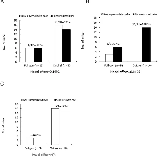
A total 42 mice (23 adult, 19 aged) were treated with hormones and when more than 25 oocyte retrieval was only considered as being superovulated. In overall group including all age of mice, no significant difference was found between hCG A treated group (Folligon) and hCG B treated group (Ovidrel)(Figure 1A). None of aged mice were superovuated (figure 1C) when sixty-seven of hCG A treated group (Folligon) and 100% of hCG B treated group (Ovidrel) were superovulated in 6 to 8 weeks old mice (Figure 1B), which yielded significant difference (p<0.0196).
A B
C
Figure 1. Superovulation of ICR mice with different gonadotrophins. Either 6 to 8 weeks old or 45 to 65 weeks old mice were treated with Folligon or Ovidrel and the mouse yielding more than 25 ovulated oocytes were considered as a superovulated mouse. Superovulation outcome of (A) total mice (n=42), (B) the mice aged 6 to 8 weeks old (n=23) and (C) the mice aged 45 to 65 weeks old (n=19).
Maturational status of oocytes retrieved from the mice superovulated with different gonadotrophins.
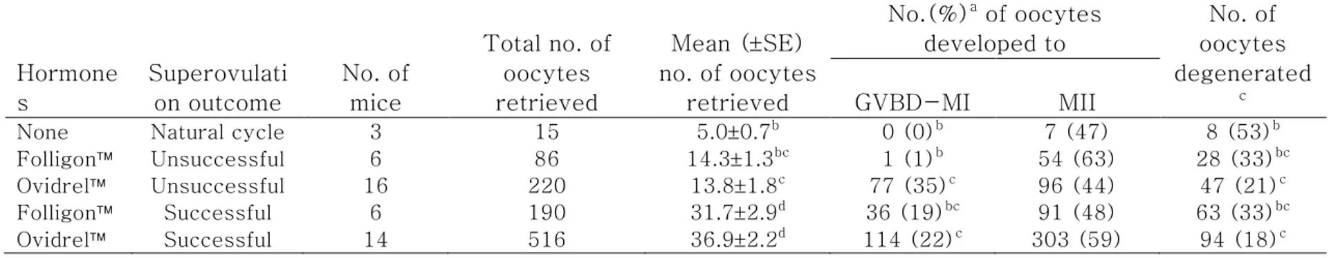
As shown in Table 1, remarkable increase in the number of oocytes retrieved was detected after superovulation treatment within overall aged mice group. Regardless of kinds of gonadotrophins, the total number of retrieved oocytes were increased in hormone treatment group. The proportion of mature oocytes (MII oocytes) retrieved showed no significant difference (44 to 63%) when there were difference in the number of maturing or degenerated oocytes among the treatment groups, while no remarkable tendency was detected.
Table 1. Maturational status of oocytes retrieved from the mice superovulated with different gonadotrophins. Hormone s Superovulati on outcome No. of mice Total no. of oocytes retrieved Mean (SE) no. of oocytes retrieved No.(%)a of oocytes developed to No. of oocytes degenerated c GVBD-MI MII
None Natural cycle 3 15 5.00.7b 0 (0)b 7 (47) 8 (53)b
Folligon Unsuccessful 6 86 14.31.3bc 1 (1)b 54 (63) 28 (33)bc
Ovidrel Unsuccessful 16 220 13.81.8c 77 (35)c 96 (44) 47 (21)c
Folligon Successful 6 190 31.72.9d 36 (19)bc 91 (48) 63 (33)bc
Ovidrel Successful 14 516 36.92.2d 114 (22)c 303 (59) 94 (18)c
GVBD=Germinal vesicle breakdown; MI=Metaphase I; MII=Metaphase II
Model effects on the mean number of oocytes retrieved, the number of oocytes developed to the stage of GVBD-MI and MII, and the number of oocytes degenerated, which were indicated as p value, were less than 0.0001, 0.0173, 0.4262 and 0.028, respectively.
Developmental status of oocytes retrieved from the mice superovulated with different gonadotrophins.
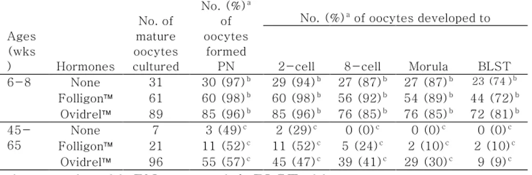
As shown in Table 2, significant difference in the capacity of oocyte activation was detected (p<0.0096). All parameters showed remarkable increase in the activation in the fertile than in aged group; 96 to 98% vs. 49 to 57% in pronuclear formation (P=0.0096), 94 to 98% vs. 29 to 52% in the first cleavage (P<0.0001), 85 to 92% vs. 0 to 41% in 8-cell development (P<0.0001), 85 to 89% vs. 0 to 30% in morula compaction (P<0.0001) and 72 to 81% vs. 0 to 10% in blastcyst formation (P<0.0001). Within the same age, however, no drug effect was detected.
Table 2. Activation of mature oocytes retrieved from 6 to 8 weeks old or 45 to 65 weeks old mice ovulated naturally or superovulated with different gonadrophins.
Ages (wks ) Hormones No. of mature oocytes cultured No. (%)a of oocytes formed PN
No. (%)a of oocytes developed to
2-cell 8-cell Morula BLST 6-8 None 31 30 (97)b 29 (94)b 27 (87)b 27 (87)b 23 (74)b Folligon 61 60 (98)b 60 (98)b 56 (92)b 54 (89)b 44 (72)b Ovidrel 89 85 (96)b 85 (96)b 76 (85)b 76 (85)b 72 (81)b 45-65 None 7 3 (49)c 2 (29)c 0 (0)c 0 (0)c 0 (0)c Folligon 21 11 (52)c 11 (52)c 5 (24)c 2 (10)c 2 (10)c Ovidrel 96 55 (57)c 45 (47)c 39 (41)c 29 (30)c 9 (9)c
wks=weeks old; PN=pronuclei; BLST=blastocyst
Model effects of treatment on the number of oocytes formed pronulcei, and the number of oocytes developed to the 2-cell, 8-cell, morula and blastocyst stages, which were indicated as p value, were 0.0096, 0.0001, 0.0001, 0.0001, and 0.0001, respectively.
aPercentage of the number of MII oocytes retrieved. bc
Different superscripts within the same parameter indicate significant differences among the ages, p<0.05.
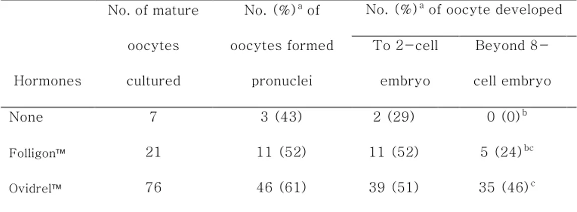
Developmental status of oocytes retrieved from the aged mice superovulated with different gonadotrophins.
As shown in table 3, the number of oocytes formed pronuclei after parthenogenetic activation (43% vs. 52% vs. 61%;
p=0.5777) and developed beyond the 2-cell stage embryos (29% vs. 52% vs. 51%; p=0.5079) were not significant different among treatments. While, the number of embryos developed beyond the 8-cell stage were significantly increased in the hCG B (Ovidrel,
p=0.0180), but not the hCG A competed with the control groups (Folligon, p=0.1667). No difference between two hCGs in the 8-cell stage (24% vs. 46%; p=0.0680).
Table 3. Activation of mature oocytes retrieved after different human chorionic gonadotrophin (hCG) injections into aged (over 45 weeks old) mice.
Hormones No. of mature oocytes cultured No. (%)a of oocytes formed pronuclei
No. (%)a of oocyte developed To 2-cell embryo Beyond 8-cell embryo None 7 3 (43) 2 (29) 0 (0)b Folligon 21 11 (52) 11 (52) 5 (24)bc Ovidrel 76 46 (61) 39 (51) 35 (46)c
Model effects of treatment on the number of oocytes formed pronulcei, and the number of oocytes developed to the 2-cell, and beyond 8-cell stage, which were indicated as p value, were 0.5777, 0.5079, and 0.0164, respectively.
aPercentage of the number of MII oocytes retrieved.
bc
Different superscripts within the same parameter indicate significant differences among the treatment, p<0.05
Discussion
Clear evidence was shown in this study showing that kind of gonadotrophin and state of hCG recipients greatly influenced hyperstimulation outcome. Care should be taken to select kind and dose of gonadotrophins especially in aged model animal, which appeared as the efficiency of superovulation treatment in young animals. Although aged mice of 45 weeks or more was reduced the capacity to ovulate oocytes, superovulation treatment still retain development competence and some of those have a capacity to overcome developmental arrest.
Superovulation influences embryonic development in vitro or
in vivo (Ertzeid and Storeng, 2001a, Van der Auwera and D'Hooghe, 2001), oocyte quality , ovarian follicle number (Choi et al., 2011), oocyte degeneration (Tarin et al., 2001), chromosome abnormality (te Velde and Pearson, 2002) and cellular or molecular processes (Miao et al., 2009).
CHAPTER 4
: Screening of integrin heterodimers functionally
expressed on the surface of undifferentiated
1. Introduction
In a previous report (Lee et al., 2012), a three-dimensional (3D) polyethylene glycol (PEG)-based non-cellular niche promoting embryonic stem cell (ESC) self-renewal was precisely defined by engineering integrin signaling in inbred 129/Ola (E14) ESCs. However, adjustment of ESCs derived from hybrid strain mice to the engineered niche revealed a lack of sustainable self-renewal (Lee et al., 2016), and the mechanical properties of the 3D PEG-based niche showed that effective maintenance of self-renewal was dependent on the genetic background of the ESCs (Lee et al., 2016). These facts emphasize the necessity of developing niches customized to each ESC line from different genetic backgrounds.
There are currently no reports on the development of a 3D PEG-based niche customized to the maintenance of self-renewal of diverse types of ESC. Recently, customization of mechanical properties promoting maintenance of hybrid ESC self-renewal was conducted in a PEG-based 3D hydrogel (Lee et al., 2016), but data on the integrin subunits presented on the surface of ESCs in the undifferentiated state were insufficient to precisely reconstruct a bio-mimicking 3D PEG-based non-cellular niche. Therefore, we examined the types of integrin heterodimers on the surface of
inbred R1 and hybrid B6D2F1 mouse ESCs in the undifferentiated state so that could be compared. The types of integrin subunits expressed in undifferentiated mouse ESCs were identified at the transcriptional and translational levels, and the combinations of integrin α and β subunits were determined by functional assays.
2. Materials and methods
Culture of ESCs
ESCs derived from blastocysts of hybrid B6D2F1 (C57BL6 × DBA2) mice (Lee et al., 2008) and R1 cell line (purchased from Nagy lab) were routinely cultured on mouse embryonic fibroblasts (MEFs) mitotically inactivated by 10 μg/mL mitomycin C (Sigma-Aldrich, St. Louis, MO, USA) in standard ESC culture medium consisting of Dulbecco’s modified Eagle’s medium (DMEM; Welgene, Daegu, Korea) supplemented with 15% (v/v) heat-inactivated fetal bovine serum (FBS; HyClone, Logan, UT, USA), 1% (v/v) nonessential amino acids (NEAA; Gibco Invitrogen, Grand Island, NY, USA), 0.1 mM β-mercaptoethanol (Gibco Invitrogen), 1% (v/v) lyophilized mixture of penicillin and streptomycin (Gibco Invitrogen), and 1,000 units/mL mouse leukemia inhibitory factor (LIF; Chemicon International, Temecula, CA, USA). Subculture was performed at 3-d intervals and the medium was changed daily during culture. The Institutional Animal Care and Use Committee (IACUC) of Seoul National University (IACUC approval No. SNU0050331-02) approved the research proposal and relevant experimental procedures of this study.
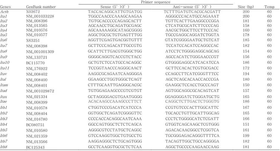
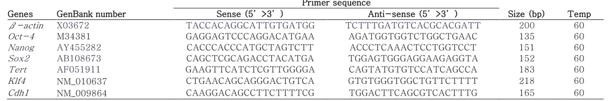
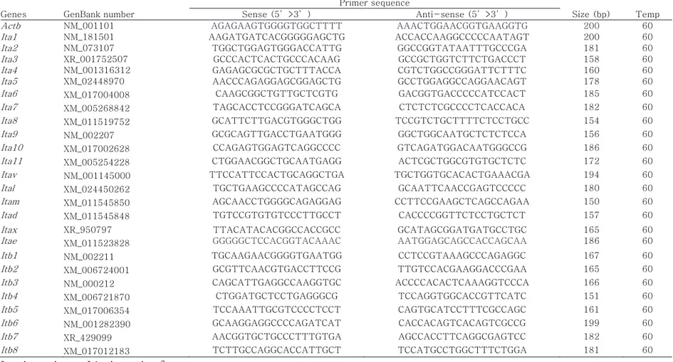
Quantitative real-time polymerase chain reaction (PCR)
An RNeasy Mini Kit (Qiagen, Valencia, CA, USA) was used to extract total mRNA from ESCs, following the manufacturer’s instructions, and cDNA synthesis from the extracted mRNA was conducted using a Superscript III first-strand synthesis system (Invitrogen, Carlsbad, CA, USA). Quantification of gene expression was performed using iQ SYBR Green Supermix (Bio-Rad Laboratories, Hercules, CA, USA) in a Bio-Rad iCycler iQ system (Bio-Rad Laboratories). Data on melting curves were collected to check PCR specificity and the β-actin gene was used as an internal control to normalize specific gene expression. mRNA levels were calculated as 2–ΔCt, where Ct = threshold cycle for target amplification and ΔCt = Cttarget gene (specific genes for each sample)
– Ctinternal reference (β-actin for each sample). Table 4 provides
general information and sequences for all specific primers designed with mouse cDNA sequences obtained from GenBank using Primer3 software (Whitehead Institute/MIT Center for Genome Research).
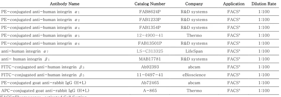
Flow cytometry
The harvested ESCs were washed with ice cold Dulbecco’s phosphate buffered saline (DPBS; Gibco Invitrogen). Then the ESCs were stained for 30 min at 4°C with PE-conjugated anti-mouse integrin α5, α6, α9, and αV, FITC-conjugated anti-mouse
integrin β1, β4, β5, and β5, and fluorescence-unconjugated
anti-mouse integrin α8 antibodies. The fluorescence-unconjugated
primary antibody was detected by incubation with Alexa Fluor 488-conjugated anti-rabbit IgG. All antibodies were diluted in DPBS supplemented with 2% (v/v) heat-inactivated FBS. Supplementary Table 3 provides detailed information and dilution rates of primary and secondary antibodies. The stained cells were washed and sorted using flow cytometry with a CyAn ADP Analyzer (Beckman Coulter, Inc., Fullerton, CA, USA). The FLOWJO Ver. 7.2.5 software program (Tree Star, Inc., Ashland, OR, USA) was used to analyze acquired data.
Attachment assay
Using a previously described method (Lee et al., 2010), we attached ESCs to extracellular matrix (ECM) ligands. Briefly,
96-well tissue culture plates were coated with the following concentrations of purified ECM ligands: 0, 40, 100, and 200 μg/mL of fibronectin (Chemicon International); 0, 100, and 200 μg/mL of laminin (Sigma-Aldrich); and 0, 5, and 50 μg/mL of vitronectin (R&D systems, McKinley Place, MN, USA) overnight at 4°C. Blocking of each well was performed by incubation with 10 mg/mL BSA (Sigma-Aldrich) at 4°C for 1 h and 1 × 10⁵ ESCs
resuspended in standard ESC culture medium were plated into each blocked well. After incubation at 37°C for 2 h, the wells were washed with DPBS to remove non-adherent ESCs from the blocked wells. Adherent ESCs were fixed in 4% (v/v) paraformaldehyde (Sigma-Aldrich) at room temperature for 10 min, stained with 0.1% (w/v) crystal violet (Sigma-Aldrich) in 20% (v/v) methanol (Sigma-Aldrich) for 5 min, and washed extensively with distilled water. Adherent levels were quantified at 570 nm using a microplate reader (VersaMax; Molecular Devices, Sunnyvale, CA, USA) after adding 50 μL of 0.2% (v/v) triton X-100 (Sigma-Aldrich) diluted with distilled water.
Antibody inhibition assay
The wells of 96-well tissue culture plates coated with 40 μg/mL fibronectin, 200 μg/mL laminin, or 5 μg/mL vitronectin overnight at 4°C were blocked with 10 mg/mL BSA for 1 h at 4°C. To inhibit integrin heterodimer function, 1 × 10⁵ ESCs were
incubated for 2 h in standard ESC culture medium containing anti-integrin α5 (5H10–27 [MFR5]), anti-integrin α6 (NKI-GoH3), or
anti-integrin αV (RMV-7) blocking antibodies at 37°C. Detailed
information and dilution rates of the antibodies used are shown in Table 5. The functionally blocked ESCs were plated in each well of 96-well plates coated with ECM proteins and incubated for 3 h at 37°C. Non-adherent ESCs were removed by washing each well with DPBS, and the adherent ESCs were fixed in 4% (v/v) paraformaldehyde for 10 min at room temperature. The fixed adherent ESCs were stained with 0.1% (w/v) crystal violet in 20% (v/v) methanol for 5 min, and each well was washed twice with distilled water. Measurement of adherent levels was conducted using a microplate reader at 570 nm after adding 50 μL of 0.2% (v/v) Triton X-100 diluted with distilled water.
Statistical analysis
All the numerical data derived from each experiment were analyzed in the Statistical Analysis System (SAS) program. Moreover, when a significant main effect was detected by analysis of variance (ANOVA) in the SAS package, the least-square or DUNCAN methods were used for comparison among treatments. Differences among treatments were considered significant when the
Table 4. Oligonucleotide primers and PCR cycling conditions of mouse integrin subunits. Genes GenBank number
Primer sequence
Size (bp) Temp Sense (5’>3’) Anti-sense (5’>3’)
Actb X03672 TACCACAGGCATTGTGATGG TCTTTGATGTCACGCACGATT 200 60
Ita1 NM_001033228 TGGCCAACCCAAAGCAAGAA AGGGCCCACATGCCAGAAAT 200 60
Ita2 NM_008396 TGTGCACCCCCAGAGCACTT TGTTCACTTGAAGGCCCGGA 181 60
Ita3 NM_013565 AGCAACCTGCAGATGCGAGC CTCATGCGCATCTTCCCCAG 158 60
Ita4 NM_010576 AGCAAAAAGGCATAGCGGGG AACGCTGGCTTCCTTCCCAC 160 60
Ita5 NM_010577 AGGCTGCGCTGTGAGTTTGG TGCCGAGGCAGGATCTGGTA 178 60
Ita6 X63251 AGGTTCGAGTGACGGTGTTT GTATCGGGGAATGCTGTCAT 185 60
Ita7 NM_008398 GCTTCCCAGACATTGCCGTG TCCATCCACATCCAGGCCAC 182 60
Ita8 NM_001001309 GCATTCTTGACGTGGGCTGG ATCCTCTGGGGAGGCAGCAG 154 60
Ita9 NM_133721 GGGGCAGGTCACCGTCTACC AGCCACATCTGGGAACCCGT 156 60
Ita10 BC115770 GCTGTCTCCATGCCACAGGC GTGGGGAGGCATCACATCCA 186 60
Ita11 NM_176922 TCCGGTAACCCAGGGCAACT GCTTCCACACTCGTGCGACC 172 60
Itav NM_008402 AAGGCGCAGAATCAAGGGGA CCAGCCTTCATCGGGTTTCC 194 60
Ital NM_008400 GGAAGCCTGGTGGGCTCAGT AGCTCAGCACAACCACCCGA 180 60
Itam NM_008401 CTTTGCAATTGAGGGCACGC GAAGGCTCCACCTGCCCAGT 150 60
Itad NM_001029872 TGTGGAGAAGCCCGTCGTGT AGTGGCAGGCGCACAGTCAT 157 60
Itax Itae NM_021334 NM_008399 GCTAGGGGACGTGAATGGGG ACACAAGCCAAAGCCCTTCT GGAGGGGATCTGGGATGCTG CAGGCTCTTGACTCTGGGTG 165 186 60 60
Itb1 NM_010578 CTGGTCCCGACATCATCCCA CCGTGTCCCACTTGGCATTC 167 60
Itb2 NM_008404 GGTGGCTCAGATCGGGGTTC TGCACCTGTTGCATTGGCAG 165 60
Itb3 NM_016780 CCCCACCACAGGCAATCAAA CCCTCTGGGGCATCTCGATT 166 60
Itb4 BC080751 GGCCAGTGGCTCTCTCAGCA GTGGTCAGCAAGCTCGTGGG 151 60
Itb5 NM_010580 AGGGCGTCCTATGCTCAGGC AGACACAACGGCCTCGGTCA 161 60
Itb6 NM_021359 GTCCAAGGTGGCTGTGCCTG TGCGGGAGACAGGGTTTTCA 199 60
Itb7 NM_013566 AAGGAGGGCTCTGCAGTGGG TACAGTTGGCTGCCAGGGGA 182 60
Itb8 BC125343 GCCTCAAGGTGCGCTCTCAA AGGCTGCCCCAAGAACCAAG 181 60
Table 5. Primary and Secondary Antibodies of mouse surface integrin.
Antibody Name Epitope Catalog
Number Company Application
Dilution Rate PE-conjugated armenian hamster anti-mouse/rat
integrin α5 HMa5-1 12-0493 eBioscience FACS
a 1:100
PE-conjugated rat anti-human/mouse integrin α6 GoH3 12-0495 eBioscience FACSa 1:100 PE-conjugated sheep anti-mouse integrin α9 NS0 FAB3827P R&D systems FACSa 1:100 PE-conjugated rat anti-mouse integrin αV RMV-7 12-0512 eBioscience FACSa 1:100 Alexa Fluor 488-conjugated chicken anti-rabbit IgG
(H+L) A-21441 Molecular Probes FACS
a 1:100
FITC-conjugated rat anti-mouse integrin β1 265917 FAB2405F R&D systems FACSa 1:100 FITC-conjugated rat anti-mouse integrin β4 308601 FAB4054F R&D systems FACSa 1:100 FITC-conjugated mouse anti-mouse integrin β5 KN52 11-0497 eBioscience FACSa 1:100 FITC-conjugated rat anti-human/mouse integrin β7 FIB504 11-5867 eBioscience FACSa 1:100 LEAFTM purified rat anti-mouse integrin α
5 5H10-27
(MFR5) 103808 BioLegend® AIA
b 1:20
Rat anti-mouse integrin α6 NKI-GoH3 MAB1378 Millipore AIAb 1:20 LEAFTM purified rat anti-mouse integrin α
V RMV-7 104108 BioLegend® AIAb 1:20
3. Results
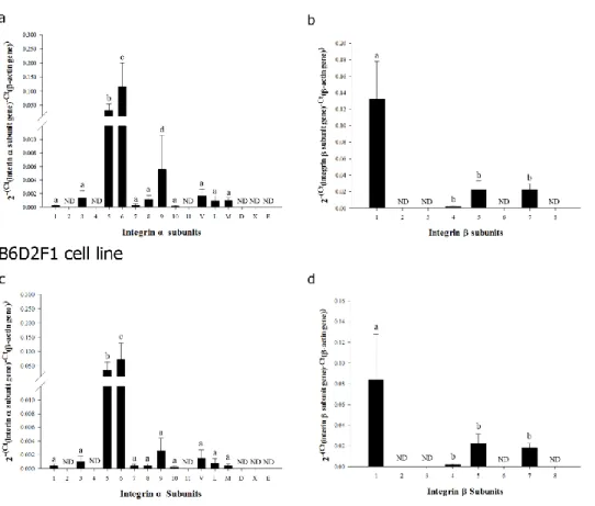
Identification of transcriptional expression of integrin subunits on the cell membrane of inbred R1 and hybrid F1 mouse ESCs in undifferentiated state
To determine the types of integrin heterodimers expressed on the membrane of inbred R1 and hybrid F1 mouse ESCs in the undifferentiated state. I monitored the expression of the individual integrin α and β subunits that make up the integrin heterodimers at the transcriptional and translational levels. In the transcriptional analysis of genes encoding 17 integrin α subunits and 8 integrin β subunits, significantly higher levels of expression were observed for integrin α5 and α6 (Figure 2a and 2c) and integrin β1 (Figure
2b and 2d) subunit genes. Minimal levels of expression were detected for integrin α1, α3, α7, α8, α9, α10, αV, αL, and αM
(Figure 2a and 2c) and integrin β4, β5, and β7 (Figure 2b and 2d)
subunit genes. I did not detect any transcriptional expression of integrin α2, α4, α11, αD, αX, and αE (Figure 2a and 2c) and
integrin β2, β3, β6, and β8 (Figure 2b and 2d) subunit genes in
Figure 2. Transcriptional expression of integrin α and β subunit genes in undifferentiated ESCs derived from inbred R1 (129X1/SvJ x 129S1/SvImJ) and hybrid (C57BL6 × DBA2) F1 mice. Messenger RNA levels of integrin α (a and c) and β (b and d) subunit genes in undifferentiated inbred R1 (a and b) and hybrid F1 (c and d) mouse ESCs were examined quantitatively using real-time PCR. All data shown are means ± SD of three independent experiments. a–cp < 0.05. ND = not detected.
Identification of translational expression of integrin subunits on the membrane of inbred R1 and hybrid F1 mouse ESCs in undifferentiated state
I measured the translational expression of integrin α5, α6,
α9, and αv and integrin β1, β4, β5, and β7 subunit genes, which
showed high levels of transcription (Figure 3). Most of the undifferentiated mouse ESCs expressed integrin α5, α6, α9, and
αv and integrin β1 subunit proteins on the cellular membrane, and
the expression of integrin β4, β5, and β7 subunit proteins were
observed in a few undifferentiated inbred R1 and hybrid F1 mouse ESCs. These results indicate that integrin α5, α6, α9, αv and β1
subunits are localized on the membrane of inbred R1 and hybrid F1 mouse ESCs in undifferentiated state.
Figure 3. Translational expression of integrin α and β subunit genes in undifferentiated ESCs derived from inbred R1 (129X1/SvJ x 129S1/SvImJ) and hybrid (C57BL6 × DBA2) F1 mice. The translational expression of integrin α (a, b, e and f) and β (c, d, g and h) subunit genes in undifferentiated inbred R1 (a, b, c and d) and hybrid B6D2F1 (e, f, g and h) mice ESCs was measured by flow cytometry. Integrin α5, α6, α9, and αv subunit proteins were
highly expressed at the transcriptional level and were detected on the surfaces of almost all undifferentiated inbred R1 and hybrid B6D2F1 mouse ESCs. Among the four integrin β subunits highly expressed at the transcriptional level, most of the undifferentiated mouse ESCs expressed an integrin β1 subunit protein on the cell
surface, while integrin β4, β5, and β7 subunit proteins were
detected on the surfaces of only a few of the undifferentiated mouse ESCs. Figure 3 a, c, e and g are representative FACS analyses of the percentage of cells stained positively with antibodies detecting integrin α or β subunit proteins, and Figure 3 b, d, f, and h are composite averages (means ± SD) of the percentage of cells stained positively with antibodies detecting integrin α or β subunit proteins from three independent experiments. a-cp < 0.05.
Determination of integrin heterodimers functionally expressed on the membrane of undifferentiated inbred R1 and hybrid F1 mouse ESCs
Based on transcriptional analysis of each integrin subunit gene, integrin combinations α5β1, α6β1, α9β1 and αVβ1
described previously (Prowse et al., 2011), were proposed as candidates for the active integrin heterodimers exhibited in the undifferentiated inbred R1 and hybrid F1 mouse ESCs. I investigated a presence of these integrin heterodimer candidates by estimating the levels of adherent mouse ESCs cultured on natural ECM proteins interacting specifically with each integrin heterodimer and the levels of adherence post-culture of mouse ESCs treated with antibodies specifically blocking each integrin function. Compared to control, both mouse ESCs cultured on fibronectin (Figure 4a, 4d), laminin (Figure 4b, 4e), and vitronectin (Figure 3d, 3f) showed significantly improved adherent levels, indicating that both mouse ESCs could express integrin α5β1, α9β1 and αVβ1
which binds specifically with fibronectin, and integrin α6β1, which
binds specifically with laminin, and αVβ1 which binds specifically
with vitronectin on the cell surface in the undifferentiated state. Specific integrin function-blocked mouse ESCs were incubated with 40 μg/mL fibronectin, 100 μg/mL laminin, or 5 μg/mL vitronectin. Significantly weakened adherent levels were
observed in both strains of mouse ESCs when integrin α6β1 and
αVβ1 were blocked (Figure 5b, 5c, 5d, and 5f, 5g, 5h), whereas
there was no significant decrease in the adherent levels when integrin α5β1 was blocked (Figure 5a and 5e) compared to the
control. Based on these results, I confirmed that both strains of mouse ESCs in the undifferentiated state show functional expression of integrin α6β1 and αVβ1 on the cellular membrane,
while integrin α5 andα9 is presented not as a heterodimer, but as a
Figure 4. Identification of integrin heterodimers interacting with fibronectin and laminin on the surfaces of undifferentiated ESCs derived from inbred R1 (129X1/SvJ x 129S1/SvImJ) and hybrid (C57BL6 × DBA2) F1 mice. Tissue culture plates (96-well) were coated with 0, 40, 100, or 200 μg/mL fibronectin (a and d) or 0, 100, or 200 μg/mL laminin (b and e) or or with 0, 5, or 50 μg/mL vitronectin (c and f). Uundifferentiated inbred R1 (a, b, and c) and hybrid F1 (d, e, and f) mouse ESCs (1 × 10⁴) were plated in each
well. After incubation for 2 h at 37°C, adherent cells were stained with crystal violet and the adhesion level was quantified using a microplate reader. The percentage of maximum adhesion is represented as the optical density of cells plated on ECM protein-free plates. The undifferentiated mouse ESCs cultured on fibronectin-, laminin-, or vitronectin-coated culture plates had significantly higher levels of adhesion than those on ECM protein-free culture plates. All data shown are means ± SD of three independent experiments. *p < 0.05.
Figure 5. Functional blocking of integrin heterodimers interacting with adhesive proteins in undifferentiated ESCs derived from inbred R1 (129X1/SvJ x 129S1/SvImJ) and hybrid (C57BL6 × DBA2) F1 mice. Undifferentiated inbred R1 (A, B, C and D) and hybrid F1 (E, F, G, and H) mice ESCs were incubated in the absence or presence of anti-integrin α5 (5H10–27 [MFR5]) (a and e), anti-integrin α6
(NKI-GoH3) (b and f), or anti-integrin αV (RMV-7)(c, d, g and
h) blocking antibodies and then plated on 40 μg/mL fibronectin-, 100 μg/mL laminin-, or 5 μg/mL vitornectin-coated wells. After incubation for 2 h at 37°C, adherent cells were stained with crystal violet, and quantification of the adhesion level was performed using a microplate reader. As a parameter of functional blocking by antibodies, the relative cell adhesion, which is represented by the optical density of cells plated on each extracellular matrix (ECM) protein-coated well in the absence of any blocking antibodies, was determined. Compared to those without blocking antibodies, undifferentiated hybrid F1 mouse ESCs treated with integrin α5
blocking antibody showed no significant difference in the rate of attachment to fibronectin (a and e), whereas significantly lower rates of attachment to laminin were observed in the integrin α6
blocking antibody-treated cells (b and f) and integrin αV blocking
(c and g) and vitronectin (d and h) compared to those not treated with blocking antibody.. All data shown are means ± SD of three independent experiments. *p < 0.05.