저작자표시-비영리-변경금지 2.0 대한민국 이용자는 아래의 조건을 따르는 경우에 한하여 자유롭게 l 이 저작물을 복제, 배포, 전송, 전시, 공연 및 방송할 수 있습니다. 다음과 같은 조건을 따라야 합니다: l 귀하는, 이 저작물의 재이용이나 배포의 경우, 이 저작물에 적용된 이용허락조건 을 명확하게 나타내어야 합니다. l 저작권자로부터 별도의 허가를 받으면 이러한 조건들은 적용되지 않습니다. 저작권법에 따른 이용자의 권리는 위의 내용에 의하여 영향을 받지 않습니다. 이것은 이용허락규약(Legal Code)을 이해하기 쉽게 요약한 것입니다. Disclaimer 저작자표시. 귀하는 원저작자를 표시하여야 합니다. 비영리. 귀하는 이 저작물을 영리 목적으로 이용할 수 없습니다. 변경금지. 귀하는 이 저작물을 개작, 변형 또는 가공할 수 없습니다.
The effect of botulinum toxin A on
transverse rectus abdominis
musculocutaneous flap survival in rats
Tae Hwan Park
Department of Medicine
The effect of botulinum toxin A on
transverse rectus abdominis
musculocutaneous flap survival in rats
Directed by Professor Dong Kyun Rah
The Doctoral Dissertation
submitted to the Department of Medicine
the Graduate School of Yonsei University
in partial fulfillment of the requirements for the degree of
Doctor of Philosophy
Tae Hwan Park
ACKNOWLEDGEMENTS
I am deeply grateful to professor Dong Kyun Rah for his
guidance, patience, and encouragement throughout my journey to
this degree. I am also greatly indebted to professors Dae Hyun Lew,
Goo Hyun Mun, Se Hoon Kim, and Young Ho Lee for their
valuable comments and suggestions on my dissertation.
I also recognize my professors Choong Hyun Chang, Sang Won
Seo, and June Kyu Kim, throughout my residency, for their
constant trust and consideration. My special thanks go to my
research partners, Prof. Sung Young Kim and Yosep Chong, for
their generous molecular and pathologic support. I would like to
express my gratitude to my parents, for whom I have the utmost
respect in every aspect of life.
Last, but not least, my wife, Yun Joo Park, deserves special
mention; her prayers, love, and patience have enabled me to
continue my research, and complete this thesis. My children, Seung
Ha and Eun Hyung, continue to inspire me and are the source of
endless joy.
December 2016
Tae Hwan Park
<TABLE OF CONTENTS>
ABSTRACT···1
I. INTRODUCTION ···3
II. MATERIALS AND METHODS···7
1. Experiment design and flap model···7
2. Flap survival area measurement··· 10
3. Histology··· 10
4. Real-time reverse transcriptase-polymerase chain reaction ··· 11
5. Western blot analysis ··· 12
6. Statistical analysis ··· 14
III. RESULTS ··· 15
1. Flap survival area measurement··· 15
2. Histologic evaluation ··· 17 3. Gene expression··· 21 4. Protein expression ··· 23 IV. DISCUSSION ··· 26 V. CONCLUSION ··· 30 REFERENCES ··· 31 ABSTRACT(IN KOREAN) ··· 38 PUBLICATION LIST ··· 40
LIST OF FIGURES
Figure 1. Transverse rectus abdominis musculocutaneous flap
design and surgical protocol··· 9
Figure 2. Comparison of flap survival areas ··· 16
Figure 3. Histologic findings for the right inferior epigastric artery
and vein in transverse rectus abdominis
musculocutaneous flaps ··· 18
Figure 4. Histologic findings of perfusion zones in transverse rectus
abdominis musculocutaneous flaps··· 19
Figure 5. Representative microvessel density counts (CD31, ×400)
··· 21
Figure 6. Relative expression of cluster of differentiation 34 (CD34),
hypoxia-inducible factor-1α (HIF-1α), and vascular
endothelial growth factor (VEGF) mRNA, as determined
using quantitative reverse-transcription polymerase chain
reaction··· 22
Figure 7. Relative expression of cluster of differentiation 34 (CD34),
hypoxia-inducible factor-1α (HIF-1α), and vascular
endothelial growth factor (VEGF), as determined using
Western blotting. ··· 24
LIST OF TABLES
Table 1. List of primers ··· 12
Table 2. Antibodies used in the experiments and its dilution
ABSTRACT
The effect of botulinum toxin A on transverse rectus abdominis musculocutaneous flap survival in rats.
Tae Hwan Park Department of Medicine
The Graduate School, Yonsei University (Directed by Professor Dong Kyun Rah)
The transverse rectus abdominis musculocutaneous (TRAM) flap is widely used in various reconstructive surgeries. Recent evidence suggests a positive effect of botulinum toxin A (BoTA) on flap survival. In this study, we hypothesized that pretreatment with BoTA augments axial-pattern TRAM flap survival in the presence of a vertical midline scar. We further hypothesized that this was due to increased angiogenesis resulting from enhanced activity of the hypoxia-inducible factor (HIF)-1α/vascular endothelial growth factor (VEGF) pathway.
Twenty-four Sprague-Dawley rats were randomly divided into 2 groups. Five days after creating vertical midline incisions, the BoTA group was pretreated with BoTA; the control group was similarly pretreated with normal saline. Ten days after the initial incision, the TRAM flaps were harvested. The final flap survival percentage, overall histologic changes, pedicle lumen areas, and microvessel densities were assessed on postoperative day 5. Quantitative reverse-transcription polymerase chain reaction and Western blotting were
performed to evaluate angiogenesis-related factors, including cluster of differentiation 34 (CD34), HIF-1α, and VEGF expression.
Flap survival was significantly higher in the BoTA group than in the control group on both the ipsilateral (91.27 ± 10.1% vs 67.91 ± 20.2%; p = 0.002) and contralateral (53.16 ± 19.2% vs 14.54 ± 8.1%; p < 0.001) sides. In the BoTA group, a significant increase in the pedicle lumen area was observed, relative to that in the control group (p < 0.001). In the control group, mild to moderate epidermal necrosis was seen, and the microvessels were relatively small, compared with those in the BoTA group. Immunohistochemically, the numbers of CD31-positive vessels were significantly higher on the contralateral sides in the control group than in the BoTA group (p < 0.001). The relative expression of CD34 and VEGF mRNA was higher in the BoTA group than in the control group, in every zone; the relative expression of HIF-1α mRNA was significantly higher in the BoTA group than in zone IV. The relative expression of CD34, VEGF, and HIF-1α proteins was higher in the BoTA group than in the control group, in every zone.
In conclusion, we showed that presurgical BoTA treatment might increase inferior-based TRAM flap survival due to increased HIF-1α/VEGF-dependent angiogenesis.
Key words: botulinum toxin A, transverse rectus abdominis musculocutaneous flap, angiogenesis, delay procedure
The effect of botulinum toxin A on transverse rectus abdominis musculocutaneous flap survival in rats.
Tae Hwan Park Department of Medicine
The Graduate School, Yonsei University (Directed by Professor Dong Kyun Rah)
I. INTRODUCTION
The transverse rectus abdominis musculocutaneous (TRAM) flap is widely
used in various reconstructive surgeries1-6. Partial necrosis of the TRAM flap
occurs more frequently in patients with several risk factors, including diabetes,
obesity, smoking, and especially previous abdominal surgery7-9. These
conditions preclude surgeons from safely performing successful TRAM flap reconstructions. In particular, the presence of scars within the flap remains a substantial challenge to many surgeons due to the high incidence of partial flap necrosis. This concept is especially true for TRAM flaps required in the presence of vertical midline abdominal scars. Tissue perfusion across a vertical midline scar is potentially compromised and unreliable. Many patients, however, favor breast reconstructions involving abdominal flaps, such as the TRAM flap, even in the presence of a midline, lower abdominal scar. The
simplest and most reliable option is to use a hemi-deep inferior epigastric perforator (DIEP) flap or hemi-TRAM flap that excludes the abdominal tissue beyond the vertical midline scar10. Unfortunately, adopting a hemi-DIEP or hemi-TRAM flap also limits the size of the reconstructed breast, thereby
limiting the aesthetic outcome in some patients with large breasts11. Therefore,
tissues crossing the midline scar might have to be included when designing a TRAM flap9. Several alternatives have been developed, including the use of bipedicled TRAM flaps, flaps designed higher in the abdomen, or anastomosing a contralateral deep inferior epigastric artery9,12,13. Although these surgical strategies may increase flap perfusion across midline abdominal scars, they are technically challenging, require long operation times, and may result in flap loss11.
Botulinum toxin is the exotoxin of Clostridium botulinum, a Gram-positive bacterium. All of the identified toxin serotypes produce muscle paralysis by inhibiting the release of acetylcholine from the presynaptic terminals of the
neuromuscular junctions of the peripheral nervous system14. Of the seven
reported toxin serotypes, botulinum toxin A (BoTA) has been widely studied and is used in many clinical fields for various therapeutic and aesthetic purposes. Acetylcholine, in axon terminals, is packaged in synaptic vesicles. Normally, these synaptic vesicles fuse with presynaptic plasma membranes, releasing the neurotransmitter (e.g., acetylcholine) into the synaptic cleft. This process is mediated by the SNARE proteins (synaptobrevin, syntaxin, and synaptosome-associated protein 25 kDa). When BoTA is injected, it is taken into vesicles where the light chain (50 kDa) of BoTA cleaves the soluble SNARE proteins, preventing assembly of the fusion complex and, thereby, blocking acetylcholine release.
On the other hand, hypoxia-inducible factor-1 (HIF-1), the first transcription factor produced in response to hypoxia, is intimately associated with angiogenesis. This transcription factor is a heterodimer composed of a novel α
subunit (HIF-1α) and a previously characterized β subunit (HIF-1β)15. HIF-1
mediates acute hypoxic responses by influencing the genes related to
angiogenesis, metabolism, cell survival, and apoptosis16. Vascular endothelial
growth factor (VEGF) is the primary angiogenesis-related cytokine and is
upregulated by HIF-117. The HIF-1α/VEGF signaling pathway is one of the
most strongly angiogenic pathways and is a recognized therapeutic target for
new anticancer drugs used to abrogate neovascularization18.
Elevated VEGF and nitric oxide synthase (NOS) expression, in the presence of hypoxia, is well-known to be regulated by HIF-1α expression. This is a molecular adaptation to hypoxia, evidenced by the onset of neoangiogenesis
and vasodilation19. Hendrickson et al. (2015) showed that NOS is both
upstream (endothelial NOS) and downstream (inducible NOS [iNOS]) of HIF-1α signaling.20 Schweizer et al. (2013) reported that BoTA raises blood flow and increases the survival of critically ischemic skin flaps due to the increased expression of the endothelial NOS and Ras homolog gene family
member, RhoA21. Kim et al. (2009) reported that BoTA increases iNOS levels
and subsequently augments flap survival in a rat random-pattern flap model22.
Thus, we hypothesized that BoTA might activate the crucial HIF-1α/VEGF angiogenesis pathway.
Although the random-pattern flap model is widely used, it lacks larger arterioles in the flap, resulting in higher rates of necrosis and limiting the
potential for compensatory mechanisms21,23. To our knowledge, no studies
angiogenic potential of BoTA. Thus, we further hypothesized that BoTA pretreatment augments axial-pattern TRAM flap survival, in the presence of vertical midline scars, by increasing HIF-1α/VEGF-dependent angiogenesis.
II. MATERIALS AND METHODS
1. Experiment design and flap modelAll animal protocols used in this study were approved by the Institutional Animal Care and Use Committee of Kangbuk Samsung Hospital and Konkuk University. All animals were cared for in compliance with the National Research Council’s criteria, as outlined in the “Guide for the Care of Laboratory Animals,” which was prepared by the Institute of Laboratory Animal Resources and published by the National Institutes of Health (Bethesda, MD, USA).
Twenty-four male Sprague-Dawley rats (8-weeks-old, 250–300 g) were individually housed, under controlled conditions (temperature, 20–22.8° C; 12-h light/dark cycle) in an animal resources facility; food and water were provided ad libitum. The rats were randomly divided into 2 groups (12 rats, each): the BoTA and control groups.
Using an induction chamber, the animals were anesthetized (5% isoflurane; Aerane, Ilsung Pharmaceuticals, Seoul, Korea) and maintained at 1.5% isoflurane, via a nasal cone, until the end of the procedure. After shaving the ventral hair, a vertical midline incision and the future 6 × 5-cm TRAM flaps were marked on each rat’s abdomen. Bilateral flaps, 5 × 3 cm in size, were designed on the abdomen of each rat, with the superior border being the costal margins and the inferior boundary located just above the horizontal line between the bilateral anterosuperior iliac spines. The TRAM zones were
designed as suggested by Holm et al.24 (Figure 1). Zone I was the area
side; zone III extended across the midline, immediately adjacent to zone I; and zone IV was lateral to zone III, on the contralateral to the pedicle. A 5-cm vertical skin incision (going down to the anterior rectus sheath) was made from the xiphoid to the symphysis pubis, without injuring the adjacent perforating vessels. Five days after creating the initial vertical midline incision (5 days before TRAM flap elevation), vials of lyophilized BoTA (Botox; Allergan, Irvine, CA) were reconstituted in 4 mL of normal saline solution to a concentration of 25 IU/mL. The BoTA group (n = 12) was pretreated with 20 IU of BoTA by subdermal injection distributed evenly in the 8 proximal and distal regions of the flap, in each zone (0.1 mL each). The control group (n = 12) was similarly pretreated with 0.8 mL of normal saline.
The encompassed skin territory was elevated toward the midline, carefully preserving all musculocutaneous perforators originating from only the right rectus abdominis muscle. This muscle was then separated from the linea alba and the lateral abdominal wall, carefully extending the dissection below the deep circumflex iliac vessels. A right, inferior-based TRAM flap was made after the superior origin was detached at the subcostal border. Any musculofascial defect in the abdominal wall was closed, primarily, using Monosyn or vicryl 4-0. To prevent revascularization of the overlying flap, a 6 × 5-cm, 0.13-mm thick silicone sheet (Bioplexus, Saticoy, CA, USA) was placed under the flaps, and the skin paddle was fixed in its bed using 4-0 nylon sutures.
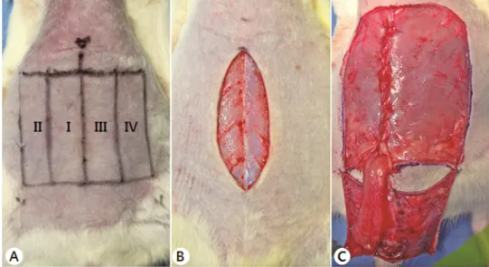
Figure 1. Transverse rectus abdominis musculocutaneous (TRAM) flap design and surgical protocol.
(A) Preoperative design of the TRAM flap.
The TRAM zones were designed as suggested by Holm et al.24 : Zone I was
the area overlying the muscle pedicle; zone II was lateral to zone I, on the ipsilateral side; zone III extended across the midline, immediately adjacent to zone I; and zone IV was lateral to zone III, on the contralateral to the pedicle.
(B) Vertical midline abdominal incision, before TRAM flap elevation. The initial vertical skin incision was made along the linea alba from the xiphoid process to the symphysis pubis
(C) Appearance immediately after flap elevation.
The encompassed skin territory was elevated toward the midline, carefully preserving all musculocutaneous perforators originating from only the right rectus abdominis muscle. This muscle was then separated from the linea alba and the lateral abdominal wall, carefully extending the dissection below the
deep circumflex iliac vessels. A right, inferior-based TRAM flap was made after the superior origin was detached at the subcostal border. Any musculofascial defect in the abdominal wall was closed, primarily, using Monosyn or vicryl 4-0.
2. Flap survival area measurement
Flap survival was evaluated on postoperative days 0, 1, 3, and 5. On each day, a digital photograph was taken; the percentage of surviving flap was calculated on postoperative day 5 using a transparent sheet and ImageJ software (National Institutes of Health). The survival area was independently assessed by 2 investigators, blinded to the treatment groups, and was expressed as a percentage of the total flap area (surviving flap area (%) = viable area/total area × 100). The calculated ratios were used for statistical analyses examining the differences between the groups.
3. Histology
Fresh tissue samples were taken from the center of each TRAM zone, 5 days after TRAM flap elevation, in both the control and BoTA groups. The samples were fixed with 10% formaldehyde, embedded in paraffin, sectioned, and stained with hematoxylin and eosin stain (H&E). Histologic changes and the blood vessels in each zone were evaluated by 2 pathologists, blinded to the treatment group.
To study whether there was pedicle dilation, a full-thickness right rectus muscle specimen (1 × 1 cm), underneath the skin flap, was harvested from
zone I, 2 cm distal to the cranial end of the TRAM flap, 5 days after TRAM flap elevation. The lumen area was measured using ImageJ software. Fresh tissue samples were taken from the centers of the 4 TRAM zones. Microvessel density was determined using the endothelial cell marker CD31 (mouse anti-rat CD31 antibody; Serotec, Dusseldorf, Germany) and a BenchMark XT immunostaining instrument (Ventana, Illkirch, France). First, the tissue area with the highest vessel density was identified, at low magnification, in each CD31-stained skin flap section. Next, high magnification (×400) pictures were taken adjacent to this area. CD31-positive vessels were counted in 25, randomly selected, high-power fields (×400) by 2 pathologists. The average microvessel density in each zone was compared between the control and BoTA groups.
4. Real-time reverse transcription-polymerase chain reaction
On postoperative day 5, we harvested 1 × 1-cm, full-thickness skin samples from above the center of each zone. These were immediately snap-frozen in liquid nitrogen and stored at -80° C. Cells in the collected tissue were dissolved by applying 1 mL of TRIzol reagent (T9424; Sigma Aldrich, St. Louis, MO, USA), at room temperature, for 5 min. Chloroform (0.2 mL) was added, and the tubes shook for 15 s, and left at room temperature for 3 minutes. Then, the solution was centrifuged (11,500 rpm) at 4°C for 15 min, and the aqueous phase was transferred to a fresh tube. RNA was precipitated by adding isopropanol (0.5 mL), mixed by inversion, and incubated at room temperature for 10 min before centrifuging (11,500 rpm) at 4°C for 10 min. After removing the supernatant and mixing it with cold (-20°C) ethanol, the
sample was inverted. The solution was centrifuged (9500 rpm) at 4°C for 5 min. After quickly removing supernatant, the precipitate was dried in a clean place. The RNA pellet was then redissolved in diethylpyrocarbonate water (40 µL). The optical densities of the samples were measured at 260 nm using a Nanodrop spectrophotometer (DaeMyung Science, Taejon, South Korea). Reverse transcription-polymerase chain reaction was performed for CD34, HIF-1α, and VEGF using specific gene primers; glyceraldehyde 3-phosphate dehydrogenase was used as a housekeeping gene control (Table 1). Table 1. List of primers
Primer Sequence CD34 F 5’ – CCGAGTGTTTGCTGATGGTC- 3’ R 5’- GGGAATAGCTCTGGTGGCTC-3’ VEGF F 5’- CTGCTGTACCTCCACCATGC-3’ R 5’- CTGGCTTTGGTGAGGTTTGA-3’ HIF-1α F 5’-GATGGCTCCCTTTTTCAAGC-3’ R 5’-TTTCTGCTGCCTTGTATGGG-3’ GAPDH F 5’-ACCACAGTCCATGCCATCAC-3’ R 5’-ACAACGGATACATTGGGGGT -3’
5. Western blot analysis
On postoperative day 5, the center tissue from each zone was immediately snap-frozen in liquid nitrogen and stored at −80°C, in RIPA buffer (50
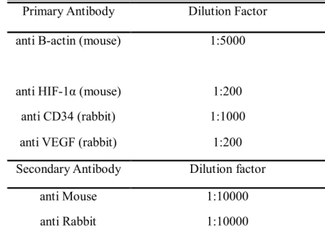
mmol/L Tris-HCl (pH 7.6), 150 mmol/L NaCl, 1% nonidet-P40, 0.5% sodium deoxycholate, 0.1% sodium dodecylsulfate [SDS]) supplemented with a protease inhibitor cocktail. At the time of analysis the thawed solution was left at −4°C for 10 minutes. Then, the solution was centrifuged (14,000 rpm) at 4°C for 30 min. The aqueous solutions were transferred to fresh tubes and boiled in SDS sample buffer, resolved by SDS polyacrylamide gel electrophoresis, and electrically transferred to nitrocellulose membranes (Bio-Rad Laboratories, Munich, Germany). Subsequently, Western blotting was performed using appropriate primary antibodies and horseradish peroxidase-conjugated secondary antibodies (Table 2). Antibody-protein complexes were visualized using Image Quant LAS 4000 (GE Healthcare, Pittsburgh, PA, USA). Band signal intensities were quantified using Multi Gauge v3.1 analysis software.
Table 2. Antibodies used in the experiments and its dilution factor
Primary Antibody Dilution Factor
anti B-actin (mouse) 1:5000
anti HIF-1α (mouse) 1:200
anti CD34 (rabbit) 1:1000
anti VEGF (rabbit) 1:200
Secondary Antibody Dilution factor
anti Mouse 1:10000
6. Statistical analysis
The results of the experiments are expressed as means ± SD. Statistical differences were determined using unpaired Student’s t-tests (2-sided) or repeated measures analysis of variance. For comparison of the flap survival percentages, pedicle lumen areas, relative mRNA expressions, and protein expressions, unpaired Student’s t-tests (2-sided) were used. Repeated measures analysis of variance was used for the statistical analysis of microvessel density results. P-values < 0.05 were considered to be significant, for all tests.
III. RESULTS
1. Flap survival area measurement
Flap survival in the BoTA group was 72.2% (11.5) and that in the control group was 41.2% (10.2); representative images of the external and inner surfaces are presented in Figure 2. The ipsilateral side (zones I + II) of the BoTA group [91.3% (10.1)] revealed increased flap survival compared with the control group [67.9% (20.2); p < 0.001). Likewise, on the contralateral side (zones III + IV), the BoTA group [53.2% (19.2%)] revealed improved flap survival compared with the control group [14.5% (8.1%); p = 0.002) (Figure 2).
Figure 2. Comparison of flap survival areas. (Upper) Gross appearance of transverse rectus abdominis musculocutaneous flaps from representative
control- (A and B) and botulinum toxin A (BoTA)-treated (C and D) animals on postoperative days 0, 1, 3, and 5. The surviving flaps in BoTA-treated (C, D) animals are significantly larger than those in similarly treated control animals (A, B), over time.
(Lower) Mean flap survival area, as a percentage. The mean percentage of surviving flap area in the BoTA group is significantly higher than that in the control group, on both the ipsilateral (p = 0.002) and contralateral (p < 0.001) sides.
2. Histologic evaluation
A. Pedicle lumen area (right inferior epigastric artery and vein)
In the BoTA group, increased pedicle lumen areas were observed, compared with the control group (H&E, ×400, Figure 3). There was a significant difference in the mean artery lumen areas between the BoTA and control groups (0.37 ± 0.02 vs 0.22 ± 0.03, respectively; p < 0.001, Figure 3). The mean vein lumen areas also showed a statistically significant difference between the groups (0.52 ± 0.03 vs 0.39 ± 0.04, respectively; p < 0.001, Figure 3].
Figure 3. Histologic findings for the right inferior epigastric artery and vein in transverse rectus abdominis musculocutaneous flaps (Upper; control group, left; botulinum toxin A [BoTA] group, right). Low-power field findings for zone I, in both groups, show that both the arterial (A) and venous (V) lumens are more dilated in the BoTA group than in the control group, considering the lumen shapes and wall thicknesses (A, B, H&E, ×40).
(Lower) Comparison of pedicle lumen areas between BoTA and control groups. For both the inferior epigastric arteries and veins, there was a highly significant difference between the two groups (p < 0.001).
B. Overall microscopic evaluation of TRAM zones
In the control group, mild to moderate epidermal necrosis was seen and the microvessels were relatively small, compared with those in the BoTA group (H&E, ×40, Figure 4).
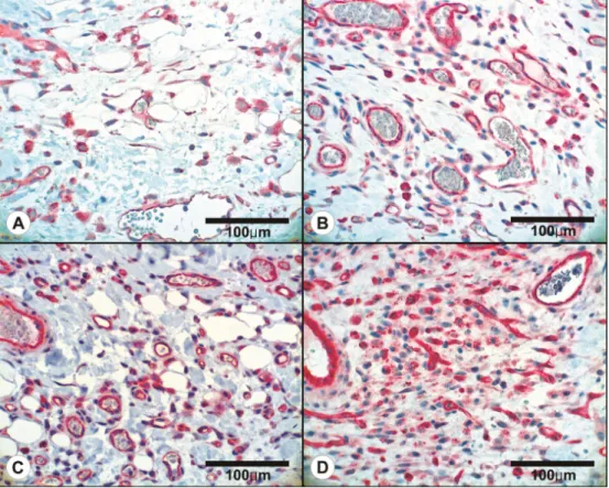
Figure 4. Histologic findings of perfusion zones in transverse rectus abdominis musculocutaneous flaps (control group, left; botulinum toxin A [BoTA] group, right; all, hematoxylin and eosin stain). In zone II of the
control group, focal ischemic necrosis of the epidermis is noted due to impaired blood supply (upper left); relatively collapsed vessels are noted (arrow) (A, ×100). In the BoTA group, the epidermal surface is mostly intact. More patent vessels are evident than in the control group (arrows) (B, ×100). A large area of necrotic surface in the epidermis is noted, along with largely collapsed vessels in the dermis (arrows), in zone III of the controls (C, ×100). In zone III of the BoTA group, intact epidermal structures with dilated microvasculature are seen (D, ×200).
C. Skin flap microvessel density
To calculate the amount of neovascularization, CD31-positive vessels were counted, and a comparative analysis was done on the number of vessels in each high-power field (×400) (Figure 5). In the control group, the numbers of CD31-positive vessels were 35.6 ± 9.1 (zone I), 53.4 ± 10.3 (zone II), 70.9 ± 14.1 (zone III), and 71.7 ± 10.0 (zone IV). Similarly, in the corresponding zones of the BoTA group, there were, respectively, 42.0 ± 11.3, 55.9 ± 11.3, 48.4 ± 12.9, and 46.5 ± 9.3 CD31-positive vessels. The differences between the numbers of CD31-stained vessels in the BoTA and control groups were significant in the contralateral halves (zones III and IV; both p < 0.001).
Figure 5. Representative microvessel density counts (CD31, ×400). The numbers of microvessels in panels A, B, C, and D are 35, 55, 70, and 115, respectively. 3. Gene expression
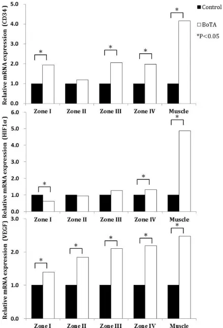
The relative expression of CD34 and VEGF mRNAs was higher in the BoTA group than in the control group, in each zone, except for CD34 in zone II. In zone IV, the relative expression of HIF-1α mRNAs was higher in the BoTA group than in the control group. Further, the relative mRNA expression of CD34, HIF-1α, and VEGF in the right rectus pedicle muscle was significantly higher in the BoTA group than in the control group (Figure 6).
Figure 6. Relative expression of cluster of differentiation 34 (CD34), hypoxia-inducible factor-1α (HIF-1α), and vascular endothelial growth factor (VEGF) mRNA, as determined using quantitative reverse-transcription polymerase chain reaction. The relative expression of CD34, HIF-1α, and
VEGF mRNA in the right rectus muscles, containing inferior epigastric vessels, is significantly higher in the botulinum toxin A (BoTA) group than in the control group. CD34 expression is significantly higher in zones I, II, and IV of the BoTA group than in the corresponding zones in the control group. Zone IV HIF-1α expression is significantly higher in the BoTA group than in the control group, whereas that in zone I of the control group is significantly higher than in the same zone in the BoTA group. VEGF expression is significantly higher in the BoTA group than in the control group. All values are expressed as fold changes.
4. Protein expression
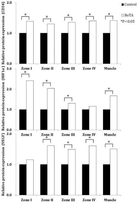
The relative expression of CD34 and VEGF proteins was higher in the BoTA group than in the control group, in each zone except for zone 1 VEGF protein expression. The relative expression of HIF-1α protein was also higher in the BoTA group than in the control group, in each zone except for zone IV. The relative expression of CD34, HIF-1α, and VEGF proteins in the right rectus muscles containing the right inferior epigastric vessels was significantly higher in the BoTA group than in the control group (Figure 7).
Figure 7. Relative expression of cluster of differentiation 34 (CD34), hypoxia-inducible factor-1α (HIF-1α), and vascular endothelial growth factor (VEGF), as determined using Western blotting. The relative expression of CD34, HIF-1α, and VEGF protein in the right rectus muscle containing the
inferior epigastric vessels was significantly higher in the botulinum toxin A (BoTA) group than in the control group. Similarly, all CD34 expression values are significantly higher in the BoTA group than in the control group. HIF-1α expression was significantly higher in zones I, II, and III in the BoTA group than in the corresponding zones in the control group; the zone IV values trend higher in the BoTA group than in the control group, despite not reaching statistical significance. VEGF expression in zones II, III, and IV is significantly higher in the BoTA group than in the control group; zone I also trends higher in the BoTA group than in the control group, but does not reach statistical significance. All values are expressed as fold changes.
IV. DISCUSSION
The purpose of the present study was to investigate the effect of BoTA on TRAM flap survival, focusing on evaluating gene and protein expression to confirm the angiogenic potential of BoTA. The TRAM flap is the workhorse flap for reconstructions occurring after breast cancer ablation2. Many plastic surgeons frequently encounter patients with abdominal scars who require TRAM flap reconstruction. However, some portions of TRAM flaps, in the presence of a scar, should be discarded to improve overall flap survival. Since its development in 1994, the rat TRAM flap model has been considered an excellent analog of the human TRAM flap and has been widely used to study
the effects of healing delays or hemodynamics affecting the flap25-55.
Therefore, our present experiment, using the rat TRAM flap with a vertical midline scar, simulates relevant clinical circumstances.
Our histologic results showed vasodilation of the pedicle and microvessels, leading to improved TRAM flap survival in the BoTA group, compared with the control group. Such vasodilation of choke vessels, between adjacent vascular territories, may help revascularization by crossing the vertical midline scar, leading to increased contralateral flap survival. In addition, higher VEGF expression in the BoTA group, regardless of the transcriptional or posttranslational level, also caused vasodilation, as VEGF is known to stimulate acute vasodilation, endothelial cell migration, and mature neovascularization. There were also significantly increased pedicle lumen areas in the BoTA group than in the control group. Vasodilation may mediate augmented pedicled TRAM survival, with effective perfusion occurring in the BoTA group.
We adopted CD34, a less sensitive or specific marker of angiogenesis, as a marker of gene and protein expression. The close relationship between CD34 and VEGF expression was demonstrated in our study, revealing that CD34 is a reliable marker of angiogenesis in the surgical flap. This is partially due to the HIF-1α/VEGF pathway, which is a strong angiogenesis-related signaling pathway. Decreased HIF-1α mRNA expression in the ipsilateral sides (zones I and II) can be interpreted in 2 ways. One suggests the covert preceding the HIF-1α peak before postoperative day 5. The second suggests the presence of an additional signaling pathway contributing to increased VEGF mRNA and protein expression.
The HIF-1α expression on the ipsilateral sides (zones I and II) is relatively low compared to that on the contralateral sides (zones III and IV). This finding is interpreted to reflect instantaneous stabilization under normoxia (zones I and II). However, relatively high expression of HIF-1α is also possibly due to HIF-1α accumulation under hypoxic conditions (zones III and IV).
Interestingly, BoTA augmented HIF-1α expression under hypoxic conditions (zones III and IV), but did not augment HIF-1α expression under normoxia conditions (zones I and II). Additionally, as this tissue was harvested on postoperative day 5, there might be an earlier, veiled peak of HIF-1α expression, especially in the ipsilateral sides (zones I and II). This interpretation is reasonable because VEGF expression is consistently higher in the BoTA group than in the control group, regardless of zones.
The pedicled right rectus abdominis muscle supplying the TRAM flap demonstrated increased expression of the CD34, HIF-1α, and VEGF genes and proteins. This finding is interesting because BoTA was only injected into the flap, and not the pedicle muscle. In other words, administering BoTA to the
flap is sufficient to stimulate the angiogenic potential, and subsequently augment flap survival. Future studies are necessary to determine in injection into the flap is sufficient to improve flap survival or if better results might be obtained with an injection into the pedicled muscle.
There are some limitations in our study. We did not document the localized hemodynamics, including blood flow and velocity, between the two groups of rats. Initially, however, we measured blood flow in each zone of each TRAM flap using a Periflux 5000 (Perimed AB, Jarfalla, Sweden). Despite this device’s well-known efficacy for measuring direct vascular flow, we could indirectly measure the skin flap’s vascular flow, above the vascular plexus. Because the interzonal flap viability is very heterogeneous, the perfusion data calculated using laser Doppler is very variable, leading us to believe that identifying blood flow or perfusion in rat TRAM flaps using this device is both inaccurate and unreliable. Second, as angiogenesis involves the orchestration of various signaling pathways, the simplification associated with using the several angiogenesis-related markers used in this study has intrinsic limitations for the accurate measurement of the angiogenesis potential of BoTA. Third, as Han et al. suggested in a previous study 56, the vertical midline incision can be considered to be the minimal procedure used to augment the vascularity of a unilateral TRAM flap. The time interval between the creation of the vertical midline incision and TRAM flap elevation would be another determinant of TRAM flap survival. Lastly, to further verify the angiogenic potential of BoTA, additional studies using an HIF-1α knockout animal model should be performed.
Nevertheless, we have discovered several important findings in the current study. First, BoTA increased TRAM flap survival, especially on the contralateral
sides (across vertical midline scars). Second, BoTA might increase HIF-1α/VEGF dependent angiogenesis, possibly involving a novel signaling pathway.
V. CONCLUSION
We demonstrated that preoperative BoTA therapy increases TRAM flap survival in a rat model by increasing HIF-1α/VEGF signaling-dependent angiogenesis.
REFERENCES
1. Kim YS, Yoo HS, Hong JW, Lew D, Roh TS. The abdominal fascial closure in a double-breasted jacket pattern following a TRAM free flap breast reconstruction. J Reconstr Microsurg 2014;30:97-102.
2. Kim EK, Eom JS, Ahn SH, Son BH, Lee TJ. Evolution of the pedicled TRAM flap: a prospective study of 500 consecutive cases by a single surgeon in Asian patients. Ann Plast Surg 2009;63:378-82.
3. McAllister E, Wells K, Chaet M, Norman J, Cruse W. Perineal reconstruction after surgical extirpation of pelvic malignancies using the transpelvic transverse rectus abdominal myocutaneous flap. Ann Surg Oncol 1994;1:164-8.
4. Qiu SS, Jurado M, Hontanilla B. Comparison of TRAM versus DIEP flap in total vaginal reconstruction after pelvic exenteration. Plast Reconstr Surg 2013;132:1020e-7e.
5. Jung JA, Kim YW, Kang SR. Reconstruction of Unexpected Huge Chest Wall Defect after Recurrent Breast Cancer Excision Using a TRAM Flap Combined with Partial Latissimus Dorsi Muscle Flap. Arch Plast Surg 2013;40:76-9.
6. Lasso JM, Uceda M, Penalver R, Moreno N, Casteleiro R, Cano RP. Large posterior chest wall defect reconstructed with a de-epithelised trans-thoracic TRAM flap. J Plast Reconstr Aesthet Surg 2010;63:e458-62.
7. Wang XL, Liu LB, Song FM, Wang QY. Meta-analysis of the safety and factors contributing to complications of MS-TRAM, DIEP, and SIEA flaps for breast reconstruction. Aesthetic Plast Surg 2014;38:681-91.
8. Lee KT, Mun GH. Effects of Obesity on Postoperative Complications After Breast Reconstruction Using Free Muscle-Sparing Transverse Rectus Abdominis Myocutaneous, Deep Inferior Epigastric Perforator, and Superficial Inferior Epigastric Artery Flap: A Systematic Review and Meta-analysis. Ann Plast Surg 2016;76:576-84.
9. Wu JD, Huang WH, Qiu SQ, He LF, Guo CP, Zhang YQ, et al. Breast reconstruction with single-pedicle TRAM flap in breast cancer patients with low midline abdominal scar. Sci Rep 2016;6:29580.
10. Henry SL, Chang CC, Misra A, Huang JJ, Cheng MH. Inclusion of tissue beyond a midline scar in the deep inferior epigastric perforator flap. Ann Plast Surg 2011;67:251-4.
11. Heller L, Feledy JA, Chang DW. Strategies and options for free TRAM flap breast reconstruction in patients with midline abdominal scars. Plast Reconstr Surg 2005;116:753-9; discussion 60-1.
12. Berrino P, Casabona F, Adami M, Muggianu M. The "parasite" TRAM flap for autogenous tissue breast reconstruction in patients with vertical midabdominal scars. Ann Plast Surg 1999;43:119-26.
13. Ohjimi H, Era K, Fujita T, Tanaka T, Yabuuchi R. Analyzing the vascular architecture of the free TRAM flap using intraoperative ex vivo angiography. Plast Reconstr Surg 2005;116:106-13.
14. Kucukkaya D, Irkoren S, Ozkan S, Sivrioglu N. The effects of botulinum toxin A on the wound and skin graft contraction. J Craniofac Surg 2014;25:1908-11.
15. Wang GL, Jiang BH, Rue EA, Semenza GL. Hypoxia-inducible factor 1 is a basic-helix-loop-helix-PAS heterodimer regulated by cellular O2 tension. Proc Natl Acad Sci U S A 1995;92:5510-4.
16. Prabhakar NR, Semenza GL. Adaptive and maladaptive cardiorespiratory responses to continuous and intermittent hypoxia mediated by hypoxia-inducible factors 1 and 2. Physiol Rev 2012;92:967-1003.
17. Lin C, McGough R, Aswad B, Block JA, Terek R. Hypoxia induces HIF-1alpha and VEGF expression in chondrosarcoma cells and chondrocytes. J Orthop Res 2004;22:1175-81.
18. Paradziej-Lukowicz J, Skwarska A, Peszynska-Sularz G,
Brillowska-Dabrowska A, Konopa J. Anticancer imidazoacridinone C-1311 inhibits hypoxia-inducible factor-1alpha (HIF-1alpha), vascular endothelial growth factor (VEGF) and angiogenesis. Cancer Biol Ther 2011;12:586-97.
19. Di Giulio C, Bianchi G, Cacchio M, Artese L, Rapino C, Macri MA, et al. Oxygen and life span: chronic hypoxia as a model for studying HIF-1alpha, VEGF and NOS during aging. Respir Physiol Neurobiol 2005;147:31-8.
20. Hendrickson MD, Poyton RO. Crosstalk between nitric oxide and hypoxia-inducible factor signaling pathways: an update. Research and Reports in Biochemistry 2015;5:147-61.
21. Schweizer DF, Schweizer R, Zhang S, Kamat P, Contaldo C, Rieben R, et al. Botulinum toxin A and B raise blood flow and increase survival of critically ischemic skin flaps. J Surg Res 2013;184:1205-13.
22. Kim TK, Oh EJ, Chung JY, Park JW, Cho BC, Chung HY. The effects of botulinum toxin A on the survival of a random cutaneous flap. J Plast Reconstr Aesthet Surg 2009;62:906-13.
23. Kim YS, Roh TS, Lee WJ, Yoo WM, Tark KC. The effect of botulinum toxin A on skin flap survival in rats. Wound Repair Regen 2009;17:411-7.
24. Holm C, Mayr M, Hofter E, Ninkovic M. Perfusion zones of the DIEP flap revisited: a clinical study. Plast Reconstr Surg 2006;117:37-43.
25. Ozgentas HE, Shenaq S, Spira M. Study of the delay phenomenon in the rat TRAM flap model. Plast Reconstr Surg 1994;94:1018-24; discussion 25-6.
26. Ozgentas HE, Shenaq S, Spira M. Development of a TRAM flap model in the rat and study of vascular dominance. Plast Reconstr Surg 1994;94:1012-7; 25-6 discussion.
27. Hallock GG. The rat TRAM flap: a human analogue? Plast Reconstr Surg 1995;96:233-4.
28. Hallock GG, Rice DC. Evidence for the efficacy of TRAM flap delay in a rat model. Plast Reconstr Surg 1995;96:1351-7.
29. Hallock GG, Rice DC. Physiologic superiority of the anatomic dominant pedicle of the TRAM flap in a rat model. Plast Reconstr Surg 1995;96:111-8.
30. Restifo RJ, Ahmed SS, Isenberg JS, Thomson JG. Timing, magnitude, and utility of surgical delay in the TRAM flap: I. Animal studies. Plast Reconstr Surg 1997;99:1211-6.
31. Hallock GG, Rice DC. Fate of the TRAM flap after abdominoplasty in a rat model. Plast Reconstr Surg 1998;101:1828-35.
32. Qiao Q, Moon W, Zhang F, Chen SG, Kunda L, Lineaweaver WC, et al. Patterns of flap loss related to arterial and venous insufficiency in the rat pedicled TRAM flap. Ann Plast Surg 1999;43:167-71.
33. Morrissey WM, Jr., Hallock GG. The increase in TRAM flap survival after delay does not diminish long term. Ann Plast Surg 2000;44:486-90.
34. Lin KY, Patterson JW, Simmons J, Long MD, Schultz RO, Amiss LR, et al. Effects of external beam irradiation on the TRAM flap: an experimental model. Plast Reconstr Surg 2001;107:1190-7; discussion 8-200.
35. Sano K, Hallock GG, Rice DC. A vertical midline scar is a 'high-risk' factor for maximum survival of the rat TRAM flap. Ann Plast Surg 2003;51:403-8.
36. Sano K, Hallock GG, Rice DC. Venous "supercharging" augments survival of the delayed rat TRAM flap. Ann Plast Surg 2003;51:398-402.
37. Sano K, Hallock GG, Rice DC. Venous interruption is unnecessary to achieve an adequate delay in the rat TRAM flap model. Plast Reconstr Surg 2003;111:300-5.
38. Seify H, Bilkay U, Jones G. Improvement of TRAM flap viability using human VEGF-induced angiogenesis: a comparative study of delay techniques. Plast Reconstr Surg 2003;112:1032-9.
39. Zhang F, Zhang J, Lin S, Oswald T, Sones W, Cai Z, et al. Small intestinal submucosa in abdominal wall repair after TRAM flap harvesting in a rat model. Plast Reconstr Surg 2003;112:565-70.
40. Hallock GG, Rice DC. Comparison of TRAM and DIEP flap physiology in a rat model. Plast Reconstr Surg 2004;114:1179-84.
41. Zhang F, Yang F, Hu EC, Sones W, Lei M, Lineaweaver WC. Vascular endothelial growth factor gene therapy in improvement of skin paddle survival in a rat TRAM flap model. J Reconstr Microsurg 2005;21:391-6. 42. Doncatto LF, da Silva JB, da Silva VD, Martins PD. Cutaneous viability
in a rat pedicled TRAM flap model. Plast Reconstr Surg 2007;119:1425-30.
43. Kim EK, Hong JP. The effect of recombinant human erythropoietin on ischemia-reperfusion injury: an experimental study in a rat TRAM flap model. Plast Reconstr Surg 2007;120:1774-81.
44. Wang H, Li Z, Liu X. Effects of various protocols of ischemic preconditioning on rat tram flaps. Microsurgery 2008;28:37-43.
45. Ely PB, Kobayashi LA, Campos JH, Gomes HC, Juliano Y, Ferreira LM. Nicotine on rat TRAM flap. Acta Cir Bras 2009;24:216-20.
46. de Freitas AL, Gomes HC, Lisboa BC, Arias V, Han SW, Ferreira LM. Effect of gene therapy with vascular endothelial growth factor after abdominoplasty on TRAM flap viability in a rat model. Plast Reconstr Surg 2010;125:1343-51.
47. Cinpolat A, Bektas G, Coskunfirat N, Rizvanovic Z, Coskunfirat OK. Comparing various surgical delay methods with ischemic preconditioning in the rat TRAM flap model. J Reconstr Microsurg 2014;30:335-42.
48. Lucca AF, Brasolin AG, Feitosa RG, Simoes e Silva Enokihara MM, Gomes HF, Ferreira LM. Histological modification in TRAM flap in rats treated with pentoxifylline. Acta Cir Bras 2014;29 Suppl 2:34-7.
49. Mericli AF, Das A, Best R, Rodeheaver P, Rodeheaver G, Lin KY. Abstract 107: Deferoxamine Mitigates Radiation-Induced Hypovascularity and Improves Tissue Elasticity in a Rat Irradiated TRAM Flap Model. Plast Reconstr Surg 2014;133:124.
50. Mericli AF, Das A, Rodeheaver P, Rodeheaver G, Lin KY. Abstract 110: The Impact of Deferoxamine on Vascularity and Soft Tissue Biomechanics in a Rat TRAM Flap Model. Plast Reconstr Surg 2014;133:127.
51. Mericli AF, Das A, Best R, Rodeheaver P, Rodeheaver G, Lin KY. Deferoxamine mitigates radiation-induced tissue injury in a rat irradiated TRAM flap model. Plast Reconstr Surg 2015;135:124e-34e.
52. Mericli AF, Das A, Rodeheaver P, Rodeheaver GT, Lin KY. The Impact of Deferoxamine on Vascularity and Soft-Tissue Biomechanics in a Rat TRAM Flap Model. Plast Reconstr Surg 2015;136:125e-7e.
53. Park TH, Rah DK, Chong Y, Kim JK. The effects of botulinum toxin A on survival of rat TRAM flap with vertical midline scar. Ann Plast Surg 2015;74:100-6.
54. Ersoy B, Cevik O, Cilingir OT. Etanercept protects myocutaneous flaps from ischaemia reperfusion injury: An experimental study in a rat tram flap model. J Plast Surg Hand Surg 2016;50:208-15.
55. Nacak U, Calis M, Atilla P, Cetin A, Aksu AE. Extracorporal Shock Wave Therapy as a Delay Procedure to Improve Viability of Zone 4: An Experimental Study in a Rat TRAM Flap Model. Ann Plast Surg 2016;77:e15-20.
56. Han S, Sup Eom J, Ho Kim D. Effects of the abdominal midline incision on the survival of the transverse rectus abdominis musculocutaneous flap in rat model. Ann Plast Surg 2003;50:171-6.
ABSTRACT(IN KOREAN) 백서의 횡복직근 피판 생존에 보툴리눔 독소 A가 미치는 영향 <지도교수 나 동 균> 연세대학교 대학원 의학과 박 태 환 본 연구에서는 백서의 횡복직근 피판 모델에 보툴리눔 독소 A 를 국소 적용하였을 때 혈관 확장 및 혈관 신생 관련 성장인자의 과발 현을 통해 횡복직근 피판 생존에 미치는 영향을 규명하고자 하였다. 24 마리의 백서를 보툴리눔 독소 국소 투여군과 생리식염수를 투여 할 대조군으로 나누었다. 먼저 복부의 칼돌기 직하방에서 5cm 길이의 직선 절개선을 가하고 봉합 시행 5 일 후에 실험군은 보툴리눔 독소 A 를 국소 투여하고 대조군에서는 같은 양의 생리 식염수를 투여하 였다. 주사 5 일 후에 횡복직근 피판을 거상하였다. 피판을 거상한 후 하방에 실리콘 시트를 적용한 후 피판을 원래 위치로 다시 위치시키 고 피판의 생존면적을 술 후 제1 일, 제 3 일, 제 5 일째 측정하였고 최 종 피판 생존면적은 술 후 5 일째 측정하였다. 헤마톡실린-에오신염 색을 통해 횡복직근 피판의 혈관경 면적의 변화 및 전체적인 조직학 적 변화의 양상, CD31 면역 화학 염색법을 통해 피판 내 미세혈관농 도를 각각 술 후 5 일째 평가하였다. 그리고 같은 시기에 혈관신생에
관여하는 인자인 CD34, HIF-1α, VEGF 의 유전자 및 단백질을 정량적 으로 측정하였다. 생존피판 면적 분석에서 보툴리눔 독소 투여군이 대조군에 비하여 통계적으로 유의하게 높은 피판 생존률을 나타내었다. 보툴리눔 독 소 투여군에서 대조군에 비해 혈관경 면적이 유의하게 확장되어 있 는 소견이 관찰되었으며, 대조군에서 보툴리눔 독소 투여군에 비해 경도에서 중증도의 표피괴사가 관찰되었으며, 미세혈관도 위축되어 있는 소견을 보였다. 미세혈관밀도 측정에서는 보툴리눔 독소 투여 군이 동측 부위에서는 높게 나타났지만 두 군간의 유의한 차이가 나 타나지 않았다. 반대측 부위에서는 오히려 보툴리눔 독소 투여군에 서 유의하게 낮게 측정되었다. CD34 와 VEGF 유전자 발현은 보툴리 눔 독소 투여군에서 대조군에 비해서 대부분 통계적으로 유의하게 증가한 소견이 관찰되었다. HIF-1α 는 허혈이 진행된 영역 IV 에서 보 툴리눔 독소 투여군에서 대조군에 비하여 발현이 증가되는 것을 관 찰하였다. CD34, VEGF, HIF-1α 의 단백질 발현은 보툴리눔 독소 투여 군에서 대조군에 비해서 통계적으로 유의하게 증가되어 있는 소견을 보였다. 본 연구를 통해서 술전 보툴리눔 독소 A 를 백서의 횡복직근 피판 모델에 국소 적용하였을 때 횡복직근피판의 혈관경을 확장시키고, 피판내 및 횡복직근의 혈관신생인자를 대조군에 비해 증가시켰으며 이를 통해 생존면적을 증가시킴을 확인할 수 있었다. 핵심 되는 말: 보툴리눔 독소 A, 횡복직근피판, 혈관 신생, 지연처치
Publication list
1. Park TH, Rah DK, Chong Y, Kim JK. The effects of botulinum toxin
A on survival of rat TRAM flap with vertical midline scar. Ann Plast Surg 2015;74:100-6.
2. Park TH, Lee SH, Park YJ, Lee YS, Rah DK, Kim SY. Presurgical
Botulinum Toxin A Treatment Increases Angiogenesis by Hypoxia-Inducible Factor-1α/Vascular Endothelial Growth Factor and Subsequent Superiorly Based Transverse Rectus Abdominis Myocutaneous Flap Survival in a Rat Model. Ann Plast Surg 2016;76:723-8.

![Figure 3. Histologic findings for the right inferior epigastric artery and vein in transverse rectus abdominis musculocutaneous flaps (Upper; control group, left; botulinum toxin A [BoTA] group, right)](https://thumb-ap.123doks.com/thumbv2/123dokinfo/5100052.78999/26.892.180.728.233.767/histologic-findings-inferior-epigastric-transverse-abdominis-musculocutaneous-botulinum.webp)
![Figure 4. Histologic findings of perfusion zones in transverse rectus abdominis musculocutaneous flaps (control group, left; botulinum toxin A [BoTA] group, right; all, hematoxylin and eosin stain)](https://thumb-ap.123doks.com/thumbv2/123dokinfo/5100052.78999/27.892.184.712.506.914/histologic-findings-perfusion-transverse-abdominis-musculocutaneous-botulinum-hematoxylin.webp)