허정식, 김성대, 박경기, 김영주, 유현욱 94 https://w cms .jejunu .ac .k r/medsci/ The Journal of M
edicine and Lif
e S cienc e https://w cms .jejunu .ac .k r/medsci/ The Journal of M
edicine and Lif
e S cienc e
서 론
방광암은 비뇨기계암 중 흔하게 발생되는 암종이며 대부분 은 표재성이며 재발율이 높다.1) 증상으로는 육안적 혈뇨가 흔 하지만 건강에 대한 관심의 증가로 인해 건강검진과 요검사가 보편화되면서 현미경적 혈뇨 등의 증상으로 병원을 방문하여 초음파검사를 통해 우연하게 방광종물이 발견되는 경우가 많 아지고 있다. 방광암의 원인은 다양하지만 흡연이 주요 원인이 되고 있으며 매년 흡연율은 감소하고 있으나 방광암의 발생률 은 줄지 않고 있다. 암의 진단으로는 비침습적인 방광과 신장 의 초음파검사가 널리 시행되고 있으나 가장 중요한 진단방법 은 방광내시경을 통한 검사와 요세포검사이다.2) 요세포검사의 경우에 암이 발견되는 경우는 많지 않으며 방광내시경은 요도 점막을 부분마취나 연성방광내시경기구를 통한 검사로 통증을 줄이기 위해 노력하고 있으나 검사할 당시의 자세와 방광내시 경 삽입 시 통증으로 인해 환자들이 검사를 꺼려하는 경우가 있고 이전에 회음부 통증 혹은 성기 부분의 통증으로 인해 간 혹 거부하는 경우도 있다. 이러한 경우에 방광종물의 재발을 발견하지 못하는 경우가 있고 암이 진행되어 침윤성방광암으 로 근치적방광적출술 혹은 수술과 항암치료 등을 하는 경우가 간혹 발생되고 있다. 이에 본 연구는 육안적 혈뇨나 혹은 현미 경적 혈뇨가 있는 환자와 표재성방광암환자의 추적관찰 중 신 장과 방광초음파검사만으로 방광종물을 발견할 수 있는지에 대하여 연구를 하였다.The Journal of Medicine and Life Science Vol. 17, No. 3, 94-97, December 2020 https://doi.org/10.22730/jmls.2020.17.3.94
eISSN: 2671-4922
방광종물에 대한 방광초음파검사의 진단적 유용성
허정식
1, 김성대
1, 박경기
1, 김영주
1, 유현욱
2,1제주대학교 의학전문대학원 비뇨기과학교실, 2유쾌한비뇨기과의원
The role of bladder sonography in patients with gross hematuria or microscopic hematuria and follow-up of patients with superficial bladder cancer by Jung-Sik Huh1, Sung Dae Kim1, Kyung Kgi Park1,
Young Joo Kim1, Hyun Wook You2 (1Department of Urology, Jeju National University Hospital, Jeju National
University, Graduate School of Medicine, Jeju, Republic of Korea; 2Happy Urologic Clinics, Jeju, Republic of Korea)
Abstract Ultrasonography is used to examine gross or microscopic hematuria without side effects. It is one of
the methods of diagnosing bladder lesions, but in some cases, the lesions are not found. We attempted to identify the problems during the ultrasonic examination by analyzing the symptoms, location of lesion, and medical history of urothelial cancer for cases of undetected bladder lesions. Thirty-three patients who underwent transurethral resection of a bladder tumor from January 1 to May 4, 2018 in one hospital were enrolled in this study. Patients who underwent preoperative ultrasonography and cystoscopy were treated. Ultrasonography did not detect bladder
lesions in five patients. The size of the lesion was 0.5~2.5cm in various locations, such as the side, front, and so on.
Ultrasonic examination requires more attention if there is gross hematuria or a history of urothelial cancer, and it is necessary to detect recurrence by conducting cystoscopy at the same time, especially when there are lesions on the anterior wall of the bladder.
Key words: Ultrasonography, Urothelial cancer, Hematuria
Received: October 20, 2020; Revised: December 7, 2020; Accepted: December 8, 2020 Correspondence to : Hyun Wook You
Happy Urologic Clinics, Jeju, Republic of Korea Tel: 82-64-805-1151, FAX: 82-64-805-1153 E-mail: [email protected]
Copyright The Journal of Medicine and Life Science
Original Article
방광초음파의 진단적 유용성 95 https://w cms .jejunu .ac .k r/medsci/ The Journal of M
edicine and Lif
e S cienc e
대상 및 방법
2018년 1월1일부터 2018년 5월 4일까지 단일 병원에서 현 미경적 혈뇨 혹은 육안적 혈뇨를 주소로 내원한 환자 혹은 이 전 표재성방광암이나 신우암 등에 대한 정기검사를 위해 내원 한 환자를 대상으로 하였다. 초음파검사는 영상의학과 전문의 에 의해 모두 시행이 되었으며 검사 전 소변을 참고 시행되었 다. 초음파의 탐촉자는 3.5MHz를 이용하여 시행하였으며 방 광의 종물을 확인하기 위해 앙와위에서 방광의 종면과 횡면을 검사하였으며 이후 앙와위 혹은 측와위에서 신장의 종물 여부 를 검사하였다. 방광에 소변이 차지 않은 경우에는 신장에 대 한 초음파검사를 먼저 시행한 이후 대상자에게 물을 마시게 한 이후 방광이 소변으로 찬 이후 다시 초음파검사를 시행하 였다. 방광종물 이외에 다른 암종에 대한 추적관찰을 위해 컴 퓨터단층촬영술을 시행하기도 하였다. 이후 방광내시경은 환 자가 동의를 한 경우에 시행하였으며 남성의 경우에는 요도에 국소마취제를 이용하여 시행하였으며 여성의 경우에는 별다 른 전처치 이후 시행하였다. 방광에 전벽과 천장부위를 잘 보 기 위해 70도의 방광경을 이용하였으며 이후 방광경부와 요도 를 관찰하기 위해 30도의 방광경을 이용하였다. 이 중 종물이 발견되어 경요도적 방광종물절제술을 시행한 33명의 환자를 대상으로 후향적으로 의무기록과 방광종물 혹은 신장의 종물 을 발견하기 위한 초음파검사와 컴퓨터단층촬영술을 확인하였 다. 방광종물이 발견되고 이후 수술이 시행되었던 환자를 대상 으로 주된 임상증상과, 종물의 위치와 종물의 크기 등에 대하 여 초음파의 한계에 대하여 분석을 하였다. 이 연구는 병원 의 학연구윤리심의위원회의 승인을 받아서 연구를 진행하였다.결 과
이 기간 내의 경요도적 방광종물절제술을 시행한 환자는 총 33명이었다(Table 1). 성별로는 남성이 29명, 여성이 4명으로 남성이 많았다. 연령은 71.94±11.48(41~92)세였다. 검사 당 시 흡연은 27.3%였고 현미경적 혈뇨 혹은 육안적 혈뇨가 주된 증상인 경우가 60.6%였다. 방광암을 포함한 비뇨기암과 다른 암으로 진단하여 치료를 받은 환자가 57.6%였으며 이 중 비뇨 기암은 45.5%였다. 방광암으로 이전 경요도적방광종물절제술 을 시행받았던 환자는 48.5%였다. 33명 중 20명은 컴퓨터단층 촬영과 방광내시경검사 이후 수술을 시행하였으며 13명은 초 음파검사와 방광내시경검사 이후 수술을 시행하였다. 13명 중 5명은 신장방광초음파에서 방광 내 종물이 발견되지 않았지 만 방광내시경검사에서 방광종물이 발견되어 수술을 시행하였 다. 신장방광초음파검사에서 방광 내 종물의 진단율은 61.54% 였다. 각 환자의 증상은 주로 육안적 혈뇨가 4례로 20%를 차 지하였으며, 표재성방광암에 대한 경요도적방광종물절제술 이 후 정기적인 검사에서 발견된 경우가 1례였다. 요로상피암의 병력이 있는 경우가 4례로 20%였다. 신장방광초음파검사 소 견으로는 1례에서 우측 후면에 0.5cm의 용종이 의심되었으나 실제 전면에 2.5cm의 종물이 발견되었다. 다른 증례로는 방광Table 1. General characteristics of subjects(N=33)
Sex(M/F) 29/4 Age (yr) 71.94±11.48(41~92) Smoking(%) 27.3 Hematuria(%) 60.6 Hypertension(%) 21.2 Diabetes mellitus(%) 12.1 All cancer (%) 57.6 Urogenital cancer(%) 45.5 TURBT(%) 48.5 Ultrasonography(%) 84.8 Computer tomography(%) 75.8 Ultrasonography+Computer tomography(%) 63.8 Hematuria: gross hematuria and microscopic hematuria
TURBT: transurethral resection of bladder tumor
Table 2. Sonographic findings and real cystoscopic findings of patient with superficial bladder cancer
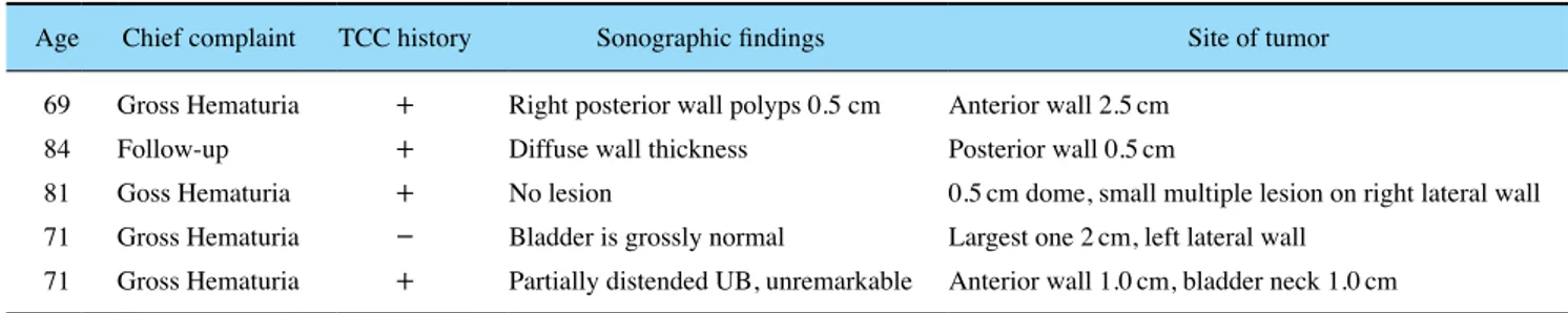
Age Chief complaint TCC history Sonographic findings Site of tumor 69 Gross Hematuria + Right posterior wall polyps 0.5 cm Anterior wall 2.5cm
84 Follow-up + Diffuse wall thickness Posterior wall 0.5cm
81 Goss Hematuria + No lesion 0.5cm dome, small multiple lesion on right lateral wall 71 Gross Hematuria - Bladder is grossly normal Largest one 2cm, left lateral wall
허정식, 김성대, 박경기, 김영주, 유현욱 96 https://w cms .jejunu .ac .k r/medsci/ The Journal of M
edicine and Lif
e S cienc e https://w cms .jejunu .ac .k r/medsci/ The Journal of M
edicine and Lif
e S cienc e 벽이 두꺼워진 것 이외에는 특이소견이 없었다. 경요도적방광 종물절제술에서 종물의 위치는 다양하였으며 전면, 후면, 최상 부, 방광경부 등 모든 부위에 다 발생이 되었다. 종물의 크기는 0.5cm부터 2.5cm까지 다양하였다. 1례의 경우 방광이 소변의 팽창이 잘 안되어 있는 경우도 있었다(Table 2, Fig. 1).
고 찰
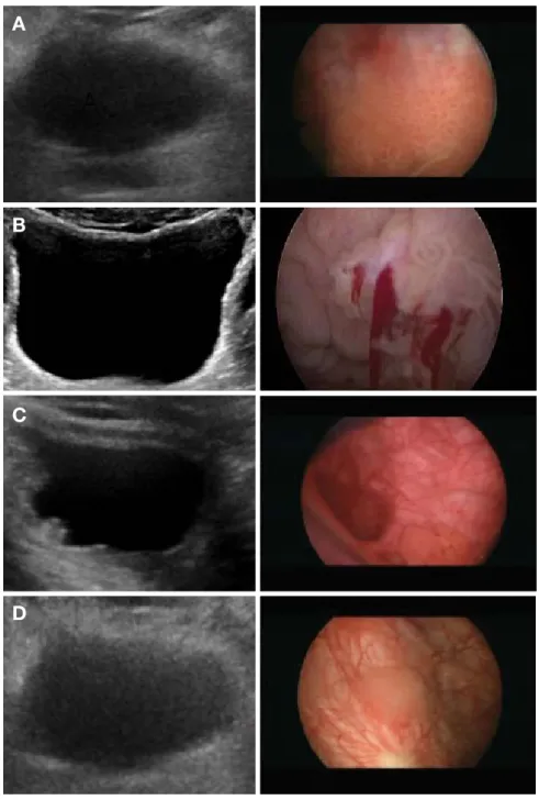
요로상피암 중 가장 많이 발생되는 방광암은 다양한 원인에 의해서 발생되고 있고 꾸준하게 유병률은 유지되고 있다. 증상 은 육안적 혈뇨가 많으며 수술 당시 대부분 표재성방광암으로Fig. 1. Bladder sonographic and cystoscopic findings. A: Sonographic findings: No bladder full filling, No lesion. Cystoscopic finding: 2.0cm
papillary lesion on left lateral wall. B: Sonographic findings: No bladder lesion. Cystoscopic finding: papillary lesion on anterior wall. C: Sono-graphic findings: moderate trabeculation of bladder wall. Cystoscopic finding: 0.5cm papillary lesion on posterior wall. D: Sonographic findings: No bladder lesion. Cystoscopic finding: papillary lesion on latreal wall.
A
B
C
방광초음파의 진단적 유용성 97 https://w cms .jejunu .ac .k r/medsci/ The Journal of M
edicine and Lif
e S cienc e 경요도방광종물절제술을 통해 완치가 되는 경우가 많지만 재 발률이나 암이 진행되어 침윤적 방광암으로 발생되기도 한다. 표재성방광암의 재발은 종물의 크기, 방광벽의 침범 정도, 종 물의 분화도와 재발 등과 연관이 있으며 재발을 방지하기 위
해 BCG나 mitomycin C, adriamycin, gemcitabine 등을 방광
내에 주입하는 경우가 있다.3) 이러한 시술 이후 재발 여부를 확인하기 위해 정기적인 추적관찰을 해야 한다. 즉 표재성방광 암의 수술 이후 1년째는 3개월에 한 차례, 2년째는 6개월에 한 차례, 이후 5년까지 1년에 한 차례 초음파검사와 방광내시경검 사, 요세포검사를 권하고 있으며 방광암의 조기진단을 위해 많 은 노력을 경주하고 있다.4) 초음파검사 이외 암의 진행 정도를 확인하기 위해 컴퓨터단층촬영을 하는 경우도 있다. 컴퓨터단 층촬영은 질병을 진단하기 위해 정확도는 높은 편이나 방사선 노출이 많고 조영제에 부작용이 있는 환자에게는 한계를 지니 고 있다. 최근 신장방광에 대한 초음파검사가 1년에 1회 보험 적용이 되고 건강보험 암 산정특례가 되면 초음파검사는 모두 보험적용이 되어 초음파검사가 늘 것으로 예상된다. 초음파는 비침습적이고 통증이 없고 부작용이 없는 것으로 표재성방광 암 환자에게는 좋은 검사방법으로 여겨지고 통증이 심한 방광 내시경검사는 꺼려하는 경우가 많고 거부하는 경우도 발생되 고 있다. 본 연구와 같이 신장방광초음파검사는 통증이 없이 시행 할 수 있는 장점은 있으나 방광종물에 대한 진단율은 61.54% 로 낮으며, 특히 방광종물의 크기가 작거나, 방광점막에 국한 되어 돌출되지 않은 경우에는 발견할 수 없는 경우가 많이 발 생한다. 또한 방광경부의 경우와 방광이 전반적으로 팽창이 되 지 않은 경우에는 방광의 전체벽을 뚜렷하게 관찰이 불가능하 기 때문에 방광의 종물인지 방광벽의 과증식을 구분하는 것은 어렵다. 즉 신장에 대한 초음파검사는 금식이나 소변을 참을 필요는 없지만 방광초음파를 시행할 경우 방광이 팽창이 되지 않으면 정확하게 병변을 발견할 수 없으며 특히 표재성방광 암이 재발된 경우에는 잦은 수술로 인해 방광의 용적이 줄어 들거나 하부요로증상 등으로 소변을 참기가 힘든 경우가 많아 정확하게 병변을 구분하기가 어렵다. 이러한 점에서 방광내시 경검사는 작은 방광 내 병변을 찾아내어 조기에 제거함으로써 병변의 진행을 막을 수 있는 진단방법으로 표재성방광암환자 나 육안적 혈뇨가 있는 환자의 경우에는 적극적으로 방광내시 경검사를 권해야 할 것이다. 또한 통증을 줄이기 위해 경성보 다는 연성 방광경을 이용하여 검사를 시도하는 것이 좋을 것 이며 연구기간이 너무 짧고 연구대상 수가 적은 제한점이 있 지만 방광과 신장초음파검사의 역할에 대한 연구가 필요하다.
결 론
육안적 혈뇨 혹은 이전에 요로상피암의 병력이 있는 경우에 초음파검사는 더욱 주의를 요하며 특히 방광의 전벽에 종물이 있는 경우와 방광경부에 종물이 있는 경우에 발견이 어려운 경우가 많아 방광내시경검사가 최우선적인 진단도구가 되어야 하며 초음파검사의 역할에 대한 연구가 필요할 것이다.REFERENCES
1. Park KK, Kim SD, Kim YJ, Huh JS. Trends in urogenital cancer incidence in Jeju(1999-2012). Korean J Urol Oncol 2016;14:27-31.
2. D’Elia C, Pycha A, Folchini DM, Mian C, Hanspeter E, Schwien-bacher C, et al. Diagnostic predictive value of Xpert Bladder Cancer Monitor in the follow-up of patients affected by non-mus-cle invasive bladder cancer. J Clin Pathol 2019;72:140-4. 3. Hori S, Miyake M, Tatsumi Y, Morizawa Y, Nakai Y, Onishi S, et
al. Intravesical treatment of chemotherapeutic agents sensitizes bacillus Calmette-Guerin by the modulation of the tumor immune environment. Oncol Rep 2019;41:1863-74.
4. Usuba W, Urabe F, Yamamoto Y, Matsuzaki J, Sasaki H, Ichikawa M, et al. Circulating miRNA panels for specific and early detec-tion in bladder cancer. Cancer Sci 2019;110:408-19.