Current Status of Laparoscopic Liver Resection in Korea
Since laparoscopic liver resection was first introduced in 2001, Korean surgeons have chosen a laparoscopic procedure as one of the treatment options for benign or malignant liver disease. We distributed and analyzed a nationwide questionnaire to members of the Korean Laparoscopic Liver Surgery Study Group (KLLSG) in order to evaluate the current status of laparoscopic liver resection in Korea. Questionnaires were sent to 24 centers of KLLSG. The questionnaire consisted of operative procedure, histological diagnosis of liver lesions, indications for resection, causes of conversion to open surgery, and postoperative outcomes. A laparoscopic liver resection was performed in 416 patients from 2001 to 2008. Of 416 patients, 59.6% had malignant tumors, and 40.4% had benign diseases. A total laparoscopic approach was performed in 88.7%. Anatomical laparoscopic liver resection was more commonly performed than non-anatomical resection (59.9% vs 40.1%). The anatomical laparoscopic liver resection procedures consisted of a left lateral sectionectomy (29.3%), left hemihepatectomy (19.2%), right hemihepatectomy (6%), right posterior sectionectomy (4.3%), central bisectionectomy (0.5%), and caudate lobectomy (0.5%). Laparoscopy-related serious complications occurred in 12 (2.8%) patients. The present study findings provide data in terms of indication, type and method of liver resection, and current status of laparoscopic liver resection in Korea.
Key Words: Laparoscopic Liver Resection; Laparoscopy; Anatomical Liver Resection; Liver Diseases; HCC
Joon Seong Park1, Ho-Seong Han2, Dae Wook Hwang2, Yoo-Seok Yoon2, Jai Young Cho2, Yang-Seok Koh3,
Choon Hyuck David Kwon4, Kyung Sik Kim5, Sang Bum Kim6, Young Hoon Kim7, Hyung Chul Kim8, Chong Woo Chu8,
Dong Shik Lee9, Hong-Jin Kim9, Sang Jae Park10, Sung-Sik Han10, Tae Jin Song11,
Young Joon Ahn12, Yung Kyung Yoo13,
Hee Chul Yu14, Dong Sup Yoon1, Min-Koo Lee15, Hyeon Kook Lee16, Seog Ki Min16,
Chi-Young Jeong17, Soon-Chan Hong17, In Seok Choi18, and Kyung Yul Hur19
Departments of Surgery, 1Yonsei University College of Medicine, Gangnam Severance Hospital, Seoul; 2Seoul National University Bundang Hospital, Seoul National University College of Medicine, Seongnam; 3Chonnam National University Medical School, Gwangju; 4Samsung Medical Center, Sungkyunkwan University School of Medicine, Seoul; 5Yonsei University College of Medicine, Seoul; 6Korea Cancer Center Hospital, Korea Institute of Radiological & Medical Sciences, Seoul; 7Dong-A University College of Medicine, Busan; 8Soonchunhyang University Bucheon Hospital, Soonchunhyang University College of Medicine, Bucheon; 9Yeung-Nam University College of Medicine, Daegu; 10Center for Liver Cancer, National Cancer Center, Goyang; 11Ansan Hospital, Korea University College of Medicine, Ansan; 12Seoul Metropolitan Government Seoul National University Boramae Medical Center, Seoul; 13Seoul St. Mary’s Hospital, The Catholic University of Korea College of Medicine, Seoul; 14Chonbuk National University Hospital and Medical School, Jeonju; 15Eulji University College of Medicine, Daejeon; 16Ewha Womans University Mokdong Hospital, Ewha Womans University School of Medicine, Seoul; 17Gyeongsang National University Hospital, Gyeongnam Regional Cancer Center, Institue of Health Sciences, Gyeongsang National University, Jinju; 18Konyang University Hospital, Konyang University College of Medicine, Daejeon; 19Soonchunhyang University College of Medicine, Seoul, Korea
Received: 19 November 2011 Accepted: 16 April 2012 Address for Correspondence: Ho-Seong Han, MD
Department of Surgery, Seoul National University Bundang Hospital, 166 Gumi-ro, Bundang-gu, Seongnam 463-707, Korea
Tel: +82.31-787-7091, Fax: +82.31-787-4055 E-mail: [email protected]
This study was supported by a grant from the Korea Healthcare Technology Research and Development Project Ministry of Health and Welfare, Korea (A102065).
http://dx.doi.org/10.3346/jkms.2012.27.7.767 • J Korean Med Sci 2012; 27: 767-771
INTRODUCTION
Since laparoscopic liver resection was first reported by Reich et
al. (1) in 1991, many reports about laparoscopic liver resection have been published. However, laparoscopic liver resection was performed only at a limited number of institutions because
of the technical difficulty associated with the procedure. In Korea, laparoscopic liver resection has been introduced and demonstrated to be safe and effective in selected patients (2). More recently, advances in laparoscopic instruments and increased experience with liver surgery have encouraged Korean surgeons to choose a laparoscopic procedure as one of the treat-ment options for benign or malignant liver diseases.
Inspired by the encouraging outcomes with this procedure, Korean surgeons who are interested in laparoscopic liver sur-gery have formed the Korean Laparoscopic Liver Sursur-gery Study Group (KLLSG). This study is a nationwide survey from mem-bers of the KLLSG to report the current status of laparoscopic liver resection in Korea.
Despite the different and evolving techniques in each center, the current status of laparoscopic liver resection in Korea was evaluated.
MATERIALS AND METHODS
A questionnaire was collected from the surgeons of the KLLSG in 24 centers. The questionnaire consists of operative procedure, histological diagnosis of liver lesions, causes of conversion to open surgery, and indications for laparoscopic liver resection. The surgeons of 19 centers who responded to questionnaires performed 416 laparoscopic liver resections in Korea from 2001 to 2008.
Ethics statement
This study was approved by the institutional review board of Yonsei University for retrospective chart review and data col-lection (3-2008-0170). Informed consent was waived by the board.
RESULTS
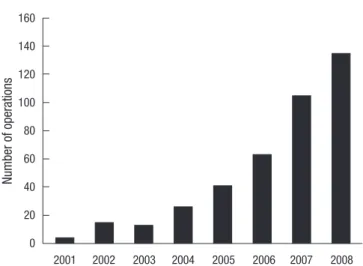
Number of patients for laparoscopic liver resection in Korea according to period
After the introduction of laparoscopic liver resection, there has been an exponential growth in the number of laparoscopic liver resections in Korea. Although there were few cases of laparo-scopic liver resection in the early period, the number of cases performed in a year increased rapidly in the late period (Fig. 1).
Pathologic diagnosis for laparoscopic liver resection
Laparoscopic liver resection including a robotic surgery was performed in 416 patients. The indications for laparoscopic liver resection included benign tumor, liver abscess, intrahepatic duct stones, hepatocellular carcinoma (HCC) and metastatic tumor (Fig. 2). Of the 416 cases, 59.6% were malignant tumors and 40.4% were benign diseases. The majority of the malignant lesions were HCC and metastatic carcinoma (38.0% and 18.5%, respectively). The remainders of the malignant lesions were chol-angiocellular carcinoma and mucinous carcinoma. The major-ity of laparoscopic liver resection for benign diseases was per-formed for hepatolithiasis (27.4%) and benign tumor (12.0%).
Laparoscopic approach to liver resection
The majority of laparoscopic liver sections were performed with a total laparoscopic approach in 369 patients (88.7%). The lapa-roscopic assisted procedures or hand assisted procedures were performed in 47 patients (11.3%). Total laparoscopic procedures included 5 robotic assisted liver resections.
Fig. 1. Exponential growth of laparoscopic liver resection in Korea from 2001 to 2008. Nu m be r o f o pe ra tio ns 2001 2002 2003 2004 2005 2006 2007 2008 160 140 120 100 80 60 40 20 0
Fig. 2. Indication for laparoscopic liver resection.
Hepatocellular carcinoma 158 38.0% Intrahepatic duct stone 114 28.0% Metastatic cancer 77 18.5% Intrahepatic abscess 1 0% Pathologic indications Mucinous carcinoma 1 0% Intrahepatic cholangiocarcinoma 14 3% Biliary stricture 1 0% Benign hepatic tumor 50 12.0%
The type of laparoscopic liver resection
The anatomical laparoscopic liver resection was more com-monly performed than the non-anatomical resection (59.9% vs 40.1%). The anatomical laparoscopic liver resection procedure consisted of a left lateral sectionectomy in 122 (29.3%) cases, left hemihepatectomy in 80 (19.2%), right hemihepatectomy in 25 (6%), right posterior sectionectomy in 18 (4.3%), central bisectionectomy in 2 (0.5%), and caudate lobectomy in 2 (0.5%) (Fig. 3).
Conversion rate to open surgery
Conversion to open surgery occurred in 42 patients (10.1%): 25 patients due to uncontrolled bleeding, 7 due to inappropriate surgical margin or an obscure location of the lesion, 5 due to anatomic difficulties, 2 due to severe adhesion, 1 due to inter-nal organ injury, and 2 due to tumor rupture and advanced carcinoma.
Laparoscopic liver resection related to serious complications
Serious complications occurred in 12 patients (2.8%). Endo-scopic stapler malfunction occurred in 8 patients (1.9%). Myo-cardial infarction during pneumoperitoneum occurred in 1 (0.2%) patient, CO2 gas embolism in 1 (0.2%), trocar port site
bleeding in 1 (0.2%), and cancer recurrence at the trocar site in 1 (0.4%). There were no mortalities.
Surgeon’s response to indications of tumor size and locations
Most surgeons reported that tumor size was an important indi-cation for laparoscopic liver resection. Of the surgeons, 21% reported that they would perform a laparoscopic liver resection
for tumors 3 cm or smaller and 37% for tumors 5 cm or smaller. However, 37% of surgeons reported that they would perform laparoscopic liver resection for HCC, irrespective of tumor size. More than half of the surgeons (79%) reported that they would perform laparoscopic liver resection for tumors in the periph-eral portion of the antero-latperiph-eral segments of the liver (segments II, III, V, and VI and the inferior part of IV). However, the others (21%) reported that they would perform laparoscopic liver re-section for lesions in the posterior or superior part of the liver (segments I, VII, and VIII and the superior part of IV).
DISCUSSION
Although laparoscopic surgery has been widely practiced in the field of abdominal surgery, such as colon and gastric surgery, laparoscopic liver resection is still limited in specialized centers. Laparoscopic liver resections demand enough experience in both laparoscopic and open surgery. In addition, it requires advanced laparoscopic surgical techniques associated with parenchymal dissection and hemostasis. With the increased experience with laparoscopic surgery and development in lap-aroscopic instruments, it has been possible to apply laparoscop-ic liver resection to various kinds of liver diseases.
Since the introduction of laparoscopic liver resection in Korea (2, 3), there has been an exponential growth of the number of cases treated with the procedure in Korea. It is likely that the numbers of surgeons who apply laparoscopic liver resection will be increasing in the future. In early stages, this procedure was performed in benign disease of the liver. With increasing reports of good outcomes of laparoscopic surgery for malignancy, appli-cation of this surgery to malignant disease of the liver has been increased accordingly.
In this study, more than half of the patients (59.6%) under-went laparoscopic surgery for HCC and metastatic cancer. Sev-eral studies have shown that survival outcomes of laparoscopic procedures are comparable to those of open surgery. In our se-ries, although there was one case of port site recurrence, there were no peritoneal seeding or perioperative mortality.
Since Azagra et al. (4) performed the first anatomical resec-tion, laparoscopic left lateral sectionectomy has become the standard treatment in some centers (5). In this study, laparo-scopic left lateral sectionectomy was the most common proce-dure of anatomical liver resection. In the near future, laparo-scopic left lateral sectionectomy may become the routine pro-cedure in most centers. However, laparoscopic major hepatec-tomy is not applied worldwide because of the complexity of procedures and fear of uncontrolled bleeding. This procedure is recommended to be performed by an experienced surgeon in the consensus meeting of laparoscopic liver surgery (6). In this study, 105 of the 416 liver resections (25.2%) were performed by major laparoscopic liver resection. This multicenter
experi-Fig. 3. Type of laparoscopic liver resection. Left hemihepatectomy 19.2% Right hemihepatectomy 6.0% Right posterior sectionectomy 4.3% Central bisectionectomy 0.5% Caudate lobectomy 0.5% Non-anatomic resection (greater or equal 1 segment) 8.7% Left lateral sectionectomy 29.3% Non-anatomic resection (less than 1 segment)
ence demonstrated that laparoscopic major hepatectomy was feasible and safe in selected patients. Although this study showed that laparoscopic major hepatectomy could be safely performed, this procedure should be reserved for centers with experience in laparoscopic liver resection.
With respect to the operative procedures, total laparoscopic procedures were performed in 88% of the patients, whereas hy-brid or hand assisted laparoscopic procedures were performed in 12%, which was lower than reports from other countries (7). Since the introduction of total laparoscopic liver resection in Korea, this procedure has shown good results when compared to open surgery. These good results have encouraged many liver surgeons to follow the pioneer’s operative methods (8). Hybrid or hand assisted laparoscopic procedures are easy to apply for surgeons who are accustomed to open surgery. There-fore, we think that hybrid or hand assisted laparoscopic proce-dures have a role as a bridge procedure of liver resection for a surgeon who is going to start a laparoscopic liver resection pro-gram.
Despite the different and evolving techniques in each center, bleeding was the most common reason for conversion. In this study, the conversion rate was 10%, which was similar to previ-ously reported rates (7, 9-12). The incidence of serious compli-cations, such as endoscopic stapler malfunction, ST depression during pneumoperitoneum, CO2 gas embolism, and cancer
recurrence at the trocar site were 2.8%. In particular, the inci-dence of endostapler malfunction was 1.9%, which was compa-rable with the previously reported rate (13). Recently, many reports have shown that using the stapler device for hepatic resection helped to reduce operation time and bleeding amount during transection of the liver (14, 15). Although we rarely en-countered stapler malfunction, the operator should use stapling devices properly and be cautious of abnormal feelings during the stapling manipulation.
In the early stage, laparoscopic liver resection applied only to lesions located in the left lateral or peripheral segments. Due to the difficulty of bleeding control and visualization of the sur-gical fields, lesions in the deep or posterior sections in the liver (segments I, VII, and VIII and the superior part of IV) were pre-viously considered to be poor indications for laparoscopic liver resection (16-18). Moreover, it would be difficult to obtain a safety tumor margin for the inferior portion of the tumor when the tumor was located in the posterior and superior parts. How-ever, recent reports have demonstrated the feasibility and safety of laparoscopic liver resection of the lesions in those locations (19, 20). In our study, over 20% of surgeons answered that they would apply the procedure even in segments I, VII, and VIII and the superior part of IV. We expect the lesions located in the posterior or superior part of the liver may be candidates for lap-aroscopic resection in the future.
In this study, we introduce a national survey about
laparo-scopic liver surgery in Korea. With the national survey, a large number of patients were included in this study, and a wide range of disease types and operation procedures could be evaluated. However, a limitation may arise. There were some centers that have not responded to this survey, and there might be a concern in terms of whether the data entirely represented the Korean status of laparoscopic liver resection. Despite the limitation, the results of this survey provide data in terms of indication, type and method of liver resection, and current status of laparoscop-ic liver resection. We believe that laparoscoplaparoscop-ic liver resection is a new treatment strategy for the treatment of liver tumors in many hospitals in Korea. The Korean Laparoscopic Liver Sur-gery Study Group plans to conduct further surveys to propose standardization of laparoscopic liver resection for HCC.
REFERENCES
1. Reich H, McGlynn F, DeCaprio J, Budin R. Laparoscopic excision of be-nign liver lesions. Obstet Gynecol 1991; 78: 956-8.
2. Min SK, Han HS, Kim YW, Choi YM. Laparoscopy-assited major liver resection. J Korean Soc Endosc Laparosc Surg 2002; 5: 75-9.
3. Hong TH, Lee SK, Park SC, Kim WW, Jeon HM, Kim EK. Laparoscopic liver resection: different methods. J Korean Soc Endosc Laparosc Surg 2002; 5: 37-43.
4. Azagra JS, Goergen M, Gilbart E, Jacobs D. Laparoscopic anatomical (hepatic) left lateral segmentectomy-technical aspects. Surg Endosc 1996; 10: 758-61.
5. Chang S, Laurent A, Tayar C, Karoui M, Cherqui D. Laparoscopy as a routine approach for left lateral sectionectomy. Br J Surg 2007; 94: 58-63. 6. Buell JF, Cherqui D, Geller DA, O’Rourke N, Iannitti D, Dagher I, Koffron
AJ, Thomas M, Gayet B, Han HS, et al. The international position on lap-aroscopic liver surgery: The Louisville Statement, 2008. Ann Surg 2009; 250: 825-30.
7. Tsuchiya M, Otsuka Y, Tamura A, Nitta H, Sasaki A, Wakabayashi G, Kaneko H. Status of endoscopic liver surgery in Japan: a questionnaire survey conducted by the Japanese Endoscopic Liver Surgery Study Group. J Hepatobiliary Pancreat Surg 2009; 16: 405-9.
8. Min SK, Han HS, Lee HK, Jie S, Yu K, Yi NJ, Choi YM. Totally laparoscopic anatomic liver resection. J Korean Surg Soc 2003; 64: 390-5.
9. Koffron AJ, Auffenberg G, Kung R, Abecassis M. Evaluation of 300 mini-mally invasive liver resections at a single institution: less is more. Ann Surg 2007; 246: 385-92.
10. Simillis C, Constantinides VA, Tekkis PP, Darzi A, Lovegrove R, Jiao L, Antoniou A. Laparoscopic versus open hepatic resections for benign and malignant neoplasms: a meta-analysis. Surgery 2007; 141: 203-11. 11. Topal B, Fieuws S, Aerts R, Vandeweyer H, Penninckx F. Laparoscopic
versus open liver resection of hepatic neoplasms: comparative analysis of short-term results. Surg Endosc 2008; 22: 2208-13.
12. Viganò L, Tayar C, Laurent A, Cherqui D. Laparoscopic liver resection: a systematic review. J Hepatobiliary Pancreat Surg 2009; 16: 410-21. 13. Chan D, Bishoff JT, Ratner L, Kavoussi LR, Jarrett TW. Endovascular
gas-trointestinal stapler device malfunction during laparoscopic nephrecto-my: early recognition and management. J Urol 2000; 164: 319-21.
14. Gumbs AA, Gayet B, Gagner M. Laparoscopic liver resection: when to use the laparoscopic stapler device. HPB (Oxford) 2008; 10: 296-303. 15. Rowe AJ, Meneghetti AT, Schumacher PA, Buczkowski AK, Scudamore
CH, Panton ON, Chung SW. Perioperative analysis of laparoscopic ver-sus open liver resection. Surg Endosc 2009; 23: 1198-203.
16. Cherqui D, Husson E, Hammoud R, Malassagne B, Stéphan F, Bensaid S, Rotman N, Fagniez PL. Laparoscopic liver resections: a feasibility study in 30 patients. Ann Surg 2000; 232: 753-62.
17. Dulucq JL, Wintringer P, Stabilini C, Berticelli J, Mahajna A. Laparoscopic liver resections: a single center experience. Surg Endosc 2005; 19: 886-91.
18. Laurent A, Cherqui D, Lesurtel M, Brunetti F, Tayar C, Fagniez PL. Lap-aroscopic liver resection for subcapsular hepatocellular carcinoma com-plicating chronic liver disease. Arch Surg 2003; 138: 763-9.
19. Cho JY, Han HS, Yoon YS, Shin SH. Experiences of laparoscopic liver resection including lesions in the posterosuperior segments of the liver. Surg Endosc 2008; 22: 2344-9.
20. Han HS, Cho JY, Yoon YS. Techniques for performing laparoscopic liver resection in various hepatic locations. J Hepatobiliary Pancreat Surg 2009; 16: 427-32.