Desktop Microtomography with A CMOS Pixel Detector
Ho Kyung Kim a,*, Min Kook Cho a, Sung Sik Lee ba School of Mechanical Eng., Pusan National University, Jangjeong-dong, Geumjeong-gu, Busan 609-735, Korea b SafeTech, Techno Park B/D, Eomgung-dong, Sasang-gu, Busan 617-729, Korea
* Corresponding Author: [email protected]
1. Introduction
Recent developments both in the microfocus X-ray source and the large-area pixel detector have brought out a cone-beam computed tomography (CBCT) with the resolving power of the order of tens of micrometers, called microtomography (micro-CT) [1-5]. Although the micro-CT shares the same technology with the conventional CT (and therefore the technology is not novel), new potential applications in biological as well as materials sciences is great [2-5].
This study is aimed at developing a cost-effective miniaturized micro-CT system. The eventual purpose is to apply it to small-animal imaging. Detailed system design and configuration are presented. The potential performance of the system is demonstrated with the initially acquired both radiographs and tomograms.
2. Design of Desktop Microtomography
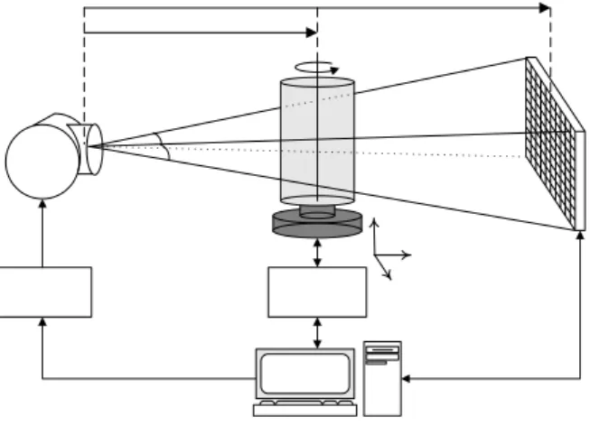
The desktop micro-CT system being developed in this study consists of an X-ray source with small focus, a rotational subject manipulator, and an X-ray imaging detector. A schematic drawing describing the system is shown in Fig. 1. The source-to-object (or subject) and source-to-detector distances, shortly SOD and SDD respectively, are variables determining the imaging parameters such as geometrical magnification ratio, actual cone-beam angles (φ1 and φ2) and field-of-view (FOV). A computer-controlled rotation system was adopted in the subject manipulator to achieve a cone-beam mode scan. The precision of the rational motion is 0.036°.
Figure 1. A sketch depicting the desktop micro-CT system being developed in this study (see text).
The X-ray source (Series 5000 Apogee, Oxford Instruments, US) is a sealed tube with a 125-µm-thick beryllium exit window. Tungsten target is incorporated to produce X rays with energies ranging from 4 to 50 kVp at 50 W. Further imaging conditions by tailoring the X-ray beam profile are possible with additional filtration. Nominal focal spot size is 35 µm. The emitted X-ray beam angle is about 22°. The X-ray source continuously irradiates the sample to be imaged without any shutters while the CMOS pixel detector acquires two-dimensional (2-D) projection data at a given frame time (or integration time).
The core of the system is an X-ray imaging detector. The detector has been constructed with a scintillation layer for converting incident X rays to optical photons and photosensitive pixel arrays for reading out the emitted optical photons, i.e. converting those to electrical signal.
A commercial phosphor screen (Min-R Medium, Eastman Kodak, US) was used as the X-ray converter. The screen is mainly made up of a terbium-doped gadolinium oxysulfide (Gd2O2S:Tb) and a polyurethane elastometer as the phosphor and binder, respectively. Therefore, it gives reduced density of about 4 g/cm3. The coverage of the screen is about 34 mg/cm2. The screen shows an emission spectrum with the main peak at 545 nm.
CMOS pixel arrays (RadEyeTM, Rad-icon Imaging Corp., US) [6], i.e. the photosensitive pixel array made by CMOS process, were employed as the optical photon readout device. The CMOS pixel array has a format of 512 × 1024 pixels with a pitch of 48 µm. In this study, two CMOS pixel arrays were tiled side by side, and therefore the FOV is about 50×50 cm2. The total conversion gain from the pixel to sensor output is about 0.5 µV/e-. The fill factor is approximately 90%. Two parallel voltage signals from two CMOS pixel arrays are simultaneously digitized to 12-bits gray level through dual ADCs (PCI-6111, National Instruments, US). It is noted that the 12-bit image data is converted into 16-bit form for further process such as post image processing and image reconstruction.
3. Image Acquisition
System magnification ratio is usually set to about 2 (SDD = 200 mm). The frame time of the CMOS pixel detector to acquire a single projection data is normally 275 ms. For the image reconstructions, we applied the
Transactions of the Korean Nuclear Society Autumn Meeting Busan, Korea, October 27-28, 2005
Feldkamp's cone-beam algorithm [7] to the projection data filtered with the Ram-Lak filter (or high-pass filter). For a typical CT scan, the size of acquired data is about 1 GByte (= 2 Bytes/pixel × 10242 pixels/projection × 500 projections).
4. Acquired Images
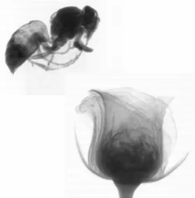
Figure 2 shows an examples of the X-ray radiographs obtained from the developed system. No digital image processing procedures were used after they were acquired. Only flat-field corrections were made. For the display, two separate images were digitally combined to one image.
For an electrolytic capacitor, a 2-D projection is shown in Fig. 3(a). In Fig. 3(b) we have shown a cross-sectional image of the capacitor. The voxel size is 24×24×24 µm3.
5. Summary and Further Study
A cost-effective desktop microtomography based on CMOS pixel detector has been constructed, and its potential performance has been demonstrated with initially obtained tomograms. For the better understanding and optimal design of the developing system, we are still trying to evaluate the detector performance in terms of MTF, NPS and DQE. The details of the results will be addressed at the conference. Quantitative investigation of tomographic imaging performances, such as voxel signal uniformity and noise characteristics, contrast-to-noise ratio, resolving power, and so on, is further study for the reliable use of the system.
Figure 2. An example of X-ray radiograph. The image was digitally synthesized with separate bee and rose images.
Figure 3. (a) A projection image of an electrolytic capacitor acquired with a magnification ratio of 2. (b) A cross-sectional image of the capacitor after applying CT reconstruction algorithm.
REFERENCES
[1] H.K. Kim, Sensor Technology for Digital Radiography, Journal of the Korean Society of Precision Engineering, Vol. 22, No. 8, pp. 7-16, 2005.
[2] S.C. Lee, H.K. Kim, I.K. Chun, M.H. Cho, S.Y. Lee, and M.H. Cho, A Flat-Panel Detector Based Micro-CT System: Performance Evaluation for Small-Animal Imaging, Physics in Medicine and Biology, Vol. 48, pp. 4173-4185. 2003.
[3] S.C. Lee, H.K. Kim, I.K. Chun, M.H. Cho, M.H. Cho, and S.Y. Lee, Development and Characterization of A Flat-Panel Detector-Based Microtomography System, Key Engineering Materials, Vols. 270-273, pp. 245-251, 2004. [4] H.K. Kim, S.C. Lee, M.H. Cho, S.Y. Lee, and G. Cho,
Use of A Flat-Panel Detector for Microtomography: A Feasibility Study for Small-Animal Imaging, IEEE Transactions on Nuclear Science, Vol. 52, No. 1, pp. 193-198, 2005.
[5] H.K. Kim, Cone-Beam Microtomography and Its
Application, Journal of the Korean Society of Precision Engineering, Vol. 22, No. 3, pp. 7-14, 2005.
[6] T. Graeve and G.P. Weckler, High Resolution CMOS imaging Detector, in Proceedings of SPIE Vol. 4320
Medical Imaging 2001: Physics of Medical Imaging,
edited by L.E. Antonuk, M.J. Yaffe, (SPIE, Bellingham, WA, 2001) pp. 68-76.
[7] L.A. Feldkamp, L.C. Davis, and J.W. Kress, Practical Cone-Beam Algorithm, Journal of the Optical Society of America A, Vol. 1, No. 6, pp. 612-619 (1984).