저작자표시-비영리-변경금지 2.0 대한민국 이용자는 아래의 조건을 따르는 경우에 한하여 자유롭게 l 이 저작물을 복제, 배포, 전송, 전시, 공연 및 방송할 수 있습니다. 다음과 같은 조건을 따라야 합니다: l 귀하는, 이 저작물의 재이용이나 배포의 경우, 이 저작물에 적용된 이용허락조건 을 명확하게 나타내어야 합니다. l 저작권자로부터 별도의 허가를 받으면 이러한 조건들은 적용되지 않습니다. 저작권법에 따른 이용자의 권리는 위의 내용에 의하여 영향을 받지 않습니다. 이것은 이용허락규약(Legal Code)을 이해하기 쉽게 요약한 것입니다. Disclaimer 저작자표시. 귀하는 원저작자를 표시하여야 합니다. 비영리. 귀하는 이 저작물을 영리 목적으로 이용할 수 없습니다. 변경금지. 귀하는 이 저작물을 개작, 변형 또는 가공할 수 없습니다.
Transgelin-2 (TAGLN2)
as a potential therapeutic target
of gemcitabine resistant-
biliary tract cancer
Joo Won Chung
Department of Medicine
The Graduate School, Yonsei University
[UCI]I804:11046-000000517579 [UCI]I804:11046-000000517579
Transgelin-2 (TAGLN2)
as a potential therapeutic target
of gemcitabine resistant-
biliary tract cancer
Joo Won Chung
Department of Medicine
Transgelin-2 (TAGLN2) as a potential
therapeutic target of gemcitabine
resistant-biliary tract cancer
Directed by Professor Si Young Song
The Doctoral Dissertation
submitted to the Department of Medicine,
the Graduate School of Yonsei University
in partial fulfillment of the requirements for the degree of
Doctor of Philosophy
Joo Won Chung
June 2018
ACKNOWLEDGEMENTS
First of all, I acknowledge my deep gratitude to Professor Si
Young Song for his supervision on this whole study. I always
think that the meeting Professor Song is one of the most
memorable events in my life. His passion and effort for research
have always been a role model for me.
I am incredibly grateful to Professor Seungmin Bang who guided
and supported this study overall. This study could not be
conducted without his guidance.
Besides, I am profoundly thankful to Professor Kyoung Tai No,
Hyun Seok Kim, and Jung Min Han who led the academic
discussion and gave keen advice on this study. Their specialist
assistance was essential and crucial in this study. And, I am
indebted to Soo Been Park for her help for my experiments.
Finally, I would like to express my love and thanks to my parents,
sister, and brothers for providing their tremendous support and
unwavering love. I was able to accomplish my research because
of their great support. And most importantly, I thank God for
giving me this loving family. I dedicate this thesis to my family
and God.
TABLE OF CONTENTS
ABSTRACT……….………..……..……....1
I. INTRODUCTION………..……...………3
II. MATERIALS AND METHODS………..………5
1. Cell culture……….……….…...5
2. Western blotting ………...….………...…….…5
3. Cell proliferation assay ……….…….………..…….6
4. Migration and invasion assays………...………...….……….…6
5. Sphere formation assay………..………7
6. Colony formation assay……….………7
7. Tissue collection and sample preparation……….….…7
8. Immunohistochemistry……….……….7 9. Transfection of TAGLN2………...8 10. Xenograft experiments………...………...……….9 11. Cytotoxic assay………...………9 12. Statistical analysis………..………..…10 III. RESULTS……….……...10
1. Suppression of TAGLN2 expression impairs cellular proliferation, migration, and invasion in BTC cells in vitro………...…..10
2. Suppression of TAGLN2 decreases the tumorigenic potential of SNU-1196 cells in vitro………..…………..12
3. Suppression of TAGLN2 expression results in inhibition of tumorigenicity and tumor growth in vivo………..13
4. The expression of TAGLN2 and Ki-67 are decreased in shTAGLN2 transfected xenograft tumors………..………....14
5. TAGLN2 suppression reduces expression of CSC and EMT markers...15
6. TAGLN2 is rarely expressed in major human organ tissue on IHC
………..………....….…………...……..17
7. TAGLN2 is highly expressed in human BTC tissues compared with normal bile duct tissues……….……….18
8. Reduced survival and the higher rate of recurrence were observed in BTC patient with highly expressed TAGLN2………..….20
9. Altered expression of CSC related markers and overexpression of TAGLN2 in gemcitabine-resistant BTC cells………...21
10. Suppression of TAGLN2 in SNU-1196/GR cells decreases drug resistance to other cytotoxic drugs……….…....24
IV. DISCUSSION………..27
V. CONCLUSION……….30
REFERENCES………..………32
ABSTRACT (IN KOREAN) ...38
LIST OF FIGURES
Figure 1. Effect of TAGLN2 knockdown on cell proliferation,
migration, and invasion of SNU-1196 cells...11
Figure 2. Effect of TAGLN2 suppression on self-renewal and
clonogenic capacities of SNU-1196 cells...12
Figure 3. Anti-proliferative effects of TAGLN2 suppression in
BTC cells in xenograft model...14
Figure 4. Illustrations of hematoxylin and eosin staining (H&E)
and IHC with antibodies against TAGLN2 antibody and the
proliferation marker Ki-67 antibody on xenograft tumor
sections………….………...………….15
Figure 5. Expression of CSC-related and EMT markers on
shTAGLN2-C4/SNU-1196 cells and shTAGLN2-C11/SNU-1196
cells...……16
Figure 6. Expression of CSC-related markers on siTAGLN2
transfected SNU-308 cells……….……….17
Figure 7. Illustrations of IHC with the anti-TAGLN2 polyclonal
antibody on multiple normal human organ tissues…...…18
Figure 8. Representative IHC of TAGLN2 and the comparison in
human BTC tissues and normal bile duct tissues within individual
patient…..………...……...…...19
Figure 9. The gemcitabine-resistant BTC (SNU-1196/GR) cells
overexpress CSC-related proteins and EMT markers………….22
Figure 10. Overexpression of ALDH, ABCG2, and MRP4 in
SNU-1196/GR cells-derived tumors by IHC…….………....…..23
Figure 11. Overexpression of TAGLN2 in gemcitabine-resistant
BTC (SNU-1196/GR) cells, and increased chemosensitivity by
downregulation of TAGLN2 using siTAGLN2 transfection
(siTAGLN2/SNU-1196/GR cells)………...24
Figure
12.
Increased
chemosensitivity
to
5-FU
in
gemcitabine-resistant BTC cells after TAGLN2 suppression…..25
Figure 13. Effect of TAGLN2 suppression on the susceptibility of
gemcitabine-resistant BTC cells to platinum-based anti-cancer
drug………..…26
Figure 14. Effect of TAGLN2 suppression on the susceptibility of
gemcitabine-resistant BTC cells to topoisomerase inhibitor...26
Figure 15. Effect of TAGLN2 suppression on the susceptibility of
gemcitabine-resistant BTC cells to EGFR inhibitor………... …27
LIST OF TABLES
Table 1. Correlations between TAGLN2 expression and
clinicopathological factors of patients with BTC………...20
1
ABSTRACT
Transgelin-2 (TAGLN2) as a potential therapeutic target of gemcitabine resistant-biliary tract cancer
Joo Won Chung Department of Medicine
The Graduate School, Yonsei University (Directed by Professor Si Young Song)
Purpose
Transgelin-2 (TAGLN2) is highly expressed in biliary tract cancer (BTC) associated cancer stem cells (CSC) compared with normal bile duct cells by genetic analysis. However, the functional role of TAGLN2 has not been determined. We investigated that the role of TAGLN2 in malignant behaviors and chemoresistance in BTC cells.
Materials and Methods
The TAGLN2 knockdown cell line was established by transfection of small hairpin TAGLN2 (shTAGLN2) into human biliary tract cancer cell line, SNU-1196 and SNU-308, and the cell proliferation, migration, invasion, and molecular characteristics were assessed in vitro and in vivo. The TAGLN2 expression on multiple human organ tissues including BTC was investigated by immunohistochemistry (IHC). Small interfering TAGLN2 (siTAGLN2) was transfected into gemcitabine-resistant BTC cells, and cell viability was assessed in chemotherapeutic agents including gemcitabine, 5-FU, cisplatin, carboplatin, oxaliplatin, irinotecan, etoposide, and erlotinib.
Results
2
migration, and invasion in shTAGLN2/SNU-1196 cells. A significant reduction of tumorigenic potential of shTAGLN2/SNU-1196 cells was observed in vitro and in vivo. Western blotting revealed decreased expression of CSC markers such as c-met, Nanog, pAKT and AKT, and mesenchymal markers such as N-cadherin, Snail, and JAG2, but an increase in the epithelial molecule, Occludin in shTAGLN2/SNU-1196 cells. TAGLN2 was rarely expressed in most of the normal human organs tissues, whereas strong expression was noted in BTC tissue. However, there was no significant association of tumor staging, differentiation, and prognosis with the level of TAGLN2 expression. Suppression of TAGLN2 in SNU-1196/GR cells by siTAGLN2 transfection increased drug sensitivity to antimetabolites, gemcitabine, and 5-FU.
Conclusion
TAGLN2 plays functional roles in the progression of BTC, and CSC markers and mesenchymal markers were downregulated in TAGLN2 suppressed BTC cells. Our data suggest that chemosensitivity may be improved by suppression of TAGLN2 in gemcitabine-resistant BTC cells.
--- Key words: biliary tract cancer, transgelin-2, cancer stem cell, epithelial-mesenchymal transition, chemoresistance
3
Transgelin-2 (TAGLN2) as a potential therapeutic target of gemcitabine resistant-biliary tract cancer
Joo Won Chung Department of Medicine
The Graduate School, Yonsei University
(Directed by Professor Si Young Song)
I. INTRODUCTION
Biliary tract cancer (BTC) is a relatively uncommon but aggressive gastrointestinal cancer with a high mortality rate.1 The surgical resection is the only curative modality for BTC,2 but the majority of the tumor has already metastasized at the time of diagnosis.3 Even though complete resection was achieved, more than 50% of surgically resected BTC recurred,4 and only 20~30% of these patients survive after 5 years due to unsuccessful treatment.5,6 For patients presenting with unresectable or metastatic BTC, there is no choice for palliative treatment except systemic chemotherapy or radiotherapy. Gemcitabine combined with cisplatin is recommended as a first-line treatment for this patient.7 This combination chemotherapy improves the progression-free and overall survival, but the median overall survival is no longer than one year in metastatic BTCs.8 Oxaliplatin might be an alternative to cisplatin,9 but no further standard regimens are established.10
The reasons for the low response to anticancer treatment and high recurrence rate are based on the cancer stem cell (CSC)-like characteristics of BTC.11 Most BTC associated CSCs show the characteristics by epithelial-mesenchymal transition (EMT) features, therefore BTCs show
4
highly desmoplastic, morphological heterogenic, aggressive and resistant to chemotherapy. An important way to improve the chemosensitivity is to understand the characteristics of CSC in BTC, especially EMT, and to develop target therapy for inhibiting EMT.
We recently identified that transgelin-2 (TAGLN2) is highly expressed in BTC associated CSCs compared with normal bile duct tissues by genetic analysis.12
TAGLN2 is an actin-binding protein that plays a role in determining cell morphology and transformation.13 This protein may be involved in cytoskeleton organization. However, we have not determined the functional role of TAGLN2 in BTC, yet. Many studies reported that TAGLN2 is highly expressed in various human malignancies, including esophageal cancer,14 colorectal cancer,15,16 breast cancer,17 renal cell carcinoma,18 neck squamous cell carcinoma,19 lung cancer,20 uterine cervical cancer,21 and bladder cancer22 suggesting that TAGLN2 has functional roles in the progression of malignant tumors. TAGLN2 may represent a metastatic potential in advanced disease rather than cancer development. TAGLN2 may influence metastasis is through direct interaction with cytoplasmic actin. There is also evidence that regulation of TAGLN2 expression affects the expression of metastasis-related genes.23 TAGLN2 may be a candidate for biomarkers not only for cancer diagnosis, but also for prediction of cancer treatment and prognosis. More importantly, TAGLN2 can be a new target for cancer treatment, which can be a clue to overcome resistance to current chemotherapy.
The roles of TAGLN2 in cancer development, metastasis and chemoresistance are unclear in BTC. Therefore, the present study was undertaken to
investigate the role of TALGN2 in malignant behaviors and chemoresistance in BTC cells.
5
II. MATERIALS AND METHODS
1. Cell Culture
SNU-1196 and SNU-308 human BTC cell lines were purchased from the Korean Cell Line Bank (Seoul, Korea), and were maintained in RPMI 1640 (Gibco, Grand Island, NY, USA) containing 10% fetal bovine serum (Hyclone, Logan, UT, USA) in a humidified incubator of 5% CO2 at 37 °C.24 NIH-3T3 mouse fibroblast cells were obtained from the American Type Culture Collection (ATCC, Manassas, VA) and were grown in DMEM (Gibco) with 10% fetal bovine serum. Gemcitabine-resistant BTC cells were established by escalating doses of gemcitabine serially in SNU-1196 as described in the previous study. 25
2. Western blotting
Cells were lysed in lysis buffer containing 70 mM -glycerophosphate (pH 7.2), 0.6 mM Na vanadate, 2 mM MgCl2, 1 mM EGTA, 1 mM DTT, 0.5% Triton X-100, 0.2 mM phenylmethylsulfonyl fluoride (PMSF), and 1x complete protease inhibitor (Roche Applied Science, Nutley, NJ, USA). Protein (20 μg) was resolved on SDS-polyacrylamide gels and transferred to polyvinylidene difluoride (PVDF) membranes (Immobilon-P, Millipore, Bedford, MA, USA). The membranes were blocked in 5% (w/v) non-fat dry milk and probed with the following primary antibodies: rabbit polyclonal anti-TAGLN2 (Sigma-Aldrich, Inc., St. Louis, MO), rabbit polyclonal anti- phospho-AKT, rabbit polyclonal anti-Nanog, rabbit polyclonal anti-Snail, rabbit polyclonal anti-JAG2 (Cell Signaling Technology, Inc., Danver, MA), rabbit polyclonal anti-Met, rabbit polyclonal anti-AKT, rabbit polyclonal anti-Occludin, rabbit polyclonal ABCG2, mouse monoclonal anti-GAPDH (Santa Cruz Biotechnology Inc, Santa Cruz, CA), mouse monoclonal anti-ALDH (BD Transduction Laboratories, San Jose, CA) and mouse monoclonal anti-N
6
cadherin (Abcam plc., Cambridge, UK). Horseradish peroxidase-conjugated secondary antibody was used for detection and immunoblots were developed with West Pico Chemiluminescent Substrate (Thermo Scientific, Rockford, IL).
3. Cell Proliferation Assay
Cell proliferation assay was performed using WST-1 reagent (Roche Applied Science) according to the manufacturer’s instructions. SNU-1196 cells transfected with TAGLN2 shRNA or control shRNA were seeded into 96-well-plates at a concentration of 1 x10³ cells/well in the 100μL culture medium. After 48 hours, WST-1 (10μL) was added to each well and incubated at 37°C for 1 hour. The absorbance of the formazan product (450 nm) against a background control (630 nm) was measured using a VersaMax ELISA microplate reader (Molecular Devices, USA).
4. Migration and Invasion Assays
Migration and invasion assays were determined in Transwell plate (Costar, USA). For invasion assays, matrigel/serum free medium (1:4, BD Biosciences) was added to Transwell membrane filter inserts with eight μm pores and incubated for 1 hour in a 5% CO2 incubator. Cells (1~2 x 10⁴ cells/well) were seeded in serum-free RPMI medium in the upper chambers, and the lower chambers were filled with conditioned media derived from NIH3T3 cultures followed by incubation for 24 hours for migration assays or 72 hours for invasion assays. Migrated and invaded cells were then fixed in 5% glutaraldehyde/PBS and stained with toluidine blue. The degree of migration/invasion was determined by counting cells that migrated through the membrane at three randomly chosen visual fields per filter.
7
5. Sphere Formation Assay
Cells were trypsinized, and single-cell suspensions were confirmed microscopically, washed with PBS, and resuspended at a concentration of 1000 cells/mL in serum-free DMEM/Ham’s F12 medium supplemented with epidermal growth factor (10 ng/mL; R&D Systems Inc, Minneapolis, Minn), basic fibroblast growth factor (10 ng/mL; R&D Systems Inc), 1x insulin transferring selenium (Invitrogen Gibco), 0.5% BSA (Invitrogen Gibco) and 0.5% FBS. Cell suspensions were seeded in 6-well ultra-low attachment plates (Corning Inc, Corning, NY) at 37°C in 5% CO2 for ten days.
6. Colony Formation Assay
Colony formation assay was performed with a total of 3 x 10² cells suspended in 0.5 mL of 0.3% Difco agar noble (Becton Dickinson Company, Sparks, MD) supplemented with sphere formation medium and plated in 24-well plates containing 0.6% agar. All samples were plated in triplicate. Cells were incubated for 2~3 weeks in a humidified incubator in 5% CO2 at 37°C, and the sphere formation medium was changed every other day.
7. Tissue collection and sample preparation
The tissue samples of biliary tract cancer and normal bile duct were obtained from those who had received surgical resection for extrahepatic bile duct cancer in Severance Hospital, Korea between 2004 and 2007. Biliary tract cancer was confirmed pathologically and did not harbor with other types of cancer. A total 46 samples from biliary tract cancer and 34 samples from normal bile duct were recruited.
8. Immunohistochemistry
Mouse tumor specimen and human bile duct specimen were embedded in paraffin and stained with hematoxylin and eosin according to standard
8
protocols. For immunohistochemistry (IHC), sections of paraffin-embedded tissue were deparaffinized in xylene and rehydrated in a graded ethanol series. Endogenous peroxidase activity was blocked by incubation in methanol containing 0.3% hydrogen peroxide at room temperature for 20 min. Antigen retrieval was performed in citrate buffer (0.01 M, pH 6.0), followed by blocking in 10% normal donkey serum for one hour at room temperature. Slides were incubated with diluted primary antibody in antibody diluent (Gibco) at 4°C; Rabbit polyclonal anti-TAGLN2 (1:500, Sigma-Aldrich, Inc., St. Louis, MO), Mouse monoclonal anti-Ki 67 (1:100, Dako, Carpinteria, CA, USA), Mouse monoclonal anti-ALDH (1:200, BD Transduction Laboratories, San Jose, CA), Rabbit polyclonal anti-ABCG2 and Mouse monoclonal anti-MRP4 (1:200, Santa Cruz Biotechnology Inc, Santa Cruz, CA). The reaction was carried out using an Envision kit (Dako, Carpinteria, CA, USA). Samples were interpreted by two certified pathologists who were blinded to patients’ clinical information from our hospital.
9. Transfection of TAGLN2
To verify the inhibitory effect of TAGLN2 expression, the TAGLN2 knockdown cell line was established by transfection of TAGLN2 short hairpin RNA (shRNA) plasmid (shTAGLN-C4 and shTAGLN2-C11) or mock shRNA plasmid from Qiagen into SNU1196 cells. The shTAGLN2-C4 plasmid, shTAGLN-C11 plasmid or mock shRNA plasmid was transfected using Lipofectamine 2000 (Invitrogen). Twenty-four hours after transfection, media were replaced by media with 2 μg/mL of puromycin and maintained for three days. Surviving cells were cultured in 100 mm plate at 20-30 cells per plate, and a single colony was selected using a cloning cylinder (Sigma-Aldrich). Western blots were performed to determine the effects of TAGLN2 knockdown.
9
10. Xenograft experiments
The 1 × 10⁶ three types of knockdown cells, shControl cells, shTAGLN2-C4, and shTAGLN2-C11 cells, were suspended in 100 μL serum-free RPMI and Matrigel (1:1 volume) respectively, and subcutaneously injected into the right flank of six-week-old female BABL/c nude mice (OrientBio, Seongnam, Korea). Tumor formation was monitored once a week by measuring the width and length. All mice were sacrificed after 5 weeks. The removed tumor volume was calculated using the formula: volume (mm3) = (length × width2)/2, and fixed in 4% paraformaldehyde. Then tumors were embedded in paraffin and stained with hematoxylin and eosin (H&E) for histological evaluation. All animal experiments were approved by the Committee for the Care and Use of Laboratory Animals of Yonsei University College of Medicine (2012-0018-1).
11. Cytotoxic assay
TAGLN2 small interfering RNA (siRNA) (Santa Cruz Biotechnology, Santa Cruz, CA; sc-106633) composed of a pool of a target-specific siRNA, and a control siRNA-A (Santa Cruz Biotechnology, Santa Cruz, CA; sc-37007) at 100 pmol/L and 150nM medium were transfected into SNU-1196/GR cells using Lipofectamine RNAiMAX (Invitrogen). The siTAGLN2/SNU-1196/GR cells and siCon/SNU-1196/GR cells were seeded at 3x 10³cells/well in 96 well plate and incubated for 24 hours under 5% CO2 and 37°C condition. Cells were then exposed to various concentrations of gemcitabine or other chemotherapeutic drugs (5-FU, cisplatin, oxaliplatin, carboplatin, irinotecan, etoposide, and erlotinib), and incubated for 72 hours. Cell viability was evaluated with the 3-(4,5-dimethylthiazol-2-yl)-2, 5-diphenyltetrazolium bromide (MTT) assay (AMRESCO, Solon, OH, USA). Half-maximal inhibitory concentration (IC50) was analyzed relative to the DMSO control. Values are shown as the means of triplicate wells from three independent experiments for each drug concentration. The absorbance was measured at
10
570 nm using tilter-Tech 96-well multi-scanner (Becton Dickinson, Heidelberg, Germany).
12. Statistical analysis
Continuous variables were expressed as the mean ± standard deviation or the mean ± standard error of the mean with 95% confidential interval depending on the normal distribution. T-test, Mann–Whitney U-test and repeated ANOVA were used to analyze the differences among groups. Pearson’s chi-squared test or Fisher’s exact test was used for categorical variables. A two-tailed p-value of <0.05 is considered to be statistically significant. The SPSS software (ver. 18.0; SPSS Inc., Chicago, IL, USA) and GraphPad Prism v.5.0 (GraphPad Inc., La Jolla, CA, USA) was used for statistical analysis.
III. RESULTS
1. Suppression of TAGLN2 expression impairs cellular proliferation, migration, and invasion in BTC cells in vitro
To evaluate whether TAGLN2 affects the cellular proliferation, migration, and invasion of BTC cells or not, TAGLN2 was down-regulated using shTAGLN2-C4 and shTAGLN2-C11 in SNU-1196 cells. Suppression of TAGLN2 in shTAGLN2-C4/SNU-1196 cells and TAGLN2-C11/SNU-1196 cells was confirmed by western blot (Figure 1a). After shTAGLN2-C4 and shTAGLN2-C11 treatment, cell proliferation was significantly suppressed by an average of 62.9% and 49.9% in SNU-1196 cells, respectively (p<0.001) (Figure 1b). In cell migration assay, cell numbers translocating across the microporous membranes after shTAGLN2-C4 and shTAGLN2-C11 treatment were significantly decreased by an average of 70.5% and 59.1%, respectively (p<0.001) (Figure 1c). Transfection of shTAGLN2 decreased the invasiveness, and shTAGLN2-C4/SNU-1196 cells were less invasive than
11
shTAGLN2-C11/SNU-1196 cells (88.3% vs. 92.2%) (Figure 1d).
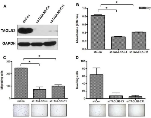
Figure 1. Effect of TAGLN2 knockdown on cell proliferation, migration, and invasion of SNU-1196 cells. (A) The expression levels of TAGLN2 in
SNU-1196 cells were measured by Western blotting after transfection with mock shRNA plasmid (shCon) or TAGLN2 shRNA plasmid (shTAGLN2-C4 and shTAGLN2-C11). GAPDH served as the loading controls. (B) Effects of TAGLN2 knockdown on cell proliferation by shRNA treatment. The absorbance was 0.821±0.022 (95% CI 0.775-0.868), 0.305± 0.006 (95% CI 0.293-0.317), and 0.411± 0.004 (95% CI 0.402-0.421) in shCon/SNU-1196, shTAGLN2-C4/SNU-1196 and shTAGLN2-C11/SNU-1196 cells, respectively (mean±SE). (C) Effects of TAGLN2 knockdown on cell migration by shRNA treatment. The migrating cells were 178.17±6.77, 63.67±10.42, and 92.33±16.7 in shCon/SNU-1196, shTAGLN2-C4/SNU-1196 and shTAGLN2-C11/SNU-1196 cells, respectively (mean±SD). (D) Invasive potential of TAGLN2 knockdown cells by shRNA treatment. Invading cells were observed 64.00±25.46, 7.50±10.61 and 5.00±4.24 in shCon/SNU-1196,
12
shTAGLN2-C4/SNU-1196 and shTAGLN2-C11/SNU-1196 cells, respectively (mean±SD). *P<0.001
2. Suppression of TAGLN2 decreases the tumorigenic potential of SNU-1196 cells in vitro
A sphere-forming assay was conducted to determine self-renewal capacity of SNU-1196 cells after shTAGLN2 treatment. The two kinds of shTAGLN2/SNU-1196 cells formed fewer tumor spheres than the shCon/SNU-1196 cells (Figure 2a). To compare the clonogenicity among the three cells, a colony forming assay was performed (Figure 2b). The colony formation was significantly reduced by 64.3% and 48.2% in shTAGLN2-C4/SNU-1196 and shTAGLN2-C11/SNU-1196 cells when compared with shCon/SNU-1196 cells (P-value <0.001, each). The shTAGLN2-C4/SNU-1196 cells formed fewer colonies than the shTAGLN2-C11/SNU-1196 (p=0.004).
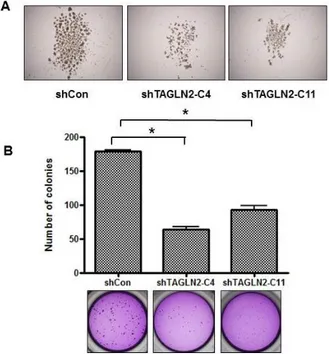
Figure 2. Effect of TAGLN2 suppression on self-renewal and clonogenic capacities of SNU-1196 cells. (A) A sphere-forming assay was conducted in
13
shCon/SNU-1196 cells, shTAGLN2-C4/SNU-1196 cells, and shTAGLN2-C11/SNU-1196 cells (×400). (B) Clonogenic assay was conducted and compared among shCon/SNU-1196, shTAGLN2-C4/SNU-1196, and shTAGLN2-C11/SNU-1196 cells. Colony formation in shTAGLN2-C4/SNU-1196 (63.67±10.42, mean±SD) and shTAGLN2-C11/SNU-1196 cells (92.33±16.17, mean ± SD) was reduced when compared to shCon/SNU-1196 cells (178.17±6.77, mean±SD). *P<0.001
3. Suppression of TAGLN2 expression results in inhibition of tumorigenicity and tumor growth in vivo
We further analyzed the tumorigenic potential of shTAGLN2 transfected SNU-1196 cells by assessing tumor development in nude mice. After the same amount of the three types of cells was injected into mice, tumors appeared in all mice from shCon/SNU-1196, shTAGLN2-C4/SNU-1196, and shTAGLN2-C11/SNU-1196 implantation groups within 5 weeks. Average tumor volume was dramatically undersized in shTAGLN2-C4/SNU-1196 (3.75±5.13mm3, mean±SD) and shTAGLN2-C11/SNU-1196 (63.81±24.5mm3, mean±SD) group, compared to the shCon/SNU-1196 group (402±233.13mm3, mean±SD) (Figure 3). All mice treated with shTAGLN2/SNU-1196 cells showed >85% decrease in volume as compared to control animals. There was also a significant decrease in volume of the shTAGLN2-C4/SNU-1196 group than the shTAGLN2-C11/SNU-1196 group (p=0.041).
14
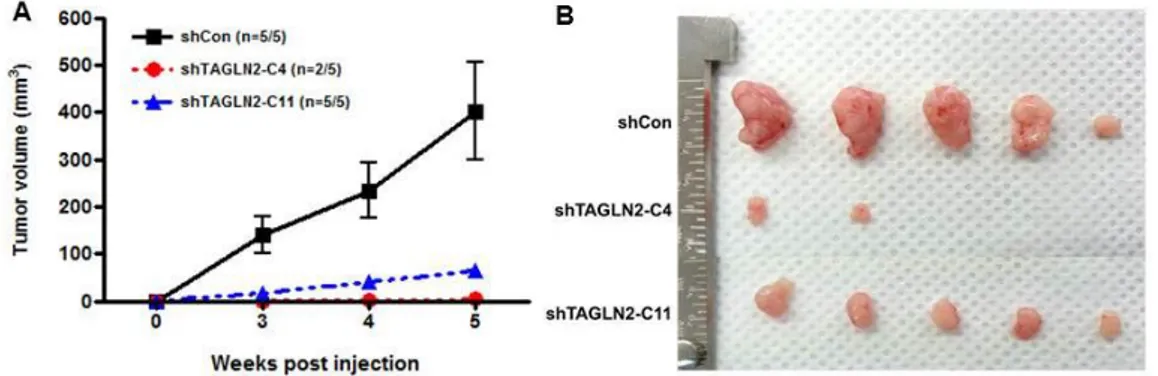
Figure 3. Anti-proliferative effects of TAGLN2 suppression in BTC cells in a xenograft model. Xenografted BTCs were established by subcutaneous
injection of shCon/SNU-1196, shTAGLN2-C4/SNU-1196, and shTAGLN2-C11/SNU-1196 cells into nude mice, respectively. (A) Tumor volume was measured weekly. (B) The size of xenograft tumors was measured at the end of the experiments on the 5th week. The average tumor volume was 402±233.13 mm3 in shCon/SNU-1196 cells injected mice, 3.75±5.13 mm3 in shTAGLN2-C4/SNU-1196 cells injected mice, and 63.81±24.5 mm3 in shTAGLN2-C11/SNU-1196 cells injected mice (mean±SD). shCon/SNU-1196 vs shTAGLN2-C4/SNU-1196; p=0.001, shCon/SNU-1196 vs shTAGLN2-C11/SNU-1196; p=0.005.
4. The expression of TAGLN2 and Ki-67 are decreased in shTAGLN2 transfected xenograft tumors
To further validate suppression of TAGLN2 after shTAGLN2 treatment, we performed IHC with antibodies against TAGLN2 antibody and Ki-67 antibody on xenograft tumors. Strong expression of TAGLN2 was observed in the shCon/SNU-1196 derived tumor, whereas rarely detected in the shTAGLN2-C4/SNU-1196 and the shTAGLN2-C11/SNU-1196 xenograft tumors (Figure 4). The expression of Ki-67 also decreased in the shTAGLN2-C4/SNU-1196 and the shTAGLN2-C11/SNU-1196 xenograft tumors.
15
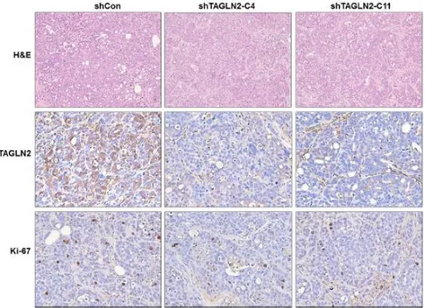
Figure 4. Illustrations of hematoxylin and eosin staining (H&E) and immunohistochemistry (IHC) with antibodies against TAGLN2 antibody and the proliferation marker Ki-67 antibody on xenograft tumor sections.
TAGLN2 were highly expressed in shCon/SNU-1196 xenograft tumors, whereas nearly observed in shTAGLN2-C4/SNU-1196 and shTAGLN2-C11/SNU-1196 xenograft tumors. (x 100)
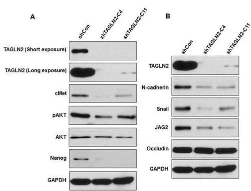
5. TAGLN2 suppression reduces expression of cancer stem cell and epithelial-mesenchymal transition markers.
To determine if the suppression of TAGLN2 induces the specific molecular changes representative of CSC and EMT, we performed western blotting in shTAGLN2-C4/SNU-1196, shTAGLN2-C4/SNU-1196, and shCon/SNU-1196 cells. In shCon/SNU-1196 cells, CSC markers (cMet, pAKT, AKT, and Nanog) and EMT markers (N-Cadherin, Snail, JAG2, and Occludin) were overexpressed. However, in shTAGLN2/SNU-1196 cells, expression of the CSC markers was significantly decreased (Figure 5-a). We found that the mesenchymal markers N-cadherin, Snail and JAC2 were significantly reduced
16
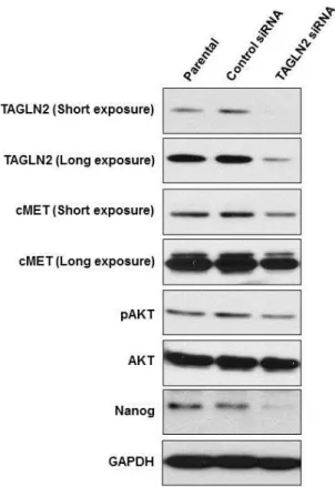
in shTAGLN2/SNU-1196 cells. Whereas the expression level of the epithelial marker, Occludin, was not changed in shTAGLN2/SNU-1196 cells (Figure 5-b). The expression patterns of CSC markers were similarly observed in another BTC cell, SNU308 after TGALN2 suppression by siTAGLN2 transfection (Figure 6).
Figure 5. Expression of CSC-related and EMT markers on shTAGLN2-C4/SNU-1196 cells and shTAGLN-C11/SNU-1196 cells. (A)
Knockdown of TAGLN2 inhibits the cMet/AKT/Nanog signaling in SNU-1196 cells. (B) Mesenchymal markers were suppressed in the TAGLN2 knockdown cells, whereas epithelial marker was highly expressed.
17
Figure 6. Expression of CSC-related markers on siTAGLN2 transfected SNU-308 cells. Knockdown of TAGLN2 inhibits the cMet/AKT/Nanog
signaling in SNU-308 cells.
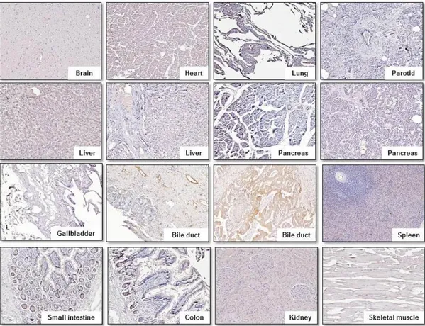
6. TAGLN2 is rarely expressed in major human organ tissue on IHC staining.
Figure 7 shows representative results of IHC staining in normal human tissue obtained using the anti-TAGLN2 polyclonal antibody. TAGLN2 is weakly expressed in small intestine and colon, but its expression was rarely observed in other major organs, which suggests that TAGLN2 can be a suitable marker for selectively treating BTC without damaging the normal human organ.
18
Figure 7. Illustrations of IHC with the anti-TAGLN2 polyclonal antibody on multiple normal human organ tissues. Heart, brain, kidney, liver, lung,
pancreas, spleen, skeletal muscle, gallbladder, parotid gland, small intestine, and colon. TAGLN2 was barely expressed in a majority of normal human organ tissue except small intestine and colon. (x 100)
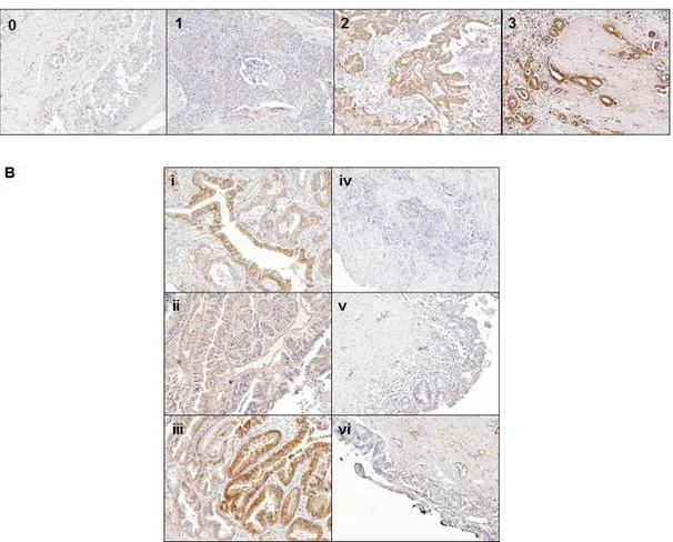
7. TAGLN2 is highly expressed in human BTC tissues compared with normal bile duct tissues
To assess the expression of TAGLN2 in human BTC tissues and normal bile duct tissues, IHC staining with the anti-TAGLN2 polyclonal antibody was performed. We scored the immunoreactivity according to the percentage of TAGLN2-positive tumor cells as follows: 0%= 0, 10-25%=1+, 26-50%=2+, and >50%= 3+, and the staining intensity as follows: 0, 0; weak=1, moderate=2, and strong=3 (Figure 8-A).26 The final scores were on a scale of
19
0-9 using multiplication of percentage and intensity. We used the following criteria to the TAGLN2 expression level of IHC results; low-level expression of TAGLN2 is under 4, and high-level expression of TAGLN2 is 4 or more. We observed the different IHC staining patterns of TAGLN2 in BTC and normal bile duct tissue. The TAGLN2 was strongly expressed in BTC tissue, whereas negative or weak expression was observed in most of the normal bile duct tissue (Fig 8-B).
Figure 8. Representative IHC staining of TAGLN2 and the comparison in human BTC tissues and normal bile duct tissues within individual patients. (A) The representative figures classified by the intensity of
TAGLN2 expression from 0 to 3 in the BTC tissue. (B) High expression of TAGLN2 was noted in biliary tract cancer tissue (i-iii), whereas negative
20
expression was noted in normal bile duct tissue (iv-vi) in each individual patient (case 1:i and vi, case 2:ii and v, case 3: iii and iv). (x 100)
8. Reduced survival and the higher rate of recurrence were observed in BTC patients with highly expressed TAGLN2.
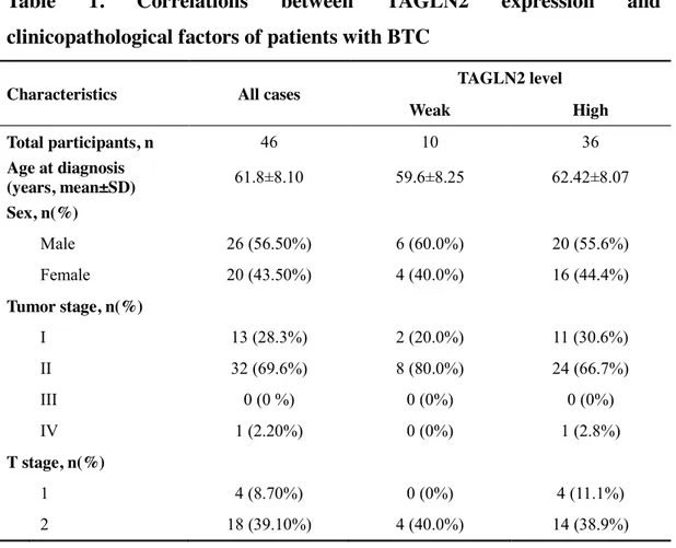
We analyzed the relationship between TAGLN2 expression on the surgical specimen and clinicopathologic factors in BTC patients (Table 1). TAGLN2 was weekly expressed in 10 patients and highly expressed in 36 patients. There was no significant difference in age, sex, tumor staging, and cell differentiation between two groups. The recurrence rate was higher, and time to progression and overall survival were shorter in the highly expressed TAGLN2 group than in the weekly expressed TAGL2 group without statistical significance.
Table 1. Correlations between TAGLN2 expression and
clinicopathological factors of patients with BTC
Characteristics All cases TAGLN2 level
Weak High Total participants, n 46 10 36 Age at diagnosis (years, mean±SD) 61.8±8.10 59.6±8.25 62.42±8.07 Sex, n(%) Male 26 (56.50%) 6 (60.0%) 20 (55.6%) Female 20 (43.50%) 4 (40.0%) 16 (44.4%) Tumor stage, n(%) I 13 (28.3%) 2 (20.0%) 11 (30.6%) II 32 (69.6%) 8 (80.0%) 24 (66.7%) III 0 (0 %) 0 (0%) 0 (0%) IV 1 (2.20%) 0 (0%) 1 (2.8%) T stage, n(%) 1 4 (8.70%) 0 (0%) 4 (11.1%) 2 18 (39.10%) 4 (40.0%) 14 (38.9%)
21
3 24 (52.20%) 6 (60.0%) 18 (50.0%)
Lymph node metastasis, n(%)
0 16 (34.80%) 6 (60.0%) 23 (63.89%) 1 1 (2.20%) 4 (40.0%) 13 (36.11%) Tumor grade, n(%) Well 12 (26.10%) 1 (10.0%) 11 (30.6%) Moderately 26 (56.50%) 6 (60.0%) 20 (55.6%) Poorly 8 (17.40%) 3 (30.0%) 5 (13.9%) Precursor lesion, n(%) Dysplasia 8 (17.40%) 1 (10.0%) 7 (19.4%) Vascular invasion, n(%) Positive 8 (17.40%) 3 (30.0%) 5 (13.9%) Perineural invasion, n(%) Positive 25 (54.30%) 8 (80.0%) 17 (47.2%) Resection margin, n(%) Positive 6 (13.00%) 2 (20.0%) 4 (11.1%) Recurrence, n(%) 23 (50.0%) 4 (40.0%) 19 (52.8%) Overall survival (days, mean±SD) 1349.33±1177.72 1428.0±1107.18 1323.11±1219.47 Time to progress (days, mean±SD) 1036.60±1178.74 1397.17±1550.18 922.74±1062.09
p value was >0.05 in all clinicopathological factors.
9. Altered expression of CSC-related markers and overexpression of TAGLN2 in gemcitabine-resistant BTC cells.
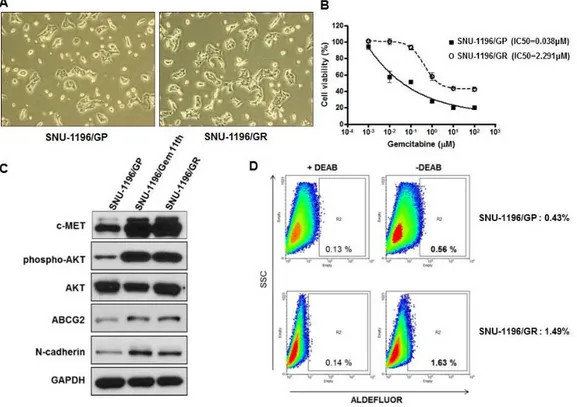
To assess the effect of TAGLN2 suppression on chemoresistant BTC cells, we established the gemcitabine resistant BTC cells (SNU-1196/GR cells) with a gemcitabine IC50 of 2.291 μM, which is approximately 60-fold higher than that in parental BTC cells (SNU-1196/GP cells, IC50=0.038μM) (Figure 9-A). There was no apparent morphologic difference between SNU-1196/GP and SNU-1196/GR (Figure 9-B) cells.25 These cells showed elevated expression of ATP-binding cassette (ABC) transporter ABCG2, as well as proto-oncogenes
22
including c-Met and AKT, the EMT marker N-cadherin, and the tumor-initiating cell surface marker aldehyde dehydrogenase (ALDH) in vitro (Figure 9-C, D) and in vivo (Figure 10). TAGLN2 was significantly overexpressed in SNU-1196/GR cells, especially in cell supernatant (Fig 11-A). After TAGLN2 was downregulated by siTAGLN2 transfection (Figure 11-B), cell viability of siTAGLN2/SNU-1196/GR cells was significantly decreased compared to that of siCon/SNU-1196/GR cells (Figure 11-C).
Figure 9. The gemcitabine-resistant BTC (SNU-1196/GR) cells overexpress CSC-related proteins and EMT markers. SNU-1196 cells
were treated with increasing doses of gemcitabine to establish a gemcitabine-resistant cell line (SNU-1196/GR). (A, B) Cell viability was assessed with the MTT assay; gemcitabine IC50 values were 0.038 and 2.291 μM in SNU-1196/GP and SNU-1196/GR cells, respectively. (C) Elevated protein levels of c-MET, phospho-AKT, N-cadherin, and ALDH were analyzed by western blotting. (D) ALDH enzymatic activity was measured
23
with the ALDEFLUOR kit; cells treated with the specific ALDH inhibitor diethylminobenzaldehyde served as a negative control. Flow cytometry also measured ALDH activity.
Figure 10. Overexpression of ALDH, ABCG2, and MRP4 in SNU-1196/GR cells-derived tumors by IHC. Multi-drug resistant proteins
differentially expressed in tumors derived from SNU-1196 and SNU-1196/GR cells, as detected by IHC using antibodies against ALDH, ABCG2, and MRP4 ( 200, H&E 100).
24
Figure 11. Overexpression of TAGLN2 in gemcitabine-resistant BTC (SNU-1196/GR) cells, and increased chemosensitivity by downregulation of TAGLN2 using siTAGLN2 transfection (siTAGLN2/SNU-1196/GR cell).
(A) TAGLN2 was highly expressed in SNU-1196/GR cells especially in cell culture supernatant. (B) SiTAGLN2 transfection in SNU-1196/GR cells resulted in a dose-dependent decrease of TAGLN2. (C) Cell viability on gemcitabine was significantly decreased in siTAGLN2/SNU-1196/GR cells compared to siCon/SNU-1196/GR cells. P<0.001
10. Suppression of TAGLN2 in SNU-1196/GR cells decreases drug resistance to other cytotoxic drugs
To evaluate whether the expression of TAGLN2 affects the chemosensitivity of anti-metabolites (5-FU), platinum-based anticancer drugs (cisplatin, oxaliplatin, and carboplatin), topoisomerase inhibitors (irinotecan and etoposide) and an EGFR inhibitor (Erlotinib) in gemcitabine-resistant BTC cells (SNU-1196/GR) or not, MTT assays were performed in siTAGLN2 transfected SNU-1196/GR cells and control (siCon/SNU-1196/GR cells). Cell
25
survival for 5-FU was significantly decreased when TAGLN2 was suppressed (p=0.026) (Figure 12). With elevating dose of platinum-based anticancer drugs (cisplatin, oxaliplatin, and carboplatin), cell viability was significantly decreased (p<0.05) in both cells, but there was no additional effect of TAGLN2 suppression (p>0.05) (Figure 13). Chemoresistance to topoisomerase inhibitors (irinotecan and etoposide) and EGFR inhibitor (Erlotinib) was rather increased after TAGLN2 suppression in SNU-1196/GR cells. (Figure 14 & 15).
Figure 12. Increased chemosensitivity to 5-FU in gemcitabine-resistant BTC cells after TAGLN2 suppression. Cell viability on 5-FU was
significantly decreased in siTAGLN2/SNU-1196/GR cells compared to siCon/SNU-1196/GR cells. P<0.001
26
Figure 13. Effect of TAGLN2 suppression on the susceptibility of gemcitabine-resistant BTC cells to the platinum-based anti-cancer drug.
Chemoresistance of siTAGLN2/SNU-1196/GR cells against cisplatin (A, P=0.185), oxaliplatin (B, p=0.201), and carboplatin (C, p=0.082) was slightly improved compared to siCon/SNU-1196/GR cells.
Figure 14. Effect of TAGLN2 suppression on the susceptibility of gemcitabine-resistant BTC cells to topoisomerase inhibitor.
Chemoresistance of siTAGLN2/SNU-1196/GR cells against irinotecan (A, p=0.333) and etoposide (B, p<0.001) was increased compared to siCon/SNU-1196/GR cell.
27
Figure 15. Effect of TAGLN2 suppression on the susceptibility of gemcitabine-resistant BTC cells to EGFR inhibitor. Chemoresistance of
siTAGLN2/SNU-1196/GR cells against erlotinib was slightly increased compared to siCon/SNU-1196/GR cells (p=0.427).
IV. DISCUSSION
In the previous study, we identified that TAGLN2 was highly expressed in BTC cell lines characterized by CSC, and significantly related with carcinogenesis of BTC by dysregulation of c-met downstream pathway.12 We undertook this study to investigate whether modulating TAGLN2 expression could improve gemcitabine-resistance in BTC cells. The suppression of TAGLN2 significantly inhibited the proliferation, migration, and invasion of gemcitabine-resistant BTC (SNU-1196/GR) cells in vitro and in vivo, and restored the chemosensitivity to anti-metabolites, gemcitabine, and 5-FU. Also, we also observed that expression of CSC- and EMT-related markers decreased when TAGLN2 was inhibited in this study.
Recently, TAGLN2 has emerged as a biomarker that plays an essential role in the development of various types of cancer.27 The alteration of TAGLN2 expression was noted in different kinds of cancer at both transcriptional and translational level, and cancer cell proliferation, invasion, and metastasis
28
might be inhibited by suppressing TAGLN2. It was suggested that tumorigenesis and tumor development might be correlated with the deregulation of TAGLN2. The tumor size, clinical stage, histological neural invasion and lymph node metastasis had a close association with upregulation of TAGLN2 in lung cancer,20 bladder cancer,22 colorectal cancer,16,28 esophageal cancer,14 and gastric cancer.29 Moreover, cancer cell proliferation and migration were decreased by downregulating TALGN2 in breast cancer,17 renal cell carcinoma,18 cervical cancer,21 and head and neck squamous cell carcinoma.19 TAGLN2 contributes cancer cells to spread out by acquiring migratory and invasive properties. Overexpression of TAGLN2 degenerates epithelial phenotype and activates mesenchymal phenotypes by altering gene expression, and EMT begins.30 On the contrary, when TAGLN2 was inhibited, the expression of mesenchymal markers, N-cadherin, Snail, and Jag2, was decreased, whereas the expression of epithelial marker, Occludin was increased (fig 5). The TAGLN2 expression may regulate EMT property.
EMT does not only contribute to cancer progression and dissemination but also enhances cancer cells to resist chemotherapy.31-33 Many studies suggested that TAGLN2 overexpression is a potential cause of chemoresistance by increasing EMT property.27,34 Paclitaxel-resistant human breast cancer cell, MCF-7/PTX, overexpressed TAGLN2, and chemosensitivity to paclitaxel was restored after TAGLN2 was inhibited.27 We could find the same result in our study that chemosensitivity to gemcitabine and 5-FU was improved in gemcitabine-resistant BTC cells after TAGLN2 suppression (Fig 11).
The mechanism of cancer development and chemoresistance by TAGLN2 was not identified. However, recent studies have reported that a few signaling pathways are correlated with TAGLN2, such as transforming growth factor-b (TGF-b)/FoxM1 pathway,35 TGF-b/SMAD4 pathway,36 IGF1R/ phosphoinositide 3-kinase (PI3K)/Akt pathway,37 and PI3K/phosphatase and tensin homolog (PTEN)/Akt pathway.17,38-40 TAGLN2 is known to be a direct
29
target of TGF-b signaling which leads to EMT associated phenotypic and morphologic alterations.41 FoxM1 is an oncogenic transcriptional factor of TGF-b signal pathway and regulates some key mediators of cell cycle progression. It was identified that TAGLN2 activates FoxM1 signaling during gliomagenesis.35 TAGLN2 overexpression was observed in glioma, and TAGLN2 knockdown led to significantly reduced levels of FoxM1.35 Activation of TGF-β/Smad4 signaling induces TAGLN expression in human bone marrow-derived stromal stem cells (hMSC)42 and colon cancer.36 TAGLN2 mediates chemoresistance in paclitaxel-resistant breast cancer by activating PI3K/PTEN/Akt pathway and inhibiting apoptosis.17,38-40 In NCSLC cells, transgelin overexpression was observed with increased resistance to chemotherapy and radiotherapy and activates the IGF1R/PI3K/Akt pathway via direct interaction with IGF1Rb.37
There have been attempts to overcome the chemoresistance by targeting TAGLN2 related signal pathway in other cancers. In paclitaxel-resistant breast cancer cells, salvianolic acid A downregulated the expression of TAGLN2 by the mechanisms of PI3K/Akt pathway activation and ABC transporter up-regulation.39 The migration and invasion abilities of human breast cancer were inhibited, and chemoresistance to paclitaxel was also restored.17 SB-T-121205, the next-generation taxoid, showed anti-tumor activity by inhibiting TAGLN2 and PI3K/Akt pathway in human breast cancer cells.43 Tumor suppressive miR-1, to which TAGLN2 contains three binding sites, directly regulates TAGLN2 and suppresses cell proliferation, invasion, and migration of head and neck SCC,19 and esophageal SCC.14 Based on previous studies, the synergism of combination therapy with anti-TAGLN2 and a cytotoxic drug is anticipated.
Increased chemosensitivity by TAGLN2 inhibition is not applied to all chemotherapeutic agents. In our study, restoration of chemosensitivity was mainly observed in anti-metabolites, gemcitabine, and 5-FU. The
30
chemosensitivity to platinum-based agents tended to be improved but was not statistically significant. On the contrary, irinotecan, and etoposide, which are topoisomerase inhibitors, have been shown to exacerbate the resistance. However, the mechanism of the different response to TAGLN2 inhibition remains unclear. Therefore, what we need to do is to identify the role of TAGLN2 in the signaling pathway and the carcinogenesis mechanism by genomic and proteomic analysis.
There are some limitations in our study. We analyzed TAGLN2 overexpression in a subset of BTC tissues. However, tissue heterogeneity complicates the identification of tumor markers and the results obtained through proteomic analysis of whole tissues may be considered controversial.44 We obtained human biliary tissues from operations in BTC patients and the normal biliary duct tissue was acquired around the BTC lesion. For a more meaningful analysis, we need to compare the expression of TAGLN2 in biliary tissue obtained from benign disease. Second, the chemoresistance was improved in gemcitabine-resistant BTC cells inhibiting TAGLN2 with siRNA transfection in our study, but the effect of TAGLN2 suppression on cell viability should be considered when analyzing the result. This is because TAGLN2 is involved in cell proliferation. However, even with this effect, the chemoresistance after TAGLN2 suppression was better than control.
V. CONCLUSION
In conclusion, we have shown here that TAGLN2 performs the specific role involving tumor proliferation, migration and invasion, and engaging in chemoresistance by inducible EMT-like changes. Targeting of TAGLN2 is expected to be successful anti-cancer therapy in advanced cancer after failed in chemotherapy. Further studies of clarifying the signaling network and
31
mechanism of TAGLN2 in involving carcinogenesis and drug resistance should be continued and make it possible to develop new therapeutic approaches for the treatment of chemo-refractory BCT in the future.
32
REFERENCES
1. Khan SA, Davidson BR, Goldin RD, Heaton N, Karani J, Pereira SP, et al. Guidelines for the diagnosis and treatment of cholangiocarcinoma: an update. Gut 2012;61:1657-69.
2. Nimura Y. Radical surgery: vascular and pancreatic resection for cholangiocarcinoma. HPB (Oxford) 2008;10:183-5.
3. Zhu AX. Future directions in the treatment of cholangiocarcinoma. Best Pract Res Clin Gastroenterol 2015;29:355-61.
4. Igami T, Nishio H, Ebata T, Yokoyama Y, Sugawara G, Nimura Y, et al. Surgical treatment of hilar cholangiocarcinoma in the "new era": the Nagoya University experience. J Hepatobiliary Pancreat Sci 2010;17:449-54.
5. Pichlmayr R, Weimann A, Klempnauer J, Oldhafer KJ, Maschek H, Tusch G, et al. Surgical treatment in proximal bile duct cancer. A single-center experience. Ann Surg 1996;224:628-38.
6. Klempnauer J, Ridder GJ, Werner M, Weimann A, Pichlmayr R. What constitutes long-term survival after surgery for hilar cholangiocarcinoma? Cancer 1997;79:26-34.
7. Valle J, Wasan H, Palmer DH, Cunningham D, Anthoney A, Maraveyas A, et al. Cisplatin plus gemcitabine versus gemcitabine for biliary tract cancer. N Engl J Med 2010;362:1273-81.
8. Valle JW, Furuse J, Jitlal M, Beare S, Mizuno N, Wasan H, et al. Cisplatin and gemcitabine for advanced biliary tract cancer: a meta-analysis of two randomised trials. Ann Oncol 2014;25:391-8. 9. Fiteni F, Nguyen T, Vernerey D, Paillard MJ, Kim S, Demarchi M, et
al. Cisplatin/gemcitabine or oxaliplatin/gemcitabine in the treatment of advanced biliary tract cancer: a systematic review. Cancer Med 2014;3:1502-11.
33
chemotherapy in advanced biliary cancer: a systematic review. Ann Oncol 2014;25:2328-38.
11. Cardinale V, Renzi A, Carpino G, Torrice A, Bragazzi MC, Giuliante F, et al. Profiles of cancer stem cell subpopulations in cholangiocarcinomas. Am J Pathol 2015;185:1724-39.
12. Park BK, Song SY. Identification and characterization of TAGLN2 as a novel biomarker for cancer stem cells in biliary tract cancers. The graduate school Yonsei University. 2013.
13. Shapland C, Hsuan JJ, Totty NF, Lawson D. Purification and properties of transgelin: a transformation and shape change sensitive actin-gelling protein. J Cell Biol 1993;121:1065-73.
14. Du YY, Zhao LM, Chen L, Sang MX, Li J, Ma M, et al. The tumor-suppressive function of miR-1 by targeting LASP1 and TAGLN2 in esophageal squamous cell carcinoma. J Gastroenterol Hepatol 2016;31:384-93.
15. Zhou HM, Fang YY, Weinberger PM, Ding LL, Cowell JK, Hudson FZ, et al. Transgelin increases metastatic potential of colorectal cancer cells in vivo and alters expression of genes involved in cell motility. BMC Cancer 2016;16:55.
16. Zhang Y, Ye Y, Shen D, Jiang K, Zhang H, Sun W, et al. Identification of transgelin-2 as a biomarker of colorectal cancer by laser capture microdissection and quantitative proteome analysis. Cancer Sci 2010;101:523-9.
17. Zheng X, Chen S, Yang Q, Cai J, Zhang W, You H, et al. Salvianolic acid A reverses the paclitaxel resistance and inhibits the migration and invasion abilities of human breast cancer cells by inactivating transgelin 2. Cancer Biol Ther 2015;16:1407-14.
18. Kawakami K, Enokida H, Chiyomaru T, Tatarano S, Yoshino H, Kagara I, et al. The functional significance of miR-1 and miR-133a in
34
renal cell carcinoma. Eur J Cancer 2012;48:827-36.
19. Nohata N, Sone Y, Hanazawa T, Fuse M, Kikkawa N, Yoshino H, et al. miR-1 as a tumor suppressive microRNA targeting TAGLN2 in head and neck squamous cell carcinoma. Oncotarget 2011;2:29-42.
20. Jin H, Cheng X, Pei Y, Fu J, Lyu Z, Peng H, et al. Identification and verification of transgelin-2 as a potential biomarker of tumor-derived lung-cancer endothelial cells by comparative proteomics. J Proteomics 2016;136:77-88.
21. Yakabe K, Murakami A, Kajimura T, Nishimoto Y, Sueoka K, Sato S, et al. Functional significance of transgelin-2 in uterine cervical squamous cell carcinoma. J Obstet Gynaecol Res 2016;42:566-72. 22. Chen CL, Chung T, Wu CC, Ng KF, Yu JS, Tsai CH, et al.
Comparative Tissue Proteomics of Microdissected Specimens Reveals Novel Candidate Biomarkers of Bladder Cancer. Mol Cell Proteomics 2015;14:2466-78.
23. Lin Y, Buckhaults PJ, Lee JR, Xiong H, Farrell C, Podolsky RH, et al. Association of the actin-binding protein transgelin with lymph node metastasis in human colorectal cancer. Neoplasia 2009;11:864-73. 24. Ku JL, Yoon KA, Kim IJ, Kim WH, Jang JY, Suh KS, et al.
Establishment and characterisation of six human biliary tract cancer cell lines. Br J Cancer 2002;87:187-93.
25. Jung DE, Park SB, Kim K, Kim C, Song SY. CG200745, an HDAC inhibitor, induces anti-tumour effects in cholangiocarcinoma cell lines via miRNAs targeting the Hippo pathway. Sci Rep 2017;7:10921. 26. Zhou L, Zhang R, Zhang L, Sun Y, Yao W, Zhao A, et al.
Upregulation of transgelin is an independent factor predictive of poor prognosis in patients with advanced pancreatic cancer. Cancer Sci 2013;104:423-30.
35
oncogenic factor. Tumour Biol 2017;39:1010428317702650.
28. Zhuo HQ, Zhang YB, Zhang H, Guo P, Lu Y, Dong LY, et al. [Expression of transgelin-2 and clinical significance in colorectal cancer]. Zhonghua Wai Ke Za Zhi 2012;50:551-4.
29. Xu XC, Zhang YH, Zhang WB, Li T, Gao H, Wang YH. MicroRNA-133a functions as a tumor suppressor in gastric cancer. J Biol Regul Homeost Agents 2014;28:615-24.
30. Puisieux A, Brabletz T, Caramel J. Oncogenic roles of EMT-inducing transcription factors. Nat Cell Biol 2014;16:488-94.
31. Park SM, Kim SM, Han JH. [The role of epithelial-mesenchymal transition in the gastroenterology]. Korean J Gastroenterol 2010;56:69-77.
32. Kajiyama H, Shibata K, Terauchi M, Yamashita M, Ino K, Nawa A, et al. Chemoresistance to paclitaxel induces epithelial-mesenchymal transition and enhances metastatic potential for epithelial ovarian carcinoma cells. Int J Oncol 2007;31:277-83.
33. Hiscox S, Jiang WG, Obermeier K, Taylor K, Morgan L, Burmi R, et al. Tamoxifen resistance in MCF7 cells promotes EMT-like behaviour and involves modulation of beta-catenin phosphorylation. Int J Cancer 2006;118:290-301.
34. Chen YX, Xie X, Cheng Q. [cDNA microarray analysis of gene expression in acquired methotrexate-resistant of human choriocarcinoma]. Zhonghua Fu Chan Ke Za Zhi 2004;39:396-9. 35. Han MZ, Xu R, Xu YY, Zhang X, Ni SL, Huang B, et al. TAGLN2 is
a candidate prognostic biomarker promoting tumorigenesis in human gliomas. J Exp Clin Cancer Res 2017;36:155.
36. Ali NA, McKay MJ, Molloy MP. Proteomics of Smad4 regulated transforming growth factor-beta signalling in colon cancer cells. Mol Biosyst 2010;6:2332-8.
36
37. Kim TR, Cho EW, Paik SG, Kim IG. Hypoxia-induced SM22alpha in A549 cells activates the IGF1R/PI3K/Akt pathway, conferring cellular resistance against chemo- and radiation therapy. FEBS Lett 2012;586:303-9.
38. Chen S, Dong Q, Hu S, Cai J, Zhang W, Sun J, et al. Proteomic analysis of the proteins that are associated with the resistance to paclitaxel in human breast cancer cells. Mol Biosyst 2014;10:294-303.
39. Cai J, Chen S, Zhang W, Zheng X, Hu S, Pang C, et al. Salvianolic acid A reverses paclitaxel resistance in human breast cancer MCF-7 cells via targeting the expression of transgelin 2 and attenuating PI3 K/Akt pathway. Phytomedicine 2014;21:1725-32.
40. Cai J, Chen S, Zhang W, Hu S, Lu J, Xing J, et al. Paeonol reverses paclitaxel resistance in human breast cancer cells by regulating the expression of transgelin 2. Phytomedicine 2014;21:984-91.
41. Christiansen JJ, Rajasekaran AK. Reassessing epithelial to mesenchymal transition as a prerequisite for carcinoma invasion and metastasis. Cancer Res 2006;66:8319-26.
42. Elsafadi M, Manikandan M, Dawud RA, Alajez NM, Hamam R, Alfayez M, et al. Transgelin is a TGFbeta-inducible gene that regulates osteoblastic and adipogenic differentiation of human skeletal stem cells through actin cytoskeleston organization. Cell Death Dis 2016;7:e2321.
43. Zheng X, Wang C, Xing Y, Chen S, Meng T, You H, et al. SB-T-121205, a next-generation taxane, enhances apoptosis and inhibits migration/invasion in MCF-7/PTX cells. Int J Oncol 2017;50:893-902.
44. Rho JH, Roehrl MH, Wang JY. Tissue proteomics reveals differential and compartment-specific expression of the homologs transgelin and
37
transgelin-2 in lung adenocarcinoma and its stroma. J Proteome Res 2009;8:5610-8.
38
ABSTRACT (IN KOREAN)
젬시타빈 저항성 담관암의 새로운 치료용 타겟으로써 transgelin-2 <지도교수 송 시 영 > 연세대학교 대학원 의학과 정 주 원 Transgelin-2 (TAGLN2)는 정상 담관 조직에 비해 암줄기세포 특성을 보이는 담관암에서 고발현 되어 있지만 그 기능에 대해서는 아직 명확히 밝혀져 있지 않다. 담관암 발생 및 항암제 내성에 미치는 TAGLN2의 역할을 확인하고 이를 담관암 치료에 적용할 수 있는 가능성을 찾고자 본 연구를 계획하였다.
인체 담관암 세포주, SNU-1196에 TAGLN2 shRNA (shTAGLN2) 형질 주입을 통하여 TAGLN2 발현 억제 세포주를 만들고, 시험관내 및 생체 내에서 세포 증식, 이동, 침윤 그리고 분자적 특징을 평가하였다. 면역 염색을 통하여 인체 다양한 정상 장기 조직에 대해 TAGLN2 발현 여부를 확인하고, 담관암에서 TAGLN2 발현 정도와 환자의 임상 양상의 차이를 비교하였다. 젬시타빈 내성 담관암 세포주에 siTAGLN2 형질 주입하여 TAGLN2를 억제 시킨 내성 세포주를 만들고, 마우스에 이식하여 형성된 종양에 대해 젬시타빈, 5-FU, 시스플라틴, 옥살리플라틴, 카보플라틴, 에토포사이드, 이리노테칸 그리고 얼로티닙 등의 항암제 감수성을 평가하였다. TAGLN2가 억제된 담관암 세포는 세포 증식, 이동성 그리고 침윤능력이 감소하였고, 종양형성능력이 현저하게 감소하는
39
것을 시험관내 및 생체 내에서 확인하였다. TAGLN2가 억제된 담관암 세포는 줄기세포 표지자인, c-met, Nanog, pAKT 와 AKT, 간엽 세포 표지자인 (mesenchymal marker) N-cadherin, Snail, 그리고 JAG2의 발현이 감소되었지만, 상피세포 표지자 (epithelial marker)인 Occludin의 발현은 증가된 것을 확인할 수 있었다. TAGLN2는 대부분의 인체 주요 장기 조직에서 매우 드물게 발현이 되었으나 담관암 조직에서는 고 발현되어 있었다. 하지만, 담관암 조직에서 TAGLN2 발현 정도는 종양 병기, 병리 분화도 그리고 환자의 예후와는 통계학적 연관성을 보이지 못했다. 젬시타빈 저항성 담관암 세포에서 TAGLN2를 억제하면 대사 길항 항암제 (anti-metabolite)인 젬시타빈과 5-FU의 감수성이 증가하는 것을 확인할 수 있었다. 본 연구 결과 TAGLN2가 담관암의 증식 및 진행에 관련된 역할을 하는 것으로 보이며, TAGLN2를 억제하였을 때 담관암 세포의 상피간엽이행 (epithelial-mesenchymal transition) 특성이 저하되는 것을 확인하였다. TAGLN2를 타겟으로 하는 약제가 개발이 된다면 젬시타빈 혹은 5-FU 등의 항암제와 병용 혹은 단독 투여 하는 방법으로 담관암 치료에 유용하게 응용될 수 있을 것으로 사료된다. --- 핵심 되는 말: 담관암, transgelin-2, 암줄기세포, 상피간엽이 행, 항암제 저항성
40
PUBLICATION LIST
Jung DE, Park SB, Kim K, Kim C, Song SY. CG200745, an HDAC inhibitor, induces anti-tumor effects in cholangiocarcinoma cell lines via miRNAs targeting the Hippo pathway. Sci Rep 2017;7:10921.supple fig 1-2.
The etablishment of gemcitabine-resistant bile duct tract cancer cells (SNU-1196/GR) and the changes of molecular characteristics were published in this article (figure 9 and 10 in our study).