428
Background : High microsatellite instability (MSI-H) colorectal carcinomas (CRCs) with
numer-ous mutations in the microsatellite sequence are characterized by a right-sided preponderance, frequent peritumoral and intratumoral lymphocytic infiltration, and frequent mucin production. However, no study has correlated anatomic site and type of genetic changes with clinicopatho-logic changes. Methods : We analyzed the histopathoclinicopatho-logic features of 135 MSI-H CRCs and compared them to 140 microsatellite stable (MSS) CRCs. Histopathologic changes in MSI-H were further analyzed according to anatomic sites and genetic changes. Results : MSI-H CRCs showed previously reported clinicopathologic findings; a right-sided preponderance, an increas-ed number of mucinous carcinomas, and peritumoral lymphoid reactions (p<0.001 for each variable). Increased serum CEA levels showed an MSS CRC preponderance (p=0.013). We further analyzed the histologic differences between right- and left-sided MSI-H tumors. We found that MSI-H CRCs on both sides had similar clinicopathologic findings, except for higher tumor stage (p=0.048) and less frequent abnormal CEA levels in left-sided MSI-H tumors (p=0.027). We found that not all clinicopathologic features were different between hereditary nonpolyposis colorectal cancers (HNPCCs) and sporadic MSI-H CRCs. Conclusions : These findings indicate that MSI-H CRCs of the left colon have similar clinicopathologic char-acteristics as right-sided MSI-H CRCs. We did not find any significant clinicopathological dif-ference between HNPCCs and sporadic MSI-H CRCs.
Key Words : Colorectal neoplasm; Microsatellite instability; Colorectal neoplasms, hereditary
nonpolyposis
Sang Kyum Kim Junjeong Choi Hyun Ki Kim Young Nyun Park Si Young Song1 Hoguen Kim
428
Clinicopathologic Characteristics of Left-Sided Colon Cancers
with High Microsatellite Instability
428 428 Corresponding Author
Hoguen Kim, M.D.
Department of Pathology, Yonsei University College of Medicine, 134 Sinchon-dong, Seodaemun-gu, Seoul 120-752, Korea
Tel: 02-2228-2643 Fax: 02-313-9028 E-mail: [email protected]
Departments of Pathology and 1Internal Medicine, Yonsei University College of Medicine, Seoul, Korea
Received : February 13, 2009 Accepted : June 4, 2009
Colorectal carcinoma (CRC) is one of the most common tumors in Western countries and the fourth most common cancer in both genders in Korea (Korean Statistical Informative Service, 2005). The molecular pathogenesis of CRC is well explained in 2 pathways: chromosomal instability (CIN) and microsatellite instability (MSI).1,2The MSI pathway begins with the
inactiva-tion of genes responsible for DNA nucleotide mismatch repair, which results in extensive mutations in DNA sequences with low frequencies of allelic losses and rare alterations of tumor DNA content.3
After the discovery of MSI in cancer of the colon,4many
stud-ies of the genetic, pathologic, and clinical features of MSI colon cancer have been carried out. A National Cancer Institute (NCI) workshop suggested that the form of genomic instability asso-ciated with defective DNA mismatch repair in tumors should be called MSI and a panel of 5 microsatellites was validated by Ruschoff and Fishel.5The panel was composed of 2
mononu-cleotide repeats (bat26 and bat25) and 3 dinumononu-cleotide repeats (D5S346, D2S123, and D7S250).6,7Tumors were divided into
3 groups according to 5 markers: high-frequency MSI (MSI-H, the majority of markers exhibit MSI); low-frequency MSI (MSI-L, only a minority of markers exhibit MSI); and a group lack-ing any apparent instability (MSS, none of the markers exhibit MSI).5Among these groups, MSI-H CRCs comprise 10-15%
of sporadic CRCs and most of hereditary nonpolyposis colorec-tal cancers (HNPCCs).5
MSI-H CRCs have characteristic clinical and pathological features compared to MSI-L and MSS CRCs such as enhanced survival, a marked preponderance of tumors proximal to the sp-lenic flexure, earlier tumor stage, increased diploid status, poor differentiation, extracellular mucin production, a Crohn’s-like lymphoid reaction, and a frequent relation to HNPCC.8-10
Until recently, although many small-scale studies had been conducted to clarify the biologic nature of MSI-H CRCs, there were no reports correlating histological changes according to anatomic site and histological comparison with HNPCC-asso-ciated MSI-H tumors versus sporadic MSI-H tumors. In order to identify any clinicopathologic characteristics related to
anatom-ic site and history of HNPCC, we analyzed the histopathologanatom-ic features of 135 MSI-H CRCs and grouped and analyzed them according to anatomic site and type of genetic change.
MATERIALS AND METHODS
Case selection
A total of 135 MSI-H CRCs and 30 MSI-L CRCs from the 1,460 colon cancers that had been operated and diagnosed at Yon-sei University Medical Center from 2003 to 2007 were includ-ed in this study. A total of 140 MSS CRCs that had been oper-ated and diagnosed between May and October 2005 were used as controls. All cases had been confirmed by histological exam-ination and molecular workup.
MSI analysis
DNA was extracted from formalin-fixed and paraffin-embed-ded tissue samples of 1,458 colon cancer cases. Tumor and adja-cent normal areas were separately marked and collected for DNA extraction. The recommended panel (bat26, bat25, D5S346, D2S123, and D7S250) was used for genetic classification. PCR
was done and differences in amplified PCR fragments between tumor and normal tissue were detected by gene analyzer. MSI-H tumors were defined as having instability in 2 or more mark-ers (Fig. 1A), MSI-L tumors as having instability in 1 marker (Fig. 1B), and MSS tumors as showing no apparent instability (Fig. 1C).5MSI-H CRCs accounted for 9.68% and MSI-L
CRCs 2.05% of the 1,460 colorectal specimens that had been accumulated over the 5 years of the study (Table 1). This study was approved by the Institutional Review Board and informed consent from all participants was obtained.
Analysis of clinical features
Clinical variables such as sex, mortality, anatomic site,
num-Molecular MSI-H MSI-L MSS markers (n=135, 9.25%) (n=30, 2.05%) (n=1,295, 88.70%) bat26 123 2 0 bat25 125 7 0 D5S346 108 5 0 D2S123 117 6 0 D7S250 121 10 0
MSI, microsatellite instability; MSI-H, high frequency MSI; MSI-L, low-frequency MSI; MSS, microsatellite stable.
Table 1. Incidence of MSI of 5 markers according to molecular type of CRCs
Fig. 1. Analysis of microsatellite instability in CRCs using the five markers (bat26, bat25, D5S346, D2S123, and D7S250) and fluo-rescence-based multiplex polymerase chain reaction show the dif-ferences of normal (first line) and tumor (second line) tissue. (A) Two mononucleotide repeats (bat26 and bat25) exhibit microsatellite instability and it is interpreted as MSI-H (arrows). (B) One mononu-cleotide repeat (bat25) exhibits microsatellite instability and this is interpreted as MSI-L (arrow). (C) None of the markers exhibits mic-rosatellite instability and it is interpreted as MSS.
A B C 1,800 1,200 800 400 0 1,920 1,280 640 0 2,240 1,680 1,120 560 0 2,660 1,920 1,280 640 0 100 150 200 100 150 200 1,800 1,200 800 400 0 1,800 1,200 800 400 0
ber of tumors, stage, patient survival status, HNPCC associa-tion, and CEA level at diagnosis of CRC patients were obtained from medical records. The anatomic site of the tumor was clas-sified as the ascending colon, transverse colon, descending colon, sigmoid colon, or rectum (Table 2) and divided into right and lest-side based on the location with regard to the splenic flexure. The number of tumor masses was determined according to endoscopic and gross findings. For diagnosis of HNPCC, we used the Amsterdam II criteria11,12 which were presented in 1999.
Preoperative serum CEA levels for all patients were measured at the time of diagnosis (normal range: 0-5 ng/mL), and we divided CRCs into 2 groups; normal (range: 0-5.0 ng/mL) and abnor-mal (≥5.0 ng/mL).
Analysis of pathologic features
The clinicopathologic features of MSI-H and MSS CRCs were further analyzed according to site (right vs left) and history of HNPCC. The histological type of tumor was subclassified by mucinous carcinoma (carcinoma containing mucin over more than 50% of the tumor area), medullary carcinoma (poorly or undifferentiated carcinoma containing scant tumor stroma with intense peritumoral and intratumoral lymphocytic infiltration) and conventional adenocarcinomas.13
Tumors were graded on the basis of glandular appearance and divided into well differentiated (glandular structures in more than 95% of the tumor), moderately differentiated (glandular struc-tures in 50-95% of the tumor), and poorly differentiated lesions (glandular structures in less than 50% of the tumor). Lympho-cytic reactions were estimated when there was more than a mod-erate degree of peritumoral or intratumoral lymphocytic infil-tration and/or lymphoid follicle formation.
Extracellular mucin pool formation and tumor necrosis was rated as absent or present. The infiltrative tumor borders were classified as irregular or pushing types.
Statistical analysis
Statistical analysis of each parameter was performed to clarify associations between MSI-H and MSS CRCs using the 2test for
categorical variables and SPSS version 13.0 (SPSS Inc., Chica-go, IL, USA). All reported p-values are 2-sided and p-values of less than 0.05 were considered to indicate statistical signifi-cance.
RESULTS
Different in clinicopathlogic features between MSI-H CRCs and MSS CRCs
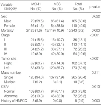
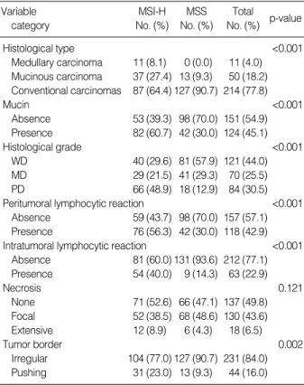
The clinicopathologic features of 135 MSI-H CRCs were com-pared to 140 MSS CRCs (Table 3, 4). The significantly different clinicopathologic features were mortality (p=0.003), anatomic site (p<0.001), pathologic tumor stage (p<0.001), CEA level (p=0.013), histological type and grade (p<0.001), peritumoral and intratumoral lymphocytic reaction (p<0.001), and mucin formation (p<0.001). However, there was no significant
differ-Variable MSI-H MSS Total
p-value
category No. (%) No. (%) No. (%)
Sex 0.622 Male 79 (58.5) 86 (61.4) 165 (60.0) Female 56 (41.5) 54 (38.6) 110 (40.0) Mortalitya 2/123 (1.6) 13/119 (10.9) 15/243 (6.2) 0.003 Stage <0.001 I 21 (15.6) 15 (10.7) 36 (13.1) II 68 (50.4) 45 (32.1) 113 (41.1) III 34 (25.2) 38 (27.1) 72 (26.2) IV 12 (8.9) 42 (30.0) 54 (19.6) Tumor site <0.001 Right 82 (60.7) 20 (14.3) 102 (37.1) Left 53 (39.3) 120 (85.7) 173 (62.9) Mass number 0.211 Single 128 (94.8) 137 (97.9) 265 (96.4) Multiple 7 (5.2) 3 (2.1) 10 (3.6) CEAb 0.013 Normal 109 (80.7) 94 (67.1) 203 (73.8) Abnormal 26 (19.3) 46 (32.9) 72 (26.2) History of HNPCC 8 (5.9) 0 (0.0) 8 (2.9) 0.003
a12 MSI-H CRC patients and 21 MSS CRC patients were missing in
clinical follow up; bPreoperative serum CEA level, normal (0.0-5.0
ng/mL), abnormal (higher than 5.0 ng/mL).
MSI-H, high frequency MSI; MSS, microsatellite stable; CRCs, col-orectal carcinomas; HNPCC, hereditary nonpolyposis colcol-orectal can-cers.
Table 3. The comparison of clinical features between MSI-H and MSS CRCs
Site MSI-H (%) MSS (%) Total (%)
Ascending 72 (53.3) 17 (12.1) 89 (32.4) Transverse 10 (7.4) 1 (0.7) 11 (8.1) Descending 15 (11.1) 9 (6.4) 24 (8.7) Sigmoid 18 (13.3) 40 (28.6) 58 (21.1) Rectum 20 (14.8) 73 (52.1) 93 (33.8) Total 135 (100.0) 140 (100.0) 275 (100.0)
Table 2. Comparison of tumor location in MSI-H and MSS CRCs
MSI-H, high frequency MSI; MSS, microsatellite stable; CRCs, col-orectal carcinomas.
ence in sex, tumor mass number and tumor necrosis between the 2 groups (p>0.05).
Comparison of clinicopathologic features between right- and left-sided MSI-H CRCs
MSI-H CRCs showed a right-sided preponderance (right-sided tumors: 82/135 [60.7%]in MSI-H CRCs vs 18/140 [12.8%]
in MSS CRCs, p<0.001). Differences between right- and left-sided MSI-H CRCs were further analyzed (Table 5). More adv-anced tumor stage was found in left-sided MSI-H CRCs (stage III and IV: 20/53 [37.7%]in left-sided MSI-H CRCs vs 26/82
[31.7%]in right-sided MSI-H CRCs, p=0.022). Serum CEA level, checked before surgery, was higher in right-than left-sided CRCs (increased CEA level: 5/53 [9.4%]in left-sided MSI-H CRCs vs 21/82 [24.7%]in right-sided MSI-H CRCs, p=0.027). There was a trend toward poor survival (mortality: 2/49 [4.1%]
in left-sided MSI-H CRCs vs 0/74 [0.0%]in right-sided MSI-H CRCs, p=0.080) and towards less mucin formation (mucin formation: 27/53 [50.9%]in left-sided MSI-H CRCs vs 55/ 82
[67.1%]in right-sided MSI-H CRCs, p=0.061). No significant differences in sex, survival rate, histologic type and grade of tu-mor, peritumoral and intratumoral lymphocytic reaction, tumor necrosis, tumor border, association with HNPCC, and 5 molec-ular markers were found (p>0.05).
Comparison of clinicopathologic features between HNPCCs and sporadic MSI-H CRCs
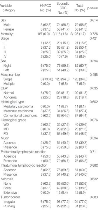
The clinicopathologic features of MSI-H CRCs were com-pared to those of sporadic tumors and HNPCCs with HNPCC being defined as a positive result on the Amsterdam II criteria. We found that not all of the clinicopathologic features were dif-ferent between CRCs from HNPCCs and sporadic MSI-H CRCs (Table 6).
Variable Right side Left side Total
p-value category No. (%) No. (%) No. (%)
Sex 0.996 Male 48 (58.5) 31 (58.5) 79 (58.8) Female 34 (41.5) 22 (41.5) 56 (41.2) Mortalitya 0/74 (0.0) 2/49 (4.1) 2/123 (1.6) 0.080 Stage 0.048 I 8 (9.8) 13 (24.5) 21 (15.4) II 48 (58.5) 20 (37.7) 68 (50.7) III 20 (24.4) 14 (26.4) 34 (25.0) IV 6 (7.3) 6 (11.3) 12 (8.8) Mass number 0.552 Single 77 (93.9) 51 (96.2) 128 (94.8) Multiple 5 (6.1) 2 (3.8) 7 (5.2) CEAb 0.027 Normal 61 (75.3) 48 (90.6) 109 (80.7) Abnormal 21 (24.7) 5 (9.4) 26 (19.3) Histological type 0.201 Medullary carcinoma 6 (7.3) 5 (9.4) 11 (8.1) Mucinous carcinoma 27 (32.9) 10 (18.9) 41 (27.4) Conventional carcinomas 49 (59.8) 38 (71.7) 87 (64.4) Mucin 0.061 Absence 27 (32.9) 26 (49.1) 53 (39.3) Presence 55 (67.1) 27 (50.9) 82 (60.7) Histologic grade 0.386 WD 22 (26.8) 18 (34.0) 40 (29.6) MD 16 (19.5) 13 (24.5) 29 (21.5) PD 44 (53.7) 22 (41.5) 66 (48.9) HNPCC 6 (7.3) 2 (3.8) 16 (11.9) 0.386
a12 MSI-H CRC patients and 21 MSS CRC patients were missing in
clini-cal follow up; bPreoperative serum CEA level: normal (0.0-5.0 ng/mL);
abnormal (higher than 5.0 ng/mL).
MSI-H, high frequency MSI; CRCs, colorectal carcinomas; WD, well differentiated; MD, moderately differentiated; PD, poorly differentiated; HNPCC, hereditary nonpolyposis colorectal cancers.
Table 5. Comparison of clinicopathologic features of right- and left-side MSI-H CRCs
Variable MSI-H MSS Total
p-value category No. (%) No. (%) No. (%) Histological type <0.001 Medullary carcinoma 11 (8.1) 0 (0.0) 11 (4.0) Mucinous carcinoma 37 (27.4) 13 (9.3) 50 (18.2) Conventional carcinomas 87 (64.4) 127 (90.7) 214 (77.8) Mucin <0.001 Absence 53 (39.3) 98 (70.0) 151 (54.9) Presence 82 (60.7) 42 (30.0) 124 (45.1) Histological grade <0.001 WD 40 (29.6) 81 (57.9) 121 (44.0) MD 29 (21.5) 41 (29.3) 70 (25.5) PD 66 (48.9) 18 (12.9) 84 (30.5) Peritumoral lymphocytic reaction <0.001
Absence 59 (43.7) 98 (70.0) 157 (57.1) Presence 76 (56.3) 42 (30.0) 118 (42.9) Intratumoral lymphocytic reaction <0.001
Absence 81 (60.0) 131 (93.6) 212 (77.1) Presence 54 (40.0) 9 (14.3) 63 (22.9) Necrosis 0.121 None 71 (52.6) 66 (47.1) 137 (49.8) Focal 52 (38.5) 68 (48.6) 130 (43.6) Extensive 12 (8.9) 6 (4.3) 18 (6.5) Tumor border 0.002 Irregular 104 (77.0) 127 (90.7) 231 (84.0) Pushing 31 (23.0) 13 (9.3) 44 (16.0) MSI-H, high frequency MSI; MSS, microsatellite stable; CRCs, colorec-tal carcinomas; WD, well differentiated; MD, moderately differentiated; PD, poorly differentiated.
Table 4. Comparison of pathologic features of MSI-H and MSS CRCs
DISCUSSION
In this study, the clinicopathologic features of MSI-H CRCs were analyzed and compared to thosed of MSS CRCs.
The incidence of MSI-H CRCs (9.25%) was slightly lower than the previously reported incidence of 12-15% in Western countries.5This difference might be due to the different preferred
location of CRCs in Korean and Western populations. It is well known that the incidence of rectal cancer in the Korean population (58.0%) is higher than in Western populations (19.3%).14,15A
very low incidence of MSI-H in rectal cancer has been reported,5
and this was confirmed in our study (incidence of rectal cancer: 20/135 [14.8%]in MSI-H CRCs vs 73/140 [52.1%]in MSS CRCs, p<0.001). Therefore, the high preponderance of rectal cancer in the Korean population would explain a slightly lower incidence of MSI-H CRCs in our study. 5
Most of the clinicopathologic findings in our patients were in concordance with previously reported characteristics of MSI-H CRCs. Our MSI-MSI-H CRCs showed similarities to previously reported clinicopathologic findings8,9: right-sided preponderance
(82/135 [60.7%]in MSI-H cancers vs 20/140 [14.3%]in MSS cancers, p<0.001); frequent mucin formation (82/135 [60.7%]
in MSI-H cancers vs 42/140 [30.0%]in MSS cancers, p< 0.001); relatively greater number of mucinous carcinomas (37/ 135 [27.4%]in MSI-H cancers vs 13/140 [9.3%]in MSS carci-nomas, p<0.001); and more peritumoral lymphoid reactions (76/135 [56.3%]in MSI-H cancers vs 42/140 [30.0%]in MSS carcinoma, p<0.001). Frequent intratumoral and peritumoral lymphoid reactions and mucin formation in MSI-H CRCs are related to the specific subtype of CRCs; medullary and mucinous carcinoma. We simplified the subclassification of histological type into medullary carcinoma, mucinous carcinoma, and other conventional adenocarcinomas,13and used this scheme to
ana-lyze clinicopathologic features. Medullary carcinomas were pre-sent in 11 of 135 (8.1%) MSI-H CRCs and mucinous carcino-mas in 37 of 135 (27.4%) MSI-H CRCs. In contrast, no me-dullary carcinoma and 13 (9%) mucinous carcinomas were pre-sent in 140 MSS CRCs. These findings indicate that the histo-logic type of MSI-H CRCs is different from MSS CRCs, and is related to different biologic features.
CEA is a useful marker in screening for early disease, aiding diagnosis, determining prognosis, predicting likely responses to specific therapies, surveillance of patients undergoing cura-tive resection, and monitoring the treatment of advanced dis-ease.16,17We found abnormal CEA level less frequently in MSI-H
CRCs than MSS CRCs (abnormal CEA level: 26/135 [19.3%]
in MSI-H CRCs vs 46/140 [32.9%]in MSS CRCs, p=0.013). In a previous report that analyzed CEA level according to MSI-H and MSS CRCs, Chang et al. compared clinicopathological parameters between 19 MSI-H and 194 MSS CRCs and showed
Variable HNPCC Sporadic Total
category No. (%) CRC No. (%) p-value
No. (%) Sex 0.814 Male 5 (62.5) 74 (58.3) 79 (58.5) Female 3 (37.5) 53 (41.7) 56 (41.5) Mortalitya 0/7 (0.0) 2/116 (1.6) 2/123 (1.7) 0.726 Stage 0.421 I 1 (12.5) 20 (15.7) 21 (15.6) II 3 (37.5) 65 (51.2) 68 (50.4) III 2 (25.0) 32 (25.2) 34 (25.2) IV 2 (25.0) 10 (7.9) 12 (8.9) Site 0.394 Right 6 (75.0) 76 (59.8) 82 (60.7) Left 2 (25.0) 51 (40.2) 53 (39.3) Mass number 0.495 Single 8 (100.0) 120 (94.5) 128 (94.8) Multiple 0 (0.0) 7 (5.5) 7 (5.2) CEAb 0.635 Normal 6 (75.0) 103 (81.7) 109 (81.3) Abnormal 2 (25.0) 23 (18.3) 26 (18.7) Histological type 0.602 Medullary carcinoma 0 (0.0) 11 (8.7) 11 (8.1) Mucinous carcinoma 3 (37.5) 34 (26.8) 37 (27.4) Conventional carcinoma 5 (62.5) 82 (64.6) 87 (64.4) Histological grade 0.076 WD 5 (62.5) 35 (27.6) 40 (29.6) MD 0 (0.0) 29 (22.8) 29 (21.5) PD 3 (37.5) 63 (49.6) 66 (48.9) Mucin 0.394 Absence 2 (25.0) 51 (40.2) 53 (39.3) Presence 6 (75.0) 76 (59.8) 82 (60.7)
Peritumoral lymphocytic reaction 0.711
Absence 4 (50.0) 55 (43.3) 59 (43.7)
Presence 4 (50.0) 72 (56.7) 76 (56.3)
Intratumoral lymphocytic reaction 0.882
Absence 5 (62.5) 76 (59.8) 81 (60.0) Presence 3 (37.5) 51 (40.2) 54 (40.0) Necrosis 0.632 None 5 (62.5) 66 (52.0) 71 (52.6) Focal 3 (37.5) 49 (38.6) 52 (38.5) Extensive 0 (0.0) 12 (9.4) 12 (8.9) Tumor border 0.883 Irregular 6 (75.0) 98 (77.2) 104 (77.0) Pushing 2 (25.0) 29 (22.8) 31 (23.0)
a12 MSI-H CRC patients and 21 MSS CRC patients were missing in
clini-cal follow up; bPreoperative serum CEA level, normal (0.0-5.0 ng/mL);
abnormal (higher than 5.0 ng/mL).
HNPCC, hereditary nonpolyposis colorectal cancers; CRCs, colorectal carcinomas; MSI-H, high frequency MSI; WD, well differentiated; MD, moderately differentiated; PD, poorly differentiated.
Table 6. Comparison of clinicopathologic features between HNPCCs and sporadic CRCs with MSI-H
that MSI-H CRCs had a greater elevation in CEA level than MSS CRCs (CEA level [>5 μg/mL]: 15/19 [78.9%]in MSI-H CRCs vs 97/194 [50%]MSS CRCs, p=0.013).18However, they
ana-lyzed a smaller number of MSI-H CRCs, and both MSI-H and MSS CRCs showed higher CEA levels than our data did.
We classified tumor location as ascending colon, transverse colon, descending colon, sigmoid colon, and rectum, and tumor distribution was divided into right- and left-sided relative to the splenic flexure. We validated a right-sided preponderance of MSI-H CRCs that was statistically significant (p<0.001). We further analyzed the difference between right- and left-sided CRCs with MSI-H. There were no significant differences in sex, mortality, histological type and grade, peritumoral and intratu-moral lymphocytic reaction, necrosis, tumor border, clinical his-tory of HNPCC, and 5 molecular markers. We found a higher tumor stage (stage III and IV: 20/53 [37.7%]in left-sided MSI-H CRCs vs 20/82 [31.7%]in right-sided MSI-H CRCs, p= 0.022) and a lower CEA level (increased CEA level: 5/53 [9.4%]
in left-sided MSI-H CRCs vs 21/82 [24.7%]in right-sided MSI-H CRCs, p=0.027). These findings suggest that closer surveillance is necessary for the detection of left-sided MSI-H CRCs.
HNPCC is a distinct autosomal dominant syndrome account-ing for approximately 5-6% of the total colorectal cancer burden with clinical and pathologic features caused by defective mis-match repair genes.11We diagnosed HNPCC cased according
to the Amsterdam II criteria and found 8 HNPCCs with MSI-H. It is well known that the majority of HNPCCs are located in the right colon.5In our study, among these 8 cases, 6 (75.0%)
were located in the right colon. However, there was no statistical significance to the difference in tumor location between MSI-H CRCs associated with HNPCCs and sporadic MSI-H CRCs. Decreased preoperative serum levels of CEA in HNPCC have been reported.19However, our study found no significant
differ-ence in serum CEA level between HNPCC and sporadic MSI-H CRCs (p=0.635). Thus, we did not find any difference in clini-copathological characteristics between HNPCC-related MSI-H CRCs and sporadic MSI-H CRCs. These findings indicate that the molecular pathways involved in the pathogenesis of MSI-H tumors are similar although the initial inactivating mechanisms of mismatch repair genes are different.
CONCLUSIONS
We found that left-sided MSI-H CRCs were infrequent. How-ever, they showed similar clinicopathological characteristics as
right-sided MSI-H CRCs, except for less frequent-mucin forma-tion, higher tumor stage and more frequent normal CEA levels. We could not find any significant clinicopathological differences between HNPCC-related MSI-H CRCs and sporadic MSI-H CRCs. These findings suggest that although the initial inacti-vating mechanisms of mismatch repair genes were different, the molecular pathways involved in the pathogenesis of MSI-H tumors are homogeneous, and show similar clinicopathological features.
REFERENCES
1. Lengauer C, Kinzler KW, Vogelstein B. Genetic instabilities in human cancers. Nature 1998; 396: 643-9.
2. Kinzler KW, Vogelstein B. Lessons from hereditary colorectal can-cer. Cell 1996; 87: 159-70.
3. Ionov Y, Peinado MA, Malkhosyan S, Shibata D, Perucho M. Ubiq-uitous somatic mutations in simple repeated sequences reveal a new mechanism for colonic carcinogenesis. Nature 1993; 363: 558-61. 4. Thibodeau SN, Bren G, Schaid D. Microsatellite instability in cancer
of the proximal colon. Science 1993; 260: 816-9.
5. Boland CR, Thibodeau SN, Hamilton SR, et al. A National Cancer Institute workshop on microsatellite instability for cancer detection and familial predisposition: development of international criteria for the determination of microsatellite instability in colorectal can-cer. Cancer Res 1998; 58: 5248-57.
6. Bocker T, Diermann J, Friedl W, et al. Microsatellite instability anal-ysis: a multicenter study for reliability and quality control. Cancer Res 1997; 57: 4739-43.
7. Dietmaier W, Wallinger S, Bocker T, Kullmann F, Fishel R, Ruschoff J. Diagnostic microsatellite instability: definition and correlation with mismatch repair protein expression. Cancer Res 1997; 57: 4749-56. 8. Kim H, Jen J, Vogelstein B, Hamilton SR. Clinical and pathological
characteristics of sporadic colorectal carcinomas with DNA replica-tion errors in microsatellite sequences. Am J Pathol 1994; 145: 148-56. 9. Thibodeau SN, French AJ, Cunningham JM, et al. Microsatellite insta-bility in colorectal cancer: different mutator phenotypes and the prin-cipal involvement of hMLH1. Cancer Res 1998; 58: 1713-8. 10. Baba S. Familial cancer: recent advances. Gan To Kagaku Ryoho 1999;
26: 735-43.
11. Ponz de Leon M. Descriptive epidemiology of hereditary non-poly-posis colorectal cancer. Tumori 1996; 82: 102-6.
12. Vasen HF, Watson P, Mecklin JP, Lynch HT. New clinical criteria for hereditary nonpolyposis colorectal cancer (HNPCC, Lynch syndro-me) proposed by the International Collaborative group on HNPCC.
Gastroenterology 1999; 116: 1453-6.
13. Jass JR, Sobin LH. Histological typing of intestinal tumours. WHO. 2nd ed. Berlin: Springer-Verlag; 1989.
14. Devesa SS, Chow WH. Variation in colorectal cancer incidence in the United States by subsite of origin. Cancer 1993; 71: 3819-26. 15. Park YJ, Park KJ, Park JG, Lee KU, Choe KJ, Kim JP. Prognostic
fac-tors in 2230 Korean colorectal cancer patients: analysis of consecu-tively operated cases. World J Surg 1999; 23: 721-6.
16. Hammarstrom S. The carcinoembryonic antigen (CEA) family: struc-tures, suggested functions and expression in normal and malignant tissues. Semin Cancer Biol 1999; 9: 67-81.
17. Duffy MJ, van Dalen A, Haglund C, et al. Clinical utility of biochem-ical markers in colorectal cancer: European Group on Tumour Mark-ers (EGTM) guidelines. Eur J Cancer 2003; 39: 718-27.
18. Chang SC, Lin JK, Yang SH, Wang HS, Li AF, Chi CW. Relation-ship between genetic alterations and prosnosis in sporadic colorec-tal cancer. Int J Cancer 2006; 118: 1721-7.
19. Schiemann U, Gunther S, Gross M, et al. Preoperative serum levels of the carcinoembryonic antigen in hereditary non-polyposis col-orectal cancer compared to levels in sporadic colcol-orectal cancer. Can-cer Detect Prev 2005; 29: 356-60.