Published Ahead of Print 29 August 2007.
10.1128/JCM.01505-07.
2007, 45(11):3759. DOI:
J. Clin. Microbiol.
Sun-Young Kim and Soon-Young Paik
Hwang-Min Kim, Young-Taek Jang, Sang-Hyuk Ma,
Kang, Jyung-Hyun Lee, Jong-Hyun Kim, Dong-Soo Kim,
Seong-Karp Hong, Sung-Geun Lee, Su-A Lee, Jin-Han
Human Rotavirus Isolated in South Korea
Characterization of a G11,P[4] Strain of
http://jcm.asm.org/content/45/11/3759
Updated information and services can be found at:
These include:
REFERENCES
http://jcm.asm.org/content/45/11/3759#ref-list-1
at:
This article cites 22 articles, 13 of which can be accessed free
CONTENT ALERTS
more»
articles cite this article),
Receive: RSS Feeds, eTOCs, free email alerts (when new
http://journals.asm.org/site/misc/reprints.xhtml Information about commercial reprint orders:
http://journals.asm.org/site/subscriptions/ To subscribe to to another ASM Journal go to:
on June 8, 2014 by YONSEI UNIV MED LIBRARY
http://jcm.asm.org/
Downloaded from
on June 8, 2014 by YONSEI UNIV MED LIBRARY
http://jcm.asm.org/
JOURNAL OFCLINICALMICROBIOLOGY, Nov. 2007, p. 3759–3761 Vol. 45, No. 11 0095-1137/07/$08.00⫹0 doi:10.1128/JCM.01505-07
Copyright © 2007, American Society for Microbiology. All Rights Reserved.
Characterization of a G11,P[4] Strain of Human Rotavirus Isolated in
South Korea
䌤
Seong-Karp Hong,
1† Sung-Geun Lee,
1† Su-A Lee,
1Jin-Han Kang,
2Jyung-Hyun Lee,
3Jong-Hyun Kim,
3Dong-Soo Kim,
4Hwang-Min Kim,
5Young-Taek Jang,
6Sang-Hyuk Ma,
7Sun-Young Kim,
8and Soon-Young Paik
1*
Department of Microbiology, College of Medicine, The Catholic University of Korea, Seoul 137-701, Republic of Korea1;
Department of Pediatry, College of Medicine, The Catholic University of Korea, Our Lady of Mercy Hospital, Incheon 403-720, Republic of Korea2; Department of Pediatry, College of Medicine, The Catholic University of
Korea, St. Vincent Hospital, Suwon 442-723, Republic of Korea3; Department of Pediatry, College of Medicine,
Yonsei University Severance Hospital, Seoul 120-752, Republic of Korea4; Department of Pediatry, College of
Medicine, Yonsei University Wonju Hospital, Wonju 220-701, Republic of Korea5; Department of
Pediatry, Jeonju Jesus Hospital, Jeonju 560-750, Republic of Korea6; Department of Pediatry,
Changwon Fatima Hospital, Changwon 641-560, Republic of Korea7; and Department of
Pediatry, College of Medicine, Chungnam National University Hospital, Daejeon 301-721, Republic of Korea8
Received 26 July 2007/Returned for modification 2 August 2007/Accepted 17 August 2007
A novel human rotavirus strain, CUK-1, containing a G11 type combined with a P[4] type was isolated from a 1-year-old female patient with fever and severe diarrhea at Our Lady of Mercy Hospital in Incheon, South Korea. This CUK-1 strain showed the highest degree of nucleic acid similarity (98.7% and 93%) to G11 Dhaka6 and P[4] RV 5, respectively. This novel combined type of CUK-1 rotavirus strain (G11,P[4]) was uncovered from humans and is reported on here for the first time.
Rotavirus infections cause acute gastroenteritis accompa-nied by watery diarrhea, fever, and vomiting in humans and a variety of animals (13) and are more likely to be associated with dehydration (22) and hospitalization (2). Rotavirus is classified into seven groups (groups A to G), based on the VP6 protein (11). Group A has been the most frequently discovered group worldwide. It is classified into G (glycoprotein) types by protein VP7 and P (protease-sensitive) types by protein VP4, and these types are considered important for vaccine develop-ment. So far, strains of at least 16 G genotypes and 27 P genotypes of rotavirus strains (11, 14) have recently been iso-lated from humans and a variety of mammalian and avian species. The major human G types are G1, G2, G3, G4, and G9, combined with the P types P[8] and P[4] (19). G11 rota-virus (strain YM, G11,P[7]) was first isolated from pigs in Mexico in 1983 (17) and was also reported in Venezuela in 1988 (3). It was subsequently identified in combination with P[6] and P[8] (15) and P[25] (21) from humans in Bangladesh. A total of 3,275 stool specimens were collected from children under 5 years of age with diarrheal disease from eight domestic hospitals (Our Lady of Mercy Hospital, Kangnam St. Mary’s Hospital, St. Vincent Hospital, Severence Hospital, Wonju Chris-tian Hospital, Jeonju Jesus Hospital, Changwon Fatima Hospital, and Chungnam National University Hospital) in South Korea from June 2005 to May 2006. Among the 3,257 stool samples
collected, 835 samples were found to be positive for rotavirus antigen. CUK-1 was isolated from 1 of those 835 samples and was from a 1-year-old female patient hospitalized with fever and se-vere diarrhea at Our Lady of Mercy Hospital in Incheon, South Korea. CUK-1 was identified as G11,P[4] by multiplex reverse transcriptase PCR (RT-PCR), and its identity was confirmed by nucleotide sequencing and alignment analysis.
RT-PCR was carried out with a One Step RT-PCR kit (QIAGEN/Westburg) for rotavirus G and P genotypes. We amplified a 1,062-bp fragment of the VP7 gene with the con-sensus forward primer Beg9 (5⬘-GGCTTTAAAAGAGAGAA TTTCCGTCTGG-3⬘) and the reverse primer End9 (5⬘-GGT CACATCATACAATTCTAATCTAAG-3⬘) (9). We also ampli-fied an 876-bp fragment of the VP4 gene with the consensus forward primer Con3 (5 ⬘-TGGCTTCGCCATTTTATAGACA-3⬘) and the reverse primer Con2 (5⬘-ATTTCGGACCATTTA TAACC-3⬘) and then performed the nested PCR (7).
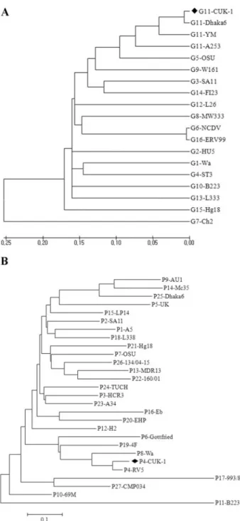
The complete open reading frame (978 bp) and deduced VP7 amino acid sequence of the CUK-1 human rotavirus strain were determined. From comparisons of the VP7 nucle-otide and amino acid sequences of CUK-1 with the corre-sponding VP7 sequences of the prototype strains (G1 to G16), the VP7 sequence of the CUK-1 strain was found to have close relationships to G11 rotavirus strains A253, Dhaka6, and YM (86.9, 98.7, and 91.97% nucleotide sequence identities, respec-tively, and 95.1, 98.5, and 95.4.5% amino acid identities, re-spectively). Overall, the CUK-1 strain showed the highest de-gree of similarity with the Dhaka6 strain. This strain had fewer corresponding identities with the other G types (types G1 to G16), with the exception of G11. We constructed the dendro-gram containing VP7 of CUK-1 and the 16 G types described above (Fig. 1A). This phylogenetic analysis indicated that
* Corresponding author. Mailing address: Department of Microbi-ology, College of Medicine, The Catholic University of Korea, 505 Banpo-dong, Seocho-go, Seoul 137-701, Republic of Korea. Phone: 82 2 590 1217. Fax: 82 2 535 6473. E-mail: paik@catholic.ac.kr.
† Seong-Karp Hong and Sung-Geun Lee contributed equally to this paper.
䌤Published ahead of print on 29 August 2007.
3759
on June 8, 2014 by YONSEI UNIV MED LIBRARY
http://jcm.asm.org/
CUK-1 was clustered with the Dhaka6 G11 type in a mono-phyletic branch.
We found that the deduced amino acid sequences of four intragenotype-conserved antigenic regions were aligned within CUK-1 and the other 16 G types. The CUK-1 strain showed a close relationship with G11 strains A253, Dhaka6, and YM in these regions. This CUK-1 strain showed only 1 amino acid change (V218I) in region C compared with the sequence of the Dhaka6 strain. Previous reports on comparative analyses of VP7 gene sequences among rotaviruses of different serotypes had identified a high degree of sequence divergence in several discrete regions, and these regions were highly conserved within the serotypes (10). Also, the A, B, C, and F antigenic regions were involved in virus neutralization (8, 12). The se-quences of the CUK-1 strain were nearly similar to those of the Dhaka6, YM, and A253 strains within these regions, with 1, 2, and 3 amino acid changes, respectively. The Asp at position 96 within region A was probably responsible for its G11 specific-ity, because Asp is also present at this position in the VP7 of the G11 strain but not in the G5 strain (4). The amino acid at this position has been reported to be critical to the conforma-tion of the major antigenic site of VP7 (4, 8, 12), although a single amino acid substitution may not be enough to com-pletely affect the serotype reactivity (4). Therefore, it can be stated that the CUK-1 strain retains its G11 specificity because it has Asp at position 96. Here, the CUK-1 strain showed a high degree of sequence divergence (more than 30%) com-pared with the antigenic region sequences strains of other serotypes, except G11.
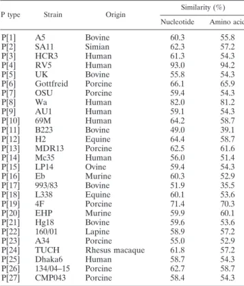
The partial 418-bp gene sequences (nucleotides [nt] 65 to 482) of the VP8* fragments within VP4 of the CUK-1 human rotavirus strain and the deduced amino acid sequences were determined. The CUK-1 strain then presented the highest degrees of identity of 93% and 94.2% with corresponding nucleotide sequences and the deduced amino acid sequences of the VP8* fragments of rotavirus type 5 (RV5) of the P[4] genotype, respectively (Table 1). In addition, phylogenetic tree analysis of the VP8* fragments of the VP4 genes of strain CUK-1 and other established rotavirus P types by the neighbor-joining method also confirmed that strain CUK-1 belonged to type P[4] (Fig. 1B). The type G11 strains A253, CUK-1, Dhaka6, and A253 and the G5 strain may have originated from a common ancestor of VP7 specificity (4), as shown in Fig. 1A. For VP4 specificity, however, the G5 viruses have been detected not only in association with P[7] but also in conjunction with P[6] and P[8] (19), while G11 was combined with P[25] (21) and type G11 strain CUK-1 was combined with P[4]. Therefore, the CUK-1 strain showed a common lineage with G11 and G5 in the molecular evolution of VP7 but a diversity from these strains in the molecular evolution of VP4. The G-type rotaviruses combined with P[4] that have been reported to date are G1,P[4] (1), G2,P[4] (19), G3,P[4] (16), G4,P[4] (1), G8,P[4] (5), G9,P[4] (20), and G12,P[4] (18). On the basis of these results, one might expect a possibility that CUK-1 occurred by a natural reassortment event.
This incidence of a novel recombinant G11,P[4] rotavirus strain in South Korea illustrates the large diversity of rotavirus strains occurring worldwide. Advanced research with this strain will pro-mote the investigation of diverse rotavirus strains in the molecu-lar, genetic, evolutionary, and epidemiological fields. Moreover, studies and surveillance of animal-to-human transmission events
FIG. 1. (A) Phylogenetic tree analysis based on the nucleotide sequences of the VP7 genes (nt 49 to 1026) of strain CUK-1 and strains of other rotavirus G types by use of the neighbor-joining method. The following strains have the indicated GenBank accession numbers: Wa, KO2033; HU5, A01028; SA11, K02028; ST3, X13603; OSU, X04613; NCDV, M12394; Ch2, X56784; MW333, AJ278257; 116E, AB180969; B223, X57852; A253, L24163; Dhaka6, AY773003; YM, M23194; L26, M58290; L338, D13549; F123, M61876; Hg18, AF237666; and Evr99, DQ981478. (B) Phylogenetic tree analysis based on nucleotide sequences (nt 65 to 482) of the VP8* fragments of the VP4 genes of strain CUK-1 and strains of other rotavirus P types by use of the neighbor-joining method. The following strains have the indicated GenBank accession numbers: A5D13395; SA11X14204; HCR3L19712; RV5M32559; UK, M22306; Gottfreid, M33516; OSU, X13190; Wa, L34161; AU1, D10970; 69 M, M60600; B223, D13394; H2, L04638; MDR13, L07886; Mc35, D14032; LP14, L11599; Eb, L18992; 993/83, D16352; L338, D13399; 4F, L10359; EHP, U08424; Hg18, AF237665; 160/01, AF526374; A34, AY174094; TUCH, AY596189; Dhaka6, AY773004; 134/04–15, DQ061053; and CMP043, DQ534016.
3760 NOTES J. CLIN. MICROBIOL.
on June 8, 2014 by YONSEI UNIV MED LIBRARY
http://jcm.asm.org/
will help to provide an understanding of and prevent rotavirus disease. Some rotavirus strains may have arisen by interspecies transmission or by reassortment between human and animal ro-taviruses (6). This enormous diversity among rotavirus strains allows the study of the evolution of rotavirus strains and creates new challenges for rotavirus vaccine development (6). Finally, the discovery of this novel combinant rotavirus strain will play an important role in future vaccine development.
Nucleotide sequence accession number.The complete open reading frame (978 bp) and deduced VP7 amino acid sequence of the CUK-1 human rotavirus strain were enrolled in GenBank under accession number EF121951.
This work was supported by MSD Korea Project (from 2004 to 2006 years) and BK21 project team for Biomedical Science.
REFERENCES
1. Abdel-Haq, N. M., R. A. Thomas, B. I. Asmar, V. Zacharova, and W. D. Lyman.2003. Increased prevalence of G1,P[4] genotype among children with rotavirus-associated gastroenteritis in metropolitan Detroit. J. Clin. Micro-biol. 41:2680–2682.
2. Brandt, C. D., H. W. Kim, J. O. Rodriguez, W. J. Arrobio, B. C. Jeffries, E. P. Stallings, C. Lewis, A. J. Miles, R. M. Chanock, A. Z. Kapikian, and R. H. Parrott.1983. Pediatric viral gastroenteritis during eight years of study. J. Clin. Microbiol. 18:71–78.
3. Ciarlet, M., M. Hidalgo, M. Gorziglia, and F. Liprandi. 1994.
Characteriza-tion of neutralizaCharacteriza-tion epitopes on VP7 surface protein of serotype G11 porcine rotaviruses. J. Gen. Virol. 75:1867–1873.
4. Ciarlet, M., Y. Hoshino, and F. Liprandi. 1997. Single point mutations may affect the serotype reactivity of serotype G11 rotavirus strains: a widening spectrum? J. Virol. 71:8213–8220.
5. Fischer, T. K., N. A. Page, D. D. Griffin, J. Eugen-Olsen, A. G. Pedersen, P. Valentiner-Branth, K. Molbak, H. Sommerfelt, and N. M. Neilsen.2003. Characterization of incompletely typed rotavirus strains from Guinea-Bissau: identification of G8 and G9 types and high frequency of mixed infections. Virology 311:125–133.
6. Gentsch, J. R., A. R. Laird. B. Bielfelt, D. D. Griffin, K. Banyai, M. Ramachandran, V. Jain, N. A. Cunliffe, O. Nakagomi, C. D. Kirkwood, T. K. Fischer, U. D. Parashar, J. S. Bresse, B. Jiang, and R. I. Glass.2005. Serotype diversity and reassortment between human and animal rotavirus strains: implication for rotavirus vaccine pro-grams. J. Infect. Dis. 192(Suppl. 1):S146–S159.
7. Gentsch, J. R., R. I. Glass, P. Woods, V. Gouvea, J. Flores, B.K. Das, and M. K. Bhan.1992. Identification of group a rotavirus gene 4 types by poly-merase chain reaction. J. Clin. Microbiol. 30:1365–1373.
8. Gorziglia, M., G. Larralde, and R. L. Ward. 1990. Neutralization epitopes on rotavirus SA11 4fM outer capsid proteins. J. Virol. 64:4534–4539. 9. Gouvea, V., R. I. Glass, P. Woods, H. F. Clack, and Z. Y. Fang. 1990.
Polymerase chain reaction amplification and typing of rotavirus nucleic acid from stool specimens. J. Clin. Microbiol. 28:276–282.
10. Green, K. Y., K. Midthun, M. Gorziglia, Y. Hoshino, A. Z. Kapikian, R. M. Chanock, and J. Flores.1987. Comparison of the amino acid sequences of the major neutralization protein of four human rotavirus serotypes. Virology 161:153–159.
11. Gulati, B. R., R. Deepa, B. K. Singh, and C. Durgo Rao. 2007. Diversity in Indian equine rotaviruses: identification of genotype G10, P6[1] and G1 strains and a new VP7 genotype (G16) strain in specimens from diarrheic foals in India. J. Clin. Microbiol. 45:972–978.
12. Hoshino, Y., K. Nishikawa, D. A. Benfield, and M. Gorziglia. 1994. Mapping of antigenic sites involved in serotype-cross-reaction neutralization on group A rotavirus outer capsid glycoprotein VP7. Virology 199:233–237. 13. Kapikian, A. Z., Y. Hoshino, and R. M. Chanock. 2001. Rotaviruses, p.
1787–1833. In P. M. Howley (ed.), Fields virology, vol. 2, 4th ed. Lippincott Williams & Wilkins, Philadelphia, PA.
14. Martella, V., M. Ciarlet, K. Banyai, E. Lorusso, A. Cavalli, M. Corrente, G. Elia, S. Arista, M. Camero, C. Desario, N. Decaro, A. Lavazza, and C. Buonavoglia.2006. Identification of a novel VP4 genotype carried by a serotype G5 porcine rotavirus strain. Virology 346:301–311.
15. Rahman, M., R. Sultana, G. Ahmed, S. Nahar, Z. M. Hassan, F. Saiada, G. Podder, A. S. G. Faruque, D. A. Sack, J. Matthijnssens, M. V. Ranst, and T. Azim. 2007. Prevalence of G2P[4] and G12P[6] rotavirus, Bangladesh. Emerg. Infect. Dis. 13:18–24.
16. Rosa e Silva, M. L., I. P. Carvalho, and V. Gouvea. 2002. 1988–1999 rotavirus seasons in Juiz de Forda, Minas Gerais, Brazil; detection of an unusual G3,P[4] epidemic strain. J. Clin. Microbiol. 40:2837–2842.
17. Ruiz, A. M., I. V. Lopez, S. Lopez, R. T. Espejo, and C. F. Arias. 1988. Molecular and antigenic characterization of porcine rotavirus YM, a possible new rotavirus serotype. J. Virol. 62:4331–4336.
18. Samajdar, S., V. Varghese, P. Barman, S. Ghosh, U. Mitra, P. Dutta, S. K. Bhattacharya, M. V. Narasimham, P. Panda, T. Krishnan, N. Kobayashi, and T. N. Naik.2006. Changing pattern of human group A rotaviruses: emergence of G12 as an important pathogen among children in eastern India. J. Clin. Virol. 36:183–188.
19. Santos, N., and Y. Hoshino. 2005. Global distribution of rotavirus serotypes/ genotypes and its implication for the development and implementation of an effective rotavirus vaccine. Rev. Med. Virol. 15:29–56.
20. Santos, N., E. M. Volotao, C. C. Soares, M. C. M. Albuquerque, F. M. da Silva, T. R. B. de Carvalho, C. F. A. Pereira, V. Chizhikov, and Y. Hoshino. 2001. Rotavirus strains bearing genotype G9 or P[9] recovered from Brazil-ian children with diarrhea from 1997 to 1999. J. Clin. Microbiol. 39:1157– 1160.
21. Uchida, R., B. D. Pandey, J. B. Sherchand, K. Ahmed, M. Yokoo, T. Nakagomi, L. E. Cuevas, N. A. Cunliffe, C. A. Hart, and O. Nakagomi.2006. Molecular epidemiology of rotavirus diarrhea among children and adults in Nepal: detec-tion of G12 strains with P[6] or P[8] and a G11,P[25] strain. J. Clin. Microbiol. 44:3499–3505.
22. Zaki, A. M., H. L. Dupont, M. A. el Alamy, R. R. Arafat, K. Amin, M. M. Awad, L. Bassiouni, I. Z. lmam, G. S. el Malih, A. el Marsafie, M. S. Mohieldin, T. Naguib, M. A. Rakha, M. Sidaros, N. Wasef, C. E. Wright, and R. G. Wyatt.1986. The detection of enteropathogens in acute diarrhea in a family cohort population in rural Egypt. Am. J. Trop. Med. Hyg. 35:1013– 1022.
TABLE 1. Nucleotide and amino acid VP4 sequence similarities of strain CUK-1 with those of strains of different P genotypesa
P type Strain Origin
Similarity (%) Nucleotide Amino acid
P[1] A5 Bovine 60.3 55.8 P[2] SA11 Simian 62.3 57.2 P[3] HCR3 Human 61.3 54.3 P[4] RV5 Human 93.0 94.2 P[5] UK Bovine 55.8 54.3 P[6] Gottfreid Porcine 66.1 65.9 P[7] OSU Porcine 59.4 54.3 P[8] Wa Human 82.0 81.2 P[9] AU1 Human 59.1 54.3 P[10] 69M Human 64.2 58.7 P[11] B223 Bovine 49.0 39.1 P[12] H2 Equine 64.4 58.7 P[13] MDR13 Porcine 62.5 61.6 P[14] Mc35 Human 56.0 51.4 P[15] LP14 Ovine 59.4 54.3 P[16] Eb Murine 60.3 52.9 P[17] 993/83 Bovine 51.9 35.5 P[18] L338 Equine 60.1 53.6 P[19] 4F Porcine 71.4 70.3 P[20] EHP Murine 59.9 60.1 P[21] Hg18 Bovine 59.6 53.6 P[22] 160/01 Lapine 58.9 57.2 P[23] A34 Porcine 55.0 52.9
P[24] TUCH Rhesus macaque 61.8 57.2
P[25] Dhaka6 Human 58.7 54.3
P[26] 134/04–15 Porcine 62.7 58.7 P[27] CMP043 Porcine 58.4 54.3
a
The gene sequences of the VP4 sequences from nt 65 to 482 were compared. The partial VP4 sequences of strain A34 (nt 69 to 481) were compared.
VOL. 45, 2007 NOTES 3761