저작자표시-비영리-변경금지 2.0 대한민국 이용자는 아래의 조건을 따르는 경우에 한하여 자유롭게 l 이 저작물을 복제, 배포, 전송, 전시, 공연 및 방송할 수 있습니다. 다음과 같은 조건을 따라야 합니다: l 귀하는, 이 저작물의 재이용이나 배포의 경우, 이 저작물에 적용된 이용허락조건 을 명확하게 나타내어야 합니다. l 저작권자로부터 별도의 허가를 받으면 이러한 조건들은 적용되지 않습니다. 저작권법에 따른 이용자의 권리는 위의 내용에 의하여 영향을 받지 않습니다. 이것은 이용허락규약(Legal Code)을 이해하기 쉽게 요약한 것입니다. Disclaimer 저작자표시. 귀하는 원저작자를 표시하여야 합니다. 비영리. 귀하는 이 저작물을 영리 목적으로 이용할 수 없습니다. 변경금지. 귀하는 이 저작물을 개작, 변형 또는 가공할 수 없습니다.
Acceleration of Bone Regeneration by
BMP-2-Loaded Collagenated Biphasic
Calcium Phosphate in Rabbit Sinus
Jung-Soo Kim
Department of Dentistry
Acceleration of Bone Regeneration by
BMP-2-Loaded Collagenated Biphasic
Calcium Phosphate in Rabbit Sinus
Directed by Professor Seong-Ho Choi
A Doctoral Dissertation
submitted to the Department of Dentistry
the Graduate School of Yonsei University
in partial fulfillment of the requirements for the degree of
Ph.D. in Dental Science
This certifies that the Doctoral Dissertation
of Jung-Soo Kim is approved.
Thesis Supervisor: Seong-Ho Choi
Jung-Kiu Chai
Kyoo-Sung Cho
Yeek Herr
Hyeong-Seob Kim
The Graduate School
Yonsei University
감사의 글
본 논문이 완성되기까지 부족한 저에게 지도와 격려를 아끼지 않
으신 최성호 교수님, 채중규 교수님, 조규성 교수님, 허익 교수님,
김형섭 교수님께 깊은 감사를 드립니다. 그리고 부족한 논문임에도
진심 어린 조언으로 격려해주시고 따뜻한 관심으로 지켜봐 주신 김
종관 교수님, 김창성 교수님, 정의원 교수님, 이중석 교수님, 김민
수 선생님, 차재국 선생님께 감사드립니다.
연구 내내 많은 도움을 준 치주과 수련의 선생님들, 그리고 때늦
은 공부에 많은 조언을 주신 대학원 선, 후배님들께 모두 진심으로
감사드립니다.
마지막으로 어려움이 있을 때마다 항상 저의 버팀목이 되어주시고,
물심양면으로 도움을 주신 아버지, 어머니와 장인, 장모님께 깊은
사랑과 감사를 드리며, 무엇보다도 말썽꾸러기 네 아이들 돌보며 늦
깎이 학생 뒷바라지 하느라 고생한 제 인생의 가장 좋은 친구이자
동반자인 아내에게 저의 온 마음을 담아 감사와 사랑을 전합니다.
아울러 논문 핑계로 많은 시간 같이하지 못했음에도 항상 아빠에게
큰 힘을 주었던 세은이, 세준이, 세연이, 세훈이 네 아이들에게도
고마움을 전합니다.
Table of contents
List of figures µµµµµµµµµµµµµµµµµµµµµµµµµµµµµµµµµµµµµµµµµµµµµµµµµµµµµµµµµµµµµµµµµµµµµµµµµµµµµ ii
List of tables µµµµµµµµµµµµµµµµµµµµµµµµµµµµµµµµµµµµµµµµµµµµµµµµµµµµµµµµµµµµµµµµµµµµµµµµµµµµµµ iii
Abstract (English) µµµµµµµµµµµµµµµµµµµµµµµµµµµµµµµµµµµµµµµµµµµµµµµµµµµµµµµµµµµµµµµµµµµµµµµ iv
I. Introduction µµµµµµµµµµµµµµµµµµµµµµµµµµµµµµµµµµµµµµµµµµµµµµµµµµµµµµµµµµµµµµµµµµµµµµµµµµµµµ 1
II. Materials and Methods µµµµµµµµµµµµµµµµµµµµµµµµµµµµµµµµµµµµµµµµµµµµµµµµµµµµµµµµµµµµµ 4
1. Preparation of the BMP-2 and BMP-2-Loaded CBCP µµµµµµµµµµµµµµµµµµµ 4
2. Study Design µµµµµµµµµµµµµµµµµµµµµµµµµµµµµµµµµµµµµµµµµµµµµµµµµµµµµµµµµµµµµµµµµµµµµµ 4
3. Animals µµµµµµµµµµµµµµµµµµµµµµµµµµµµµµµµµµµµµµµµµµµµµµµµµµµµµµµµµµµµµµµµµµµµµµµµµµµµ 5
4. Surgical Procedures µµµµµµµµµµµµµµµµµµµµµµµµµµµµµµµµµµµµµµµµµµµµµµµµµµµµµµµµµµµµµµ 5
5. Radiographic Analysis µµµµµµµµµµµµµµµµµµµµµµµµµµµµµµµµµµµµµµµµµµµµµµµµµµµµµµµµµµµ
6
6. Histologic and Histomorphometric Analysis µµµµµµµµµµµµµµµµµµµµµµµµµµµµµµµ
7
7. Statistics µµµµµµµµµµµµµµµµµµµµµµµµµµµµµµµµµµµµµµµµµµµµµµµµµµµµµµµµµµµµµµµµµµµµµµµµµµµµ
8
III. Results µµµµµµµµµµµµµµµµµµµµµµµµµµµµµµµµµµµµµµµµµµµµµµµµµµµµµµµµµµµµµµµµµµµµµµµµµµµµµµµµµµ 9
1. Clinical Observations µµµµµµµµµµµµµµµµµµµµµµµµµµµµµµµµµµµµµµµµµµµµµµµµµµµµµµµµµµµµ
9
2. Radiographic Analysis µµµµµµµµµµµµµµµµµµµµµµµµµµµµµµµµµµµµµµµµµµµµµµµµµµµµµµµµµµµ
9
3. Histologic Analysis µµµµµµµµµµµµµµµµµµµµµµµµµµµµµµµµµµµµµµµµµµµµµµµµµµµµµµµµµµµµµµ
11
4. Histometric Analysis µµµµµµµµµµµµµµµµµµµµµµµµµµµµµµµµµµµµµµµµµµµµµµµµµµµµµµµµµµµµ
13
IV. Discussion µµµµµµµµµµµµµµµµµµµµµµµµµµµµµµµµµµµµµµµµµµµµµµµµµµµµµµµµµµµµµµµµµµµµµµµµµµµµ 15
References µµµµµµµµµµµµµµµµµµµµµµµµµµµµµµµµµµµµµµµµµµµµµµµµµµµµµµµµµµµµµµµµµµµµµµµµµµµµµµµµµ 19
Figure Legends µµµµµµµµµµµµµµµµµµµµµµµµµµµµµµµµµµµµµµµµµµµµµµµµµµµµµµµµµµµµµµµµµµµµµµµµµµµ 25
Figures µµµµµµµµµµµµµµµµµµµµµµµµµµµµµµµµµµµµµµµµµµµµµµµµµµµµµµµµµµµµµµµµµµµµµµµµµµµµµµµµµµµµµ 29
Abstract (Korean) µµµµµµµµµµµµµµµµµµµµµµµµµµµµµµµµµµµµµµµµµµµµµµµµµµµµµµµµµµµµµµµµµµµµµµµ 36
List of Figures
Figure 1. (A) The ROI for radiographic analysis. (B) The ROI for
histomorphometric analysis.
Figure 2. 3D-reconstructed micro-computed tomographic (μCT) view of the
augmented area.
Figure 3. μCT views of the augmented area.
Figure 4. Composition of the three segmental regions in the radiographic
analysis.
Figure 5. Histologic photomicrographs after the 2 weeks of healing.
Figure 6. Histologic photomicrographs after the 4 weeks of healing.
Figure 7. Composition of the three segmental regions in the
List of Tables
Table 1. Composition of The Total Augmented Volume.
Table 2. Composition of the Total Augmented Area.
Abstract
Acceleration of Bone Regeneration by BMP-2-Loaded
Collagenated Biphasic Calcium Phosphate in Rabbit Sinus
Jung-Soo Kim, D.D.S., M.S.D.
Department of Dentistry
The Graduate School, Yonsei University
(Directed by Professor Seong-Ho Choi, D.D.S., M.S.D., PhD.)
Objective: The objective of this study was to determine the effectiveness of
collagenated biphasic calcium phosphate (CBCP) as a carrier for bone morphogenetic protein-2 (BMP-2) at the early stage of healing in rabbit sinus.
Material and Methods: In 16 rabbits, BMP-2-loaded CBCP was grafted into one
sinus (the BMP group) and saline-soaked CBCP was grafted into another sinus (the CTL group). The groups were assigned randomly. After 2 weeks (n = 8) or 4 weeks (n = 8), radiographic and histologic analysis was performed.
found in the BMP groups at the early healing period. At 4 weeks, evenly distributed new bone was observed in the BMP group, whereas the new bone was sparsely distributed in the central portion in the CTL group.
Conclusion: It can be concluded that the addition of BMP-2 to CBCP resulted in a
greater initial augmented volume as a result of postoperative swelling, which is replaced by early bone formation, and it was prominent near the Schneiderian membrane.
Acceleration of Bone Regeneration by BMP-2-Loaded
Collagenated Biphasic Calcium Phosphate in Rabbit Sinus
Jung-Soo Kim, D.D.S., M.S.D.
Department of Dentistry
The Graduate School, Yonsei University
(Directed by Professor Seong-Ho Choi, D.D.S., M.S.D., PhD.)
I. Introduction
A suitable carrier matrix is required to act as a scaffold for the effective application and release of growth factors. In the case of a maxillary sinus, the volume stability of the carrier against further repneumatization should also be considered. The absorbable collagen sponge (ACS), which has been approved by the US Food and Drug Administration, is known to have a limited capacity with respect to maintaining volume.1 On the other hand, particulate graft materials are difficult to manipulate at
collagen has recently been developed with a view to reinforcing the mechanical durability while simultaneously improving the manageability.2,3
The present study attempted sinus augmentation using collagenated biphasic calcium phosphate (CBCP) as a carrier for bone morphogenetic protein-2 (BMP-2). It was hypothesized that the slowly resorbing hydroxyapatite (HA) core would offer good osteoconductivity for new bone growth, while the rapidly resorbing beta-tricalcium phosphate (β-TCP) would be a good substrate for bone-forming cells due to its excellent biocompatibility.4,5 Moreover, the resorption rate of this biomaterial
can be controlled by modifying the mixing ratios of HA and β-TCP without altering its osteoconductive properties,6,7 and the collagen matrix should stabilize the bone
particles and help with the adsorption of blood coagulum at the initial stage of healing.8 A recent study found that CBCP composite blocks loaded with BMP-2
facilitate 3D bone regeneration in a vertical augmentation model, yielding bone with a
mature appearance.9
In terms of the release kinetics of BMP-2, ACS exhibited an initial burst release pattern of BMP-2, whereas allografts and ceramics inefficiently released BMP-2.10
HA particles are poorly bioresorbable, whereas TCP particles progressively undergo bioresorption.11The bioresorption properties of the biphasic calcium phosphate (BCP)
ceramics could provide optimum conditions for BMP-2-induced bone formation, rendering CBCP an excellent carrier system for BMP-2.12 The controlled release of
BCP showed biocompatible and osteoconductive properties in sinus
augmentation,13however it is known that adequate levels of new bone formation are
generally achieved only after 6–8 months of healing following the sinus augmentation procedure.14In order to overcome this shortcoming, various studies have investigated
the use of various growth factors such as BMPs.15,16
The objective of this study was to determine the effectiveness of CBCP as a carrier system for BMP-2 at the early stage of healing for sinus augmentation in the standardized rabbit sinus model.
II. Materials and Methods
1. Preparation of the BMP-2 and BMP-2-Loaded CBCP
The E. coli expressed BMP-2 and the CBCP were generously supplied by Genoss institute (Suwon, Korea). BMP-2 at the concentration of 0.1mg/ml was reconstituted and diluted in a buffer, and then CBCP (Osteon Collagen®, Genoss, Suwon, Korea)
with 0.3–0.5 mm particle size, was soaked with 200ml of BMP-2 or saline. The CBCP blocks were porous in structure and had a uniform volume of ø6.0 x 5.0 mm. The cylindrical shaped bone filler composed of synthetic bone containing 70% HA and 30% β-TCP and a natural type I collagen. After allowing a binding period of 10 min, the CBCPs soaked with BMP-2 or saline were placed into the maxillary sinuses.
2. Study Design
In total, 16 rabbits were subjected to bilateral sinus augmentation. The animals were divided into two groups according to the healing period: 2 weeks (n = 8) and 4 weeks (n = 8). One sinus of each rabbit was augmented using BMP-2-loaded CBCP (BMP group), and the other sinus was augmented using saline-soaked CBCP (CTL group); the groups were assigned randomly. Thus, there were ultimately four groups:
BMP groups with 2- and 4-week healing periods (2wBMP and 4wBMP groups), and CTL groups with 2- and 4-week healing periods (2wCTL and 4wCTL groups).
3. Animals
Male New Zealand white rabbits weighing 2.5–3.0 kg were selected for the experimental model. In total, 32 maxillary sinuses in 16 rabbits were used for sinus augmentation. Animals were housed in separate cages under standard laboratory conditions, with ad libitum access to water and a standard diet. The selection, management, and preparation of animals, and the surgical protocol were approved by the Institutional Animal Care and Use Committee of Yonsei Medical Center, Seoul, Korea (approval no. 2011-0262).
4. Surgical Procedures
The surgical procedure has been described in our previous report.17In brief, a
full-thickness flap was elevated laterally under general anesthesia and local infiltration after a straight incision along the midline along the nasal bone. Standardized circular windows were prepared bilaterally with a 5.5-mm-diameter trephine bur (C-reamer, Neobiotech, Seoul, Korea). Drilling was performed until the grayish membrane was seen through the trephined bone, at which point the circular bony disk was carefully
the sides for the CTL and BMP groups being chosen at random). After grafting, the flaps were sutured layer by layer with 4–0 Monosyn (glyconate absorbable monofilament, B-Braun, Aesculap, Center Valley, PA, USA), which was removed after 7 days. The rabbits were sacrificed at either 2 or 4 weeks postoperatively.
5. Radiographic Analysis
All collected samples, including the augmented sinus and the surrounding tissue, were fixed in 10% formalin for 10 days. They were scanned using a µCT system (high-energy spiral scan µCT, Skyscan 1173, Bruker-microCT, Kontich, Belgium) at a resolution of 14.91 µm (achieved using 130 kV and 60 µA). The scanned data were reconstructed using NRecon software (Bruker-microCT), and the region of interest (ROI) was established and analyzed using CTAn software (Bruker-microCT). The mineralization of bone can be assessed by the comparison of x-ray attenuation with that of HA.
Analysis of the 3D images allowed measurement of the total augmented volume (TAV; in mm3) and the volumes of the following components: newly formed bone
(NBV; in mm3), residual particles of graft materials (RMV; in mm3), and
nonmineralized tissue (NMV; in mm3); the volume ratios of each of the latter three
components to TAV were also calculated (designated as %NBV, %RMV and %NMV, respectively). In addition, the same measurements were performed at three segmental
ROIs; window, center, and membrane. The volume of each sectional ROI was
4.94 mm3(cylinder form, ø2.5 mm × 1.0 mm; Figure 1A).
6. Histologic and Histomorphometric Analysis
After radiographic analysis, the sections were decalcified in 5% formic acid for 10 days and then embedded in paraffin. Serial sections were cut at a thickness of 5 µm coronally along the center of the window. The two most central sections of each block were selected and stained with Masson’s trichrome. Histologic analysis was performed using a light microscope (BX-50, Olympus Optical, Tokyo, Japan).
Histomorphometric measurements were made using an automated image-analysis system (Image-Pro Plus, Media Cybernetics, Silver Spring, MD, USA). The composition of the total augmented area (TAA; in mm2) was identified, and the
relative areas of new bone, residual material, non-mineralized tissue were separately
detected manually and calculated (NBA, RMA, and NMA, respectively; in mm2). The
proportions of each composite (i.e., %NBA, %RMA, and %NMA) out of the TAA were also obtained. To evaluate the homogeneity of regenerated bone in the grafted sinus area as a secondary outcome variable, the above-mentioned areas and proportions of each composite were calculated in specific standardized ROI; near window, at central, and near membrane (square form, 1.0 mm × 1.0 mm; Figure 1B).
7. Statistics
The statistical analyses were performed using the Statistical Analysis System (SAS v9.2, SAS Institute, Cary, NC, USA). The Shapiro-Wilk normality test was performed prior to the statistical analysis and all variables were found to be normally distributed. Independent t-tests were carried out to compare the results obtained at 2 and 4 weeks, and paired t-tests were used to evaluate differences between the BMP-2-treated and control groups (p < 0.05).
III. RESULTS
1. Clinical Observations
Only one rabbit in the 4wBMP group was excluded from the results (due to maxillary sinusitis). The wound healing process was generally uneventful, although small tears in the sinus membranes did occur in five sinuses (one of the BMP group and four of the CTL group).
2. Radiographic Analysis
The 3D-reconstructed views revealed that all window areas of augmented sinuses were partially externally closed in all groups and the augmented sinus had a dome-shaped appearance in all groups. The volume of the augmented area appeared to be greater in the BMP groups than in the CTL groups (Figure 2). The sinus cavity was augmented in all groups with radiopaque substance, including remnant bone substitute and regenerated mineralized tissue. In the BMP groups, a large amount of new bone was evident in the overall area of the sinus at the 4-week healing period, although newly formed bone was mainly observed near the window and at the anterior portion of sinus at 2 weeks. On the other hand, newly formed bone in the CTL groups was mainly observed only near the window at 2 weeks; it did not expand
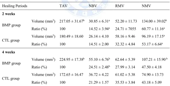
The mean TAV, NBV, RMV, and NMV of each group are given in Table 1. TAV was significantly larger in the BMP group than in the CTL group at both 2 weeks
(217.1 ± 31.7 mm3vs 180.5 ± 18.6 mm3, p = 0.01) and 4 weeks (225.0 ± 17.4 mm3vs
172.7 ± 16.5 mm3; p < 0.0001). NBV was significantly larger in the BMP group than
in the CTL group at 4 weeks (p < 0.0001), did it did not differ significantly between these groups at 2 weeks. Both NBV and %NBV were significantly greater in the 4wBMP group than in the 2wBMP group (p < 0.05). NMV was significantly larger in the BMP groups than in the CTL groups at both 2 and 4 weeks (p = 0.03 and 0.002, respectively). %NMV of both the BMP group (p = 0.0120) and the CTL group (p = 0.0066) decreased significantly with healing time.
Table 1. Composition of the Total Augmented Volume. (mean ± standard deviation)
Healing Periods TAV NBV RMV NMV
2 weeks BMP group Volume (mm 3) 217.05 ± 31.67b 30.85 ± 6.31a 52.20 ± 11.73 134.00 ± 39.02b Ratio (%) 100 14.52 ± 3.94a 24.71 ± 7055 60.77 ± 11.16a CTL group Volume (mm 3) 180.49 ± 18.60 26.14 ± 4.10 58.16 ± 9.46 96.19 ± 17.15a Ratio (%) 100 14.51 ± 2.00 32.32 ± 4.84 53.17 ± 6.64a 4 weeks BMP group Volume (mm 3) 224.95 ± 17.38b 55.10 ± 6.76b 62.64 ± 5.39 107.21 ± 15.90b Ratio (%) 100 24.51 ± 2.48b 27.99 ± 3.14 47.50 ± 4.18 CTL group Volume (mm 3) 172.65 ± 16.47 36.72 ± 4.22 61.02 ± 5.38 74.90 ± 13.73 Ratio (%) 100 21.29 ± 1.57 35.53 ± 3.84 43.18 ± 5.09
a Significantly different from the group with the same protocol at 4 weeks (p < 0.05). b Significantly different from the CTL group at the same observation period (p < 0.05)
TAV = total augmented volume; NBV = volume of newly formed bone; RMV = volume of residual particles of graft material; NMV = volume of non-mineralized tissue.
Statistically significant differences between the groups are shown in Figure 4. %NBV was significantly larger in the 4wBMP group than in the 4wCTL group at the three segmental regions (p < 0.05). %NMV was significantly smaller in the 4wBMP group than in the 2wBMP group in the membrane region (p < 0.01).
3. Histologic Analysis
The maxillary sinus cavity was surrounded by the respiratory mucosa and a thin layer of cortical bone. Lining of the Schneiderian membrane was intact and numerous serous glands were seen within lamina propria between the epithelium and the periosteum-like layer. The overall morphology of the five sinuses which experienced membrane tearing during surgical procedure was similar with to that of the normal sinus without perforation of the Schneiderian membrane.
At 2 weeks, cross-sectional shape of the augmented sinus was convex and the window region was not yet completely regenerated in both groups. The collagen matrix which acts as a cohesive granule stabilizer in the CBCP carrier was completely resorbed. In the 2wCTL group, only a small amount of new bone could be detected all around the augmented area, and most extensively at the periphery of window region
mostly near the Schneiderian membrane and window regions, and the residual biomaterials were closer to each other than at the BMP group (Figure 5C, D).
In 2wBMP group, the newly formed bone was mainly observed along the outer surface of CBCP, close to the parent bone wall and the raised Schneiderian membrane (Figure 5E). The new bone was woven bone containing extensive blood vessels and viable osteocytes were visible inside lacunae. The surface of new bone was lined by osteoblasts and several active multinucleated osteoclasts were also observed (Figure 5F, G, H).
Compared with 2 weeks, the new bone area increased visibly at 4 weeks in both groups and the augmented spaces remained convex. In 4wCTL group, the quantity of new bone was increased in the window and membrane regions, however the new bone was sparsely observed in the central portion of the augmented area (Figure 6A, B, C, D). The window region was partially closed and the augmented surface of sinus window was rather depressed.
In 4wBMP group, a considerable amount of new bone was evenly distributed throughout the entire augmented sinus (Figure 6E). The window region was almost continuous with the newly formed bone from the sinus cavity, and the original curvature of the nasal bone was restored. The new bone showed evidence of maturation in part being lamellar but mostly represented woven bone with osteoclastic activity. Most of residual particles were tightly attached to new bone even at the central region of the sinus and the bone regeneration and remodeling was still in
progressed with a blended appearance (Figure 6F, G, H).
4. Histometric Analysis
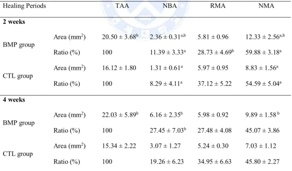
The differences in the TAA confirmed the radiographic volumetric analysis. At both 2 and 4 weeks, the TAA and NBA were significantly larger in the BMP group than in the CTL group (Table 2). In accordance to the healing period, the NBA of 4wBMP group was significantly larger than those of 2wBMP group, while the NMA of 4wBMP group was significantly less than those of 2wBMP group (p = 0.001, p = 0.005, respectively).
Table 2. Composition of the Total Augmented Area. (mean ± standard deviation)
Healing Periods TAA NBA RMA NMA
2 weeks BMP group
Area (mm2) 20.50 ± 3.68b 2.36 ± 0.31a,b 5.81 ± 0.96 12.33 ± 2.56a,b
Ratio (%) 100 11.39 ± 3.33a 28.73 ± 4.69b 59.88 ± 3.18a CTL group Area (mm2) 16.12 ± 1.80 1.31 ± 0.61a 5.97 ± 0.95 8.83 ± 1.56a Ratio (%) 100 8.29 ± 4.11a 37.12 ± 5.22 54.59 ± 5.04a 4 weeks BMP group Area (mm2) 22.03 ± 5.89b 6.16 ± 2.35b 5.98 ± 0.92 9.89 ± 1.58b Ratio (%) 100 27.45 ± 7.03b 27.48 ± 4.08 45.07 ± 3.86 CTL group Area (mm2) 15.34 ± 2.22 3.07 ± 1.27 5.24 ± 0.30 7.03 ± 1.12
b Significantly different from the CTL group at the same observation period (p < 0.05)
TAA = total augmented area; NBA = area of newly formed bone; RMA = area of residual particles of graft material; NMA = area of non-mineralized tissue.
The compositions of specific standardized regions are summarized in Figure 7. Interestingly, at the 2 week healing point, only membrane region showed a statistical difference of %NBA between BMP and CTL group (p = 0.016), and then the greater %NBA was observed not only in the membrane area, but also in the center area at 4 weeks (p = 0.043, p = 0.001, respectively). We observed that the %NBA had increased in all specific regions (i.e. widow, center and membrane) by the end of the observation period with the exception of the center region of the CTL group.
IV. DISCUSSION
The burst release of BMP-2 adsorbed on ACS generates a supraphysiologic concentration of the drug at a very early phase of healing, which is associated with potential complications such as bone resorption, graft resorption, hematoma formation, and heterotopic ossification.18,19It was thus expected that the combination
CBCP carrier would allow a more controlled and sustained release of the protein, since it has been shown that collagen is associated with the early release of growth factors,20and BCP is responsible for the relatively late protein release.10
The overall volume analysis revealed that TAV and NMV were significant larger in the BMP groups than in the CTL groups. It can be assumed that the initial increase in TAV at 2 weeks was associated with the increase in NMV, which can be attributed to postoperative swelling. This finding was also supported by the histometric analysis revealing greater TAA value in the 2wBMP groups than the 2wCTL group along with the corresponding difference in the NMA values showing statistical significance. Such soft-tissue swelling might be the combined result of inflammation of the surrounding tissues and sterile seroma and hematoma formation.21-23Lu et al. reported
considerable initial swelling in the study using BMP-2-loaded ACS or
collagen/β-TCP/HA on supra-alveolar, peri-implant defects.24 In addition, Lee et al.
On the other hand, the late and remarkable increase in TAV at 4 weeks was attributable to not only the increase in NMV but also the increase in NBV, which differed significantly between the 2wBMP and 4wBMP groups. Moreover, the statistically significant decrease in NMV between the 2wCTL and 4wCTL groups implies a reduction in postoperative swelling, which was also observed in the BMP groups. However, maintenance of the increased volume or even greater increased volume was observed in the BMP group (unlike in the CTL group). An explanation for this is that new bone formation was accelerated by BMP-2 before the volumetric increase caused by postoperative swelling had resolved between the 2- and 4-week healing periods. This phenomenon could be explained by different histologic healing pattern observed between the 2wBMP and 2wCTL groups: especially in terms of the regional distribution of the newly formed bone, the %NBA at the membrane regions of the sinus was significantly greater in the BMP group than in the CTL group. This is concurrent with the previous study representing that osteoinductive potential of the Schneiderian membrane is provoked at the early stage of healing with BMP-2.17,26
Early corticalization of the outer surface of augmented sinus may provide support for the maintenance of augmented volume by resisting the positive respiratory pressure. Therefore, the volumetric increase caused by postoperative swelling may be considered a positive effect in terms of providing a space for new bone to augment the insufficient bone volume in the maxillary sinus, if it is utilized appropriately with growth factors such as BMP-2.
Similar studies have been conducted using BMP-2. Choi et al. found no difference in new bone formation between their experimental and control groups in the rabbit
sinus model using BMP-2-coated-BCP and BCP alone after 8 weeks of healing.17The
rabbit sinus model has a strong osteogenic potential, therefore the effect of BMP-2 would not appear pronounced in the late stage of healing. Hence, healing periods of 2 and 4 weeks were used in the present study to determine the characteristics of bone substitutes with BMP-2 at the early stage of healing. The findings of this study demonstrate that, BMP-2 could result in earlier bone regeneration in greater quantity in the maxillary sinus, offering improved implant stability at the early stage of healing, achieving reduced healing time.
One particularly interesting observation in the present study was that an improvement in bone quality was achieved using BMP-2, which effected homogeneous bone formation even at the central region of the augmented sinus. The central region is distant from the osteogenic sources such as native bone; hence resulting in a relative lack of new bone formation. In the present study, BMP-2/CBCP seemed to facilitate angiogenesis in areas distant from the parental bone. BMP-2 stimulates adult mesenchymal stem cells to differentiate into osteogenic cells and causes angiogenesis through chemotaxis of osteoblasts and endothelial cells.27,28
Moreover, the porous structure of CBCP could also provide an appropriate environment for rapid revascularization to avoid an ischemic condition.29
BMP-Large ranges of BMP-2 concentrations have been used in rabbit sinus model thus far (0.1–2 mg/ml).30-32The high doses of BMP-2 in clinical use may contribute to the
numerous adverse effects that are observed in humans but not in animal experiments.33 Therefore, the clinical usage of BMP-2 would be improved by the
minimum dose of BMP-2 that would both form high-quality bone and reduce side effects.34Toward this end, in the present study, we used the minimal concentration of
BMP-2 (0.1 mg/ml, corresponding to 0.02 mg of BMP-2) which has been the lowest concentration of BMP-2 to induce the osteogenic effect in rabbit sinus model, and it was shown to successfully accelerate the regeneration of bone. Therefore, consistent to the aim of minimizing the dosage of BMP-2 for bone regeneration, our study suggests the necessity to further reduce the concentration of BMP-2 in future studies to less than 0.1 mg/ml.
It can be concluded that the addition of BMP-2 to CBCP resulted in a greater initial augmented volume as a result of postoperative swelling, which is replaced by early bone formation, and that bone formation was prominent near the Schneiderian membrane in the rabbit sinuses.
REFERENCES
1. Razzouk S, Sarkis R. BMP-2: biological challenges to its clinical use. N Y State
Dent J 2012;78:37-9.
2. Fontana F, Rocchietta I, Dellavia C, Nevins M, Simion M. Biocompatibility and
manageability of a new fixable bone graft for the treatment of localized bone defects: preliminary study in a dog model. Int J Periodontics Restorative Dent 2008;28:601-7.
3. Jung UW, Lee JS, Park WY, Cha JK, Hwang JW, Park JC, et al. Periodontal regenerative effect of a bovine hydroxyapatite/collagen block in one-wall intrabony defects in dogs: a histometric analysis. J Periodontal Implant Sci 2011;41:285-92.
4. Gauthier O, Bouler JM, Aguado E, Pilet P, Daculsi G. Macroporous biphasic calcium phosphate ceramics: influence of macropore diameter and macroporosity percentage on bone ingrowth. Biomaterials 1998;19:133-9.
5. Karabuda C, Ozdemir O, Tosun T, Anil A, Olgac V. Histological and clinical evaluation of 3 different grafting materials for sinus lifting procedure based on 8 cases. J Periodontol 2001;72:1436-42.
6. Yamada S, Heymann D, Bouler JM, Daculsi G. Osteoclastic resorption of
1997;37:346-7. Nery EB, LeGeros RZ, Lynch KL, Lee K. Tissue response to biphasic calcium phosphate ceramic with different ratios of HA/beta TCP in periodontal osseous defects. J Periodontol 1992;63:729-35.
8. Tsai CH, Chou MY, Jonas M, Tien YT, Chi EY. A composite graft material containing bone particles and collagen in osteoinduction in mouse. J Biomed Mater Res 2002;63:65-70.
9. Kim JW, Jung IH, Lee KI, Jung UW, Kim CS, Choi SH, et al. Volumetric bone
regenerative efficacy of biphasic calcium phosphate-collagen composite block loaded with rhBMP-2 in vertical bone augmentation model of a rabbit calvarium. J Biomed Mater Res A 2012;100:3304-13.
10. Hsu EL, Ghodasra JH, Ashtekar A, Nickoli MS, Lee SS, Stupp SI, et al. A comparative evaluation of factors influencing osteoinductivity among scaffolds designed for bone regeneration. Tissue Eng Part A 2013;19:1764-72.
11. Metsger DS, Driskell TD, Paulsrud JR. Tricalcium phosphate ceramic--a resorbable bone implant: review and current status. J Am Dent Assoc 1982;105:1035-8.
12. Alam I, Asahina I, Ohmamiuda K, Enomoto S. Comparative study of biphasic calcium phosphate ceramics impregnated with rhBMP-2 as bone substitutes. J Biomed Mater Res 2001;54:129-38.
13. Artzi Z, Weinreb M, Carmeli G, Lev-Dor R, Dard M, Nemcovsky CE. Histomorphometric assessment of bone formation in sinus augmentation
phosphate graft materials: at 6 and 9 months in humans. Clin Oral Implants Res 2008;19:686-92.
14. Wallace SS, Froum SJ. Effect of maxillary sinus augmentation on the survival of endosseous dental implants. A systematic review. Ann Periodontol 2003;8:328-43.
15. Triplett RG, Nevins M, Marx RE, Spagnoli DB, Oates TW, Moy PK, et al. Pivotal, randomized, parallel evaluation of recombinant human bone morphogenetic protein-2/absorbable collagen sponge and autogenous bone graft for maxillary sinus floor augmentation. J Oral Maxillofac Surg 2009;67:1947-60. 16. Boyne PJ, Lilly LC, Marx RE, Moy PK, Nevins M, Spagnoli DB, et al. De novo
bone induction by recombinant human bone morphogenetic protein-2 (rhBMP-2) in maxillary sinus floor augmentation. J Oral Maxillofac Surg 2005;63:1693-707. 17. de Sanctis M, Goracci C, Zucchelli G. Long-term effect on tooth vitality of regenerative therapy in deep periodontal bony defects: a retrospective study. Int J Periodontics Restorative Dent 2013;33:151-7.
18. McClellan JW, Mulconrey DS, Forbes RJ, Fullmer N. Vertebral bone resorption after transforaminal lumbar interbody fusion with bone morphogenetic protein (rhBMP-2). J Spinal Disord Tech 2006;19:483-6.
19. Wong DA, Kumar A, Jatana S, Ghiselli G, Wong K. Neurologic impairment from ectopic bone in the lumbar canal: a potential complication of off-label
20. Seeherman H, Wozney J, Li R. Bone morphogenetic protein delivery systems. Spine (Phila Pa 1976) 2002;27:S16-23.
21. Wikesjo UM, Sorensen RG, Wozney JM. Augmentation of alveolar bone and dental implant osseointegration: clinical implications of studies with rhBMP-2. J Bone Joint Surg Am 2001;83-A Suppl 1:S136-45.
22. Lee KB, Taghavi CE, Song KJ, Sintuu C, Yoo JH, Keorochana G, et al. Inflammatory characteristics of rhBMP-2 in vitro and in an in vivo rodent model. Spine (Phila Pa 1976) 2011;36:E149-54.
23. Garrett MP, Kakarla UK, Porter RW, Sonntag VK. Formation of painful seroma and edema after the use of recombinant human bone morphogenetic protein-2 in posterolateral lumbar spine fusions. Neurosurgery 2010;66:1044-9; discussion 9. 24. Lu SX, Fiorini T, Lee J, Prasad HS, Buxton AN, Bisch FC, et al. Evaluation of a
compression resistant matrix for recombinant human bone morphogenetic protein-2. J Clin Periodontol 2013;40:688-97.
25. Lee KB, Taghavi CE, Murray SS, Song KJ, Keorochana G, Wang JC. BMP induced inflammation: a comparison of rhBMP-7 and rhBMP-2. J Orthop Res 2012;30:1985-94.
26. Gruber R, Kandler B, Fuerst G, Fischer MB, Watzek G. Porcine sinus mucosa holds cells that respond to bone morphogenetic protein (BMP)-6 and BMP-7 with increased osteogenic differentiation in vitro. Clin Oral Implants Res 2004;15:575-80.
2004;22:233-41.
28. Li G, Cui Y, McIlmurray L, Allen WE, Wang H. rhBMP-2, rhVEGF(165), rhPTN and thrombin-related peptide, TP508 induce chemotaxis of human osteoblasts and microvascular endothelial cells. J Orthop Res 2005;23:680-5.
29. Potier E, Ferreira E, Meunier A, Sedel L, Logeart-Avramoglou D, Petite H. Prolonged hypoxia concomitant with serum deprivation induces massive human mesenchymal stem cell death. Tissue Eng 2007;13:1325-31.
30. Choi Y, Yun JH, Kim CS, Choi SH, Chai JK, Jung UW. Sinus augmentation using absorbable collagen sponge loaded with Escherichia coli-expressed recombinant human bone morphogenetic protein 2 in a standardized rabbit sinus model: a radiographic and histologic analysis. Clin Oral Implants Res 2012;23:682-9.
31. Park JB. Use of bone morphogenetic proteins in sinus augmentation procedure. J Craniofac Surg 2009;20:1501-3.
32. Xia L, Xu Y, Wei J, Zeng D, Ye D, Liu C, et al. Maxillary sinus floor elevation using a tissue-engineered bone with rhBMP-2-loaded porous calcium phosphate cement scaffold and bone marrow stromal cells in rabbits. Cells Tissues Organs 2011;194:481-93.
33. Kaneko H, Arakawa T, Mano H, Kaneda T, Ogasawara A, Nakagawa M, et al. Direct stimulation of osteoclastic bone resorption by bone morphogenetic protein
34. Cha JK, Lee JS, Kim MS, Choi SH, Cho KS, Jung UW. Sinus augmentation using BMP-2 in a bovine hydroxyapatite/collagen carrier in dogs. J Clin Periodontol 2014;41:86-93.
Figure Legends
Figure 1. (A) The ROI for radiographic analysis. The sinus window is indicated by
two arrowheads (W, near window; C, at center; M, near membrane). The red dotted line represents the overall augmented area, and the yellow lines represent the three segmental parts. Each segment is a cylinder form (ø2.5 mm × 1.0 mm). (B) The ROI for histomorphometric analysis. Each segment is a square form (1.0 mm × 1.0 mm). L is the line between the edges of the window. L’ is the line contacting the outline of the Schneiderian membrane; the slope of line L’ equals that of line L. The center of region C is at the center point between L and L’. The centers of regions W and M are 1 mm (*) from L and L’, respectively, and lie on the line between L and L’ (Masson’s trichrome stain).
Figure 2. 3D-reconstructed micro-computed tomographic (μCT) view of the
augmented area. The green and red colors represent areas augmented with BMP-2-treated CBCP (the BMP group) and saline-soaked CBCP (the CTL group), respectively. Partial overflow of the augmented materials was observed in the sinus windows of the BMP group (arrow). Conversely, a slight depression and partial closure of the sinus window was observed in the CTL group at the 4-week healing
augmented areas was more evident in oblique views (B and E) than in upper views (A and D). (C and F) Inside views of the augmented area of the sinus clearly revealed the volumetric expansion in the BMP groups, in contrast with the CTL groups.
Figure 3. μCT views of the augmented area. Each color in the augmented area
represents a different phase of bone formation. The blue, light-green, purple, and yellow areas indicate original particles, actively absorbing particles, loose new bone, and dense new bone, respectively. (A and C) The 2-week groups. (B and D) The 4-week groups. A and B are coronal views, while C and D are axial views. (ant, anterior; post, posterior.)
Figure 4. Composition of the three segmental regions in the radiographic analysis:
(A) W, (B) C, and (C) M. *Significantly different from the CTL group at the same observation period. **Significantly different between the 2- and 4-week groups with the same protocol. (NB, newly formed bone; RM, residual particles of graft material; NM, nonmineralized tissue.)
Figure 5. Histologic photomicrographs after the 2 weeks of healing. The surgically
created windows were clearly distinguished (arrowheads, stained with Masson’s trichrome). (A) The CTL group. A small amount of NB was detected all around the augmented area. The highlighted areas are presented in D) (scale bar = 2 mm).
(B-D) W, C and M regions, respectively (scale bar = 500mm). (E) The BMP group. The NB was mainly observed along the outer surface of CBCP, close to the pristine bone (PB) and the raised Schneiderian membrane (SM). The highlighted areas are presented in (F-H) (scale bar = 2 mm). (F) W region, The NB projection from the PB
was observed (scale bar = 500mm). (G) C region (scale bar = 500 mm). (H) M region,
The NB was found along the SM and RMs were directly attached with the NB (scale
bar = 500mm).
Figure 6. Histologic photomicrographs after the 4 weeks of healing. The surgically
created windows were indicated (arrowheads, stained with Masson’s trichrome). (A) The CTL group. The quantity of NB was increased in the W and M regions compared with 2wCTL group. The highlighted areas are presented in (B-D) (scale bar = 2 mm).
(B) W region (scale bar = 500mm). (C) C region, The RMs were encapsulated with
dense non-mineralized tissue and the NB was sparsely visible (scale bar = 500mm).
(D) M region (scale bar = 500mm). (E) The BMP group. Evenly distributed new bone
was observed throughout the sinus. The highlighted areas are presented in (F-H) (scale bar = 2 mm). (F) W region, The window area was almost regenerated and the
PB was hardly distinguishable from the NB (scale bar = 500mm). (G) C region, A
large number of blood vessels were found and bone regeneration was still in progress mm). (H) M region, The NB was found in direct contact along the
Figure 7. Composition of the three segmental regions in the histomorphometric
analysis: (A) W, (B) C, and (C) M. *Significantly different from the CTL group at the same observation period. **Significantly different between the 2- and 4-week groups with the same protocol.
Figures
국문요약
토끼 상악동에서 BMP-2-loaded collagenated biphasic
calcium phosphate에 의한 골재생 촉진
< 지도교수 최성호 >
연세대학교 대학원 치의학과
김 정 수
목적: 이 연구의 목적은 토끼 상악동의 초기 치유단계에서 골형성 유도
단백질-2 (BMP-2)의 carrier로서 collagenated biphasic calcium
phosphate (CBCP)의 효과를 결정하는 것이다. 재료 및 방법: 16마리의 토끼의 한쪽 상악동에는 BMP-2를 첨가한 CBCP (the BMP group)를, 그리고 다른 한쪽 상악동에는 생리식염수를 첨 가한 CBCP (the CTL group)를 이식했다. 2주와 4주 후, 각각 8마리씩 방 사선학적 분석 및 조직학적 분석을 시행하였다. 결과: 총 증대된 부피는 2주, 4주 모두 실험군에서 통계적 유의성 있게 증대되었다. 그리고 새로운 골부피는 4주째에 실험군에서 통계적 유의성 있게 가장 크게 나타났다. 상악동막 가까이에서 상당한 골형성이 초기 치
유 기간 동안 실험군에서 관찰되었다. 4주 후, 고르게 분포된 새로운 골이 실험군에서 발견되었으나, 대조군에서는 새로운 골이 상악동 중앙 부분에 드문드문 분포되었다. 결론: CBCP에 BMP-2의 첨가는 술후 부종의 결과로서 많은 초기 부피 증대량을 보였고, 이는 빠른 골형성에 의해 대체되었으며, 이러한 현상은 상악동막 부분에서 현저하게 나타났다.