Brief Report
Vol. 29, No. 2, 2017 251 Received December 3, 2015, Revised April 18, 2016, Accepted for publication April 27, 2016
Corresponding author: Young Suck Ro, Department of Dermatology, Hanyang University Seoul Hospital, 222-1 Wangsimni-ro, Seongdong-gu, Seoul 04763, Korea. Tel: 82-2-2290-8441, Fax: 82-2-2291-9619, E-mail: [email protected]
This is an Open Access article distributed under the terms of the Creative Commons Attribution Non-Commercial License (http://creativecommons.org/ licenses/by-nc/4.0) which permits unrestricted non-commercial use, distribution, and reproduction in any medium, provided the original work is properly cited.
Copyright © The Korean Dermatological Association and The Korean Society for Investigative Dermatology
gists should thoroughly evaluate eruptive hemangiomas when they are accompanied by polyneuropathy without other underlying causes. Early identification is important to allow a multidisciplinary management and avoid complica-tions associated with thrombotic diatheses, including embo-lism, vascular dissection or necrosis, and volume overload including ascites, pleural effusion, and pericardial effusion1.
CONFLICTS OF INTEREST
The authors have nothing to disclose.
REFERENCES
1. Dispenzieri A. POEMS syndrome: 2014 update on diagnosis,
risk-stratification, and management. Am J Hematol 2014;89: 214-223.
2. Miest RY, Comfere NI, Dispenzieri A, Lohse CM, el-Azhary RA. Cutaneous manifestations in patients with POEMS syndrome. Int J Dermatol 2013;52:1349-1356.
3. Rho NK, Park SJ, Lee DY, Lee IS. A case of glomeruloid hemangioma in a patient with multicentric Castleman's disease. Ann Dermatol 2002;14:220-225.
4. Chan JK, Fletcher CD, Hicklin GA, Rosai J. Glomeruloid hemangioma. A distinctive cutaneous lesion of multicentric Castleman's disease associated with POEMS syndrome. Am J Surg Pathol 1990;14:1036-1046.
5. Gupta J, Kandhari R, Ramesh V, Singh A. Glomeruloid hemangioma in normal individuals. Indian J Dermatol 2013;58:160.
https://doi.org/10.5021/ad.2017.29.2.251
Hair Mineral Analysis in Children with Atopic Dermatitis
Jeong Eun Kim, Jae Min Shin, Joo Yeon Ko, Young Suck Ro
Department of Dermatology, Hanyang University College of Medicine, Seoul, KoreaDear Editor:
Minerals and essential elements are important compo-nents of nutrition1. These elements play crucial roles in the normal functioning of the immune system and anti-oxidant mechanisms which are related to the pathogenesis of atopic dermatitis (AD)2. Previous studies have hypothe-sized that AD is associated with a non-specific decrease concerning trace metals3,4. Furthermore, we previously re-ported that zinc (Zn) supplementation led to clinical im-provement in AD patients with low hair Zn levels5. However, there is little data on other hair mineral levels in AD. Therefore, the aim of this study was to analyze the concentrations of trace elements in hair and to evaluate
their relevance to disease severity in children with AD. A total of 66 children (37 boys, 29 girls; mean age, 5.88 years; range, 1∼14 years) with confirmed diagnoses of mild to moderate AD (eczema area and severity index [EASI] scores <26) were enrolled. A sex- and age-matched control group consisted of 25 children (15 boys, 10 girls; mean age, 6.12 years; range, 2∼12 years) without derma-tological disorders. The study protocol was approved by the ethics committee at Hanyang University Seoul Hospital (IRB no. 2011-R-34).
Participants were asked not to chemically process their hair for at least 8 weeks prior to mineral analysis. Mineral measurements were performed using a microwave
tem-Brief Report
252 Ann Dermatol
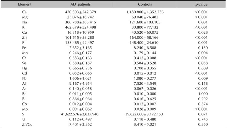
Table 1. Comparison of hair mineral contents in atopic dermatitis (AD) patients and controls
Element AD patients Controls p-value Ca 470.303±242.379 1,180.800±1,352.756 <0.001 Mg 25.076±18.247 69.040±76.482 <0.001 Na 308.788±365.415 121.600±103.105 0.001 K 462.879±524.498 80.800±77.132 <0.001 Cu 16.318±10.959 40.520±60.075 0.028 Zn 101.515±38.280 164.000±58.166 <0.001 P 133.485±22.497 148.400±24.610 0.001 Fe 7.652±3.165 8.240±6.508 0.130 Mn 0.246±0.177 0.179±0.144 0.004 Cr 0.583±0.163 0.412±0.088 <0.001 Se 0.580±0.187 0.584±0.528 0.058 Hg 0.665±0.236 0.708±0.355 0.809 Cd 0.052±0.065 0.015±0.012 <0.001 Pb 1.606±1.021 1.080±0.277 0.009 Al 9.167±4.954 7.520±3.549 0.158 As 0.140±0.058 0.067±0.026 <0.001 Be 0.011±0.005 0.010±0.000 1.000 B 0.864±0.964 0.616±0.625 0.292 Co 0.012±0.004 0.012±0.007 0.574 Mo 0.091±0.062 0.028±0.009 <0.001 S 41,622.576±3,837.940 39,822.000±3,172.150 0.071 U 0.112±0.497 0.118±0.480 0.745 Zn/Cu 7.401±3.362 8.410±5.021 0.360
Table 2. The correlation between clinical parameters and hair mineral levels in AD patients
Nutritional
element EASI TEWL
VAS for pruritus VAS for sleep disturbance Ca 0.102 −0.006 −0.015 −0.043 Mg 0.108 −0.053 0.014 −0.080 Cu −0.100 −0.026 −0.031 −0.048 Zn 0.008 0.099 0.025 −0.016 P 0.124 0.238 0.126 −0.009 Na −0.099 0.027 −0.027 −0.031 K −0.245* −0.071 −0.222 −0.168 Mn −0.165 0.111 −0.130 −0.117 Cr −0.072 −0.210 0.007 −0.004 Cd 0.059 0.405† 0.188 0.114 Pb −0.165 0.045 −0.063 0.104 As −0.037 −0.210 0.110 0.039 Mo 0.204 −0.138 0.263* 0.153 AD: atopic dermatitis, EASI: eczema area and severity index, TEWL: trans-epidermal water loss, VAS: visual analogue scale. Analyzed by Pearson’s or Spearman’s correlation, *p<0.05, †
p
<0.01.
perature-controlled digestion technique and Perkin-Elmer Mass Spectrometer (SciexElan 6100; Perkin-Elmer Corpo-ration, Foster City, CA, USA)6. The mineral concentrations are expressed as mg% (mg/100 g of hair). The reference
ranges determined by US Trace Elements Inc. (TEI), which has been derived comprehensively from numerous data and widely used in the several studies, were used in this study.
The EASI score, trans-epidermal water loss (TEWL) using a Tewameter TM210Ⓡ
(Courage & Khazaka, Cologne, Ger-many), and visual analogue scales (VAS) for pruritus and sleep disturbance were assessed.
Fifteen nutritional elements and seven toxic elements were analyzed. The nutritional elements included calcium (Ca), magnesium (Mg), sodium (Na), potassium (K), copper (Cu), Zn, phosphate (P), iron (Fe), manganese (Mn), chro-mium (Cr), selenium (Se), boron (B), cobalt (Co), molybde-num (Mo), and sulfur (S). The toxic elements included ura-nium (U), arsenic (As), beryllium (Be), mercury (Hg), cad-mium (Cd), lead (Pb), and aluminum (Al).
Among the nutritional elements, Ca, Mg, Cu, Zn, P levels were statistically lower and Na, K, Mn, Cr, and Mo levels were statistically higher in the AD patients compared to those in the control group. However, the mean levels of all these minerals were within the TEI reference ranges, except for K, which was higher than the reference range. Among the toxic minerals, Cd, Pb and As were sig-nificantly higher in AD patients than in control patients. In both groups, the mean levels of Cd and Pb were below the lower reference limit. Similarly, the mean level of As
Brief Report
Vol. 29, No. 2, 2017 253 was within the reference range in the AD patients (Table 1).
Next, we analyzed the significance of the relationship be-tween numerical value of minerals that showed difference between two groups and the parameters of clinical severity and found a statistical significance for a few minerals. The increased levels of K in AD patients were negatively lated with EASI scores. The Cd level was positively corre-lated with TEWL, but the mean level of Cd in AD patients was lower than the normal reference range. Although there was a statistical significance between Mo level and VAS for pruritus, they showed a weak correlation (Table 2). Based on the clinical parameters used, we did not find that these minerals were clinically relevant to AD.
There is growing interest in the role of minerals and mi-cronutrients in AD. There have been several studies on mineral concentrations in AD patients, but the reported re-sults have been contradictory. Zn is the most widely stud-ied mineral that has clinical relevance in AD patients3,4. In this study, we found that the hair Zn level was sig-nificantly lower in AD patients as in our previous study5. Ca is involved in cellular physiology and is a major com-ponent of bone and teeth mineralization. Mg is an essen-tial mineral that plays a critical role in the immune response. The lower levels of Ca and Mg in AD patients may reflect an avoidance of dairy products and nuts. Cu is an essential trace element in antioxidant systems and DNA synthesis7. Hon et al.4 suggested that the Cu/Zn ratio were increased in AD patients and positively correlated with the clinical severity3,4,8. However, in this study, there was no difference in the Cu/Zn ratio between the two groups or correlation with the clinical severity. This dis-crepancy between studies may result from differences in nutritional habits, age, or study methodology. K was the only element whose level exceeded the upper limit of the reference range in AD patients. However, since essential minerals interact with one another, there could be an im-balance in AD patients9.
There seems to be low risk of environmental hazards from heavy metal intoxication in Korean children with AD, be-cause levels of toxic elements which were significantly high-er in AD patients than in normal controls whigh-ere below or within the reference ranges. Regardless, caution must be tak-en to prevtak-ent intoxication with such elemtak-ents in AD patitak-ents. To our knowledge, no previous studies have evaluated the hair levels of various mineral levels in AD patients and es-tablished a relationship between the clinical severity and the mineral levels. Despite some differences between the two groups with regard to mineral levels, we did not find meaningful clinical relevance between the severity of AD and the micronutrient levels.
Although we did not evaluate the subjects’ diets, the
lev-els of trace elements may have differed due to nutritional imbalances in atopic patients. Nevertheless, most mineral levels were within the reference ranges. This finding in-dicates that atopic children did not have severe mineral deficiencies. Based on our findings, mildly reduced miner-al levels are likely insufficient to produce clinicminer-al changes10. Large supplementation trials are needed to assess the clin-ical efficacy of replacing depleted minerals in AD patients. Indiscriminate commercial exploitation of mineral supple-ment product should be sublated before the confirmation of therapeutic effects of mineral supplementation.
CONFLICTS OF INTEREST
The authors have nothing to disclose.
REFERENCES
1. Wang IJ, Guo YL, Weng HJ, Hsieh WS, Chuang YL, Lin SJ, et al. Environmental risk factors for early infantile atopic der-matitis. Pediatr Allergy Immunol 2007;18:441-447.
2. Puertollano MA, Puertollano E, de Cienfuegos GÁ, de Pablo MA. Dietary antioxidants: immunity and host defense. Curr Top Med Chem 2011;11:1752-1766.
3. David TJ, Wells FE, Sharpe TC, Gibbs AC, Devlin J. Serum levels of trace metals in children with atopic eczema. Br J Dermatol 1990;122:485-489.
4. Hon KL, Wang SS, Hung EC, Lam HS, Lui HH, Chow CM, et al. Serum levels of heavy metals in childhood eczema and skin diseases: friends or foes. Pediatr Allergy Immunol 2010; 21:831-836.
5. Kim JE, Yoo SR, Jeong MG, Ko JY, Ro YS. Hair zinc levels and the efficacy of oral zinc supplementation in patients with atopic dermatitis. Acta Derm Venereol 2014;94:558-562. 6. Miekeley N, de Fortes Carvalho LM, Porto da Silveira CL,
Lima MB. Elemental anomalies in hair as indicators of endocrinologic pathologies and deficiencies in calcium and bone metabolism. J Trace Elem Med Biol 2001;15:46-55. 7. Arredondo M, Núñez MT. Iron and copper metabolism. Mol
Aspects Med 2005;26:313-327.
8. Tasaki M, Hanada K, Hashimoto I. Analyses of serum copper and zinc levels and copper/zinc ratios in skin diseases. J Dermatol 1993;20:21-24.
9. Hataguchi Y, Tai H, Nakajima H, Kimata H. Drinking deep- sea water restores mineral imbalance in atopic eczema/ dermatitis syndrome. Eur J Clin Nutr 2005;59:1093-1096. 10. Namkoong S, Hong SP, Kim MH, Park BC. Reliability on
intra-laboratory and inter-laboratory data of hair mineral analysis comparing with blood analysis. Ann Dermatol 2013;25:67-72.