Fungal infections by filamentous fungi, molds, are occasionally seen in especially among the immunocompromised patients. Culturing the clinical specimens for fungus in the diagnosis of fungal infection is still a gold standard in clinical mycology laboratory, but, the process can require up to 4 weeks of incubation and often fail without identification. Recently, owing to the new active diagnostic methods such as sequencing and matrix assisted laser desorption ionization time-of-flight mass spectrometry (MALDI-TOF MS), large and heterogenous groups of molds are identified and those are revealed as infection causing pathogens in both immunocompromised and immunocompetent patients. Molds exist ubiquitously in the environment and often transiently colonize the respiratory, integument, and gastrointestinal systems of healthy hosts. So, it is difficult to conclude the
identified mold as clinically relevant pathogens and enable the timely initiation of appropriate antifungal treatment.
The Eutypella is a genus of fungi in the family Diatrypaceae1), and well-known plant pathogens, is capable of infecting many species of maple trees and produces a large, distinguishable canker on the main trunk of the tree, causing the infection with the release of ascospores from perithecia, but, human infections are still rarely reported.
We introduce here a case of Eutypellaspecies isolated from a bronchoalveolar lavage in an immunocompromised patient with small cell lung cancer initially not suspected by SDA and LPCB stain and revealed by PCR and 18S rDNA sequencing as the Eutypellaspecies.
A 63-year-old man who had received surgery for rectal cancer in other hospital four years ago, and had taken the adjuvant radiotherapy in our hospital and has followed per six months in our oncology outpatient clinic, admitted due to the mass in the right upper lobe of lung on the chest X-ray. Further work up with chest computer tomography (CT)
Eutypella species Isolated from a BronchoAlveolar Lavage
in a Patient with Small Cell Lung Cancer
Seung Joo Yeo
1, Young Ree Kim
2,3,4, Bong Chan Kim
4, Sung Ha Kang
2,4 1From medical course in Jeju National University School of Medicine,2Department of Labortory Medicine in Jeju National University School of Medicine, 3Jeju National University Institute of Medical Science, 4Department of Labortory
Medicine in Jeju National University Hospital, Jeju, Korea
(Received May 15, 2017; Revised May 22, 2017; Accepted May 29, 2017)
The Eutypella is a genus of fungi in the family Diatrypaceae, and well-known plant pathogens, is capable of infecting many species of maple trees. We introduce here a case of Eutypella species isolated from a bronchoalveolar lavage in an immunocompromised patient with small cell lung cancer initially not suspected by SDA and LPCB stain and revealed by PCR and 18S rDNA sequencing. But, in this case, we couldn't judge whether this fungus was the pathogen for inflammation of lung and additional treatment was not needed for fungus isolation with no clinical problems. The human mycobiome includes 390 fungal species detected on the skin, in the vagina, in the oral cavity, and in the digestive tract that includes 335 species and 158 genera. Among these, 221 species are found only in the digestive tract, 88 only in the oral cavity, and 26 in both. These species belong to 126 genera of yeast and filamentous fungi, of the Ascomycota, Basidiomycota, and Zygomycota phyla. So, no matter how sensitive the diagnosis is, the results of molecular diagnosis method for fungal infection must be carefully considered with clinical conditions.. (J Med Life Sci 2017;6(1):4-7)
Key Words
: Eutypella species, BronchoAlveolar Lavage, 18S rDNA sequencingIntroduction
Correspondence to : Young Ree Kim
Department of Laboratory Medicine, Jeju National University School of Medicine, Aran 13gil 15, Jeju-si, Jeju Special Self-governing Province, 63241, Republic of Korea
E-mail : [email protected] ([email protected])
Abstract
The Journal of Medicine and Life Science Vol. 14, No. 1(June), 2017
4
-Case Report
Figure 1A. Front image after 12 days incubation
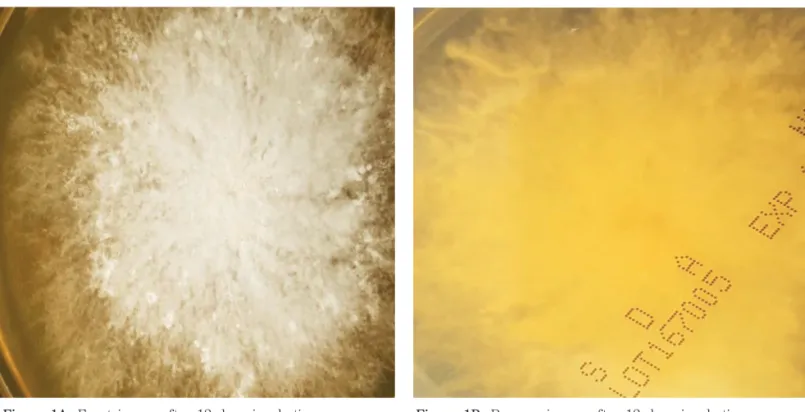
Figure 1A, 1B. The front surface of the Sabouraudes Dextrose Agar showed white cottony fungus within 12 days (Fig. 1A).
The reverse surface of the Sabouraudes Dextrose Agar had yellow color pigmentation after 12 days incubations (Fig. 1B).
Figure 1B. Reverse image after 12 days incubation
Eutypella species Isolated from a BronchoAlveolar Lavage in a Patient with Small Cell Lung Cancer
5 -showed 3.3cm lobulated and speculated mass in the right upper lobe of lung, obliteration of the apical segmental bronchus, subtle ground-glass attenuation in the posterior basal segment of right lower lobe, and mild chronic obstructive pulmonary disease change with emphysema and diffuse bronchial wall thickening in both lungs.
On the fungus culture of the sample from bronchoalveolar lavage, molds are noted, so, the specimen was cultured in Sabouraud's Dextrose Agar (SDA, Hanil KOMED, Seoul,
Korea) at 30℃. On the 5th culture day, the isolate started to grow. The front surface of the Sabouraudes Dextrose Agar showed white cottony fungus within 12 days (Fig. 1A). The reverse surface of the Sabouraudes Dextrose Agar had yellow color pigmentation after 12 days incubations (Fig. 1B). Lacto-Phenol Cotton Blue stain for microscopic examination showed coiled filamentous fungus (Fig. 2A, 2B). Based on the above results, the definite diagnosis and the differentiation of the organism couldn't be possible.
Figure 2A. (LPCB stain, x200)
Figure 2A, 2B. Lacto-Phenol Cotton Blue stain for microscopic examination showed coiled filamentous
fungus (Fig. 2A, 2B).
Figure 2B. (LPCB stain, x400)
We extracted DNAs of fungus using the bead beater-phenol extraction method. We suspended a loopful of culture of each isolate in 200 μL of TEN buffer (10m M Tris - HCl, 1 mM EDTA, 100 mM NaCl: pH 8.0), placed in a 2.0 mL screw-cap microcentrifuge tube filled with 200 μL (packed volume) of glass beads (diameter, 0.1 mm: Biospec Products, Bartlesville, Okla.) and 200 μL of Phenol:Chloroform: Isoamyl alcohol (25:24:1) (SIGMA chemical co. P-2069). We oscillated the tube on a Mini-Bead Beater (Biospec Products) for 1 min to disrupt the fungus. We centrifuged the tube at 15,000 rpm, for 15 min to separate the phases. After the aqueous phase was transferred into another clean tube, we added 10 μL of 3 M sodium acetate and 250 μL of ice-cold ethanol; to enable the DNA to precipitate, we kept the mixture at -20℃ for 30 min. We washed the DNA pellet with 70% ethanol, dissolved in 60 μL of TE buffer (10 mM Tris - HCl, 1 mM EDTA, 100 mM NaCl: pH 8.0) and used it as a template for PCR. We performed PCR with a set of primers (Forward primer (0817) 5'-TTAGCATGGAATAATRRAATAGGA-3' and Reverse primer (1536) 5'-ATTGCAATGCYCTATCCCCA-3') for amplification of the partial 18S rDNA sequences. We added 3 μL of template DNA and 20 μL of each primer to a PCR mixture tube (AccuPower PCR PreMix; Bioneer, Daejeon, Korea), which contained 1 U of Taq DNA polymerase, each deoxynucleoside triphosphate at a concentration of 250 μL, 50 mM Tris-HCl (pH 8.3), 40 mM KCl, 1.5 mM MgCl2, and gel loading dye. The reaction mixture was subjected to 35 cycles of amplification (2 min at 94℃, 10 s at 56℃, and 30s at 72℃), followed by a 5-min extension at 72℃ (model 9600 thermocycler; Perkin Elmer Cetus). We electrophoresed PCR products on a 1.2% agarose gel and purified with a QIAEX II gel extraction kit (QIAGEN, Hilden, Germany). We used Applied Biosystems model 373A automatic sequencer and a BigDye Terminator Cycle Sequencing kit (Perkin-Elmer Applied Biosystems, Warrington, United Kingdom) for the 18S rDNA analysis. Sequencing revealed Eutypella species with forward and reverse sequencing showing 99% homology.
Pathology report of sample from endobronchial ultrasound-guided transbronchial needle aspiration (EBUS-TBNA) confirmed small cell lung cancer. Though slight inflammation signs such as CT finding with subtle ground-glass attenuation in the posterior basal segment of right lower lobe existed, other infections signs were not observed. The patient had discharged after evaluation of mass in right upper lobe of lung and after then, have taken the palliative chemotherapy with etoposide, steroid, carboplatin, and palonosetron in our oncology department.
Eutypella species, a genus of fungi in the family Diatrypaceae1), are known plant pathogens2-6), is capable of infecting many species of maple trees, but, human infections by Eutypellaspecies are still rarely reported.
Eutypella parasitica among Eutypella species is characterized as an ascomycete fungus that produces fruiting bodies called perithecia7), and it has been known that the perithecium will produce sexual spores called ascospores which are dispersed primarily by the wind and may enter through a wound and establish themselves in the xylem tissue of the susceptible host.
By conventional mycological methods, identification of fungus species is often difficult. Several cases8-10) of identification by molecular methods such as ITS rDNA and LSU rDNA D1-D2 sequencing have been reported for reliable and rapid identification of human pathological fungus. Fungal infections are increasing, particularly among immunocompromised hosts, and a rapid, accurate diagnosis is essential for the initiation of targeted antifungal therapy11).
We introduce here a case of Eutypella species isolated from a bronchoalveolar lavage in an immunocompromised patient with small cell lung cancer. But, in this case, we couldn't judge whether this fungus was the pathogen for inflammation of lung and additional treatment was not needed for fungus isolation with no clinical problems. So, no matter how sensitive the diagnosis is, the results of molecular diagnosis method for fungal infection must be carefully considered with clinical conditions.
1) Lumbsch TH, Huhndorf SM. "Outline of Ascomycota 2007". Myconet. Chicago, USA: The Field Museum, Department of Botany. 13: 158. Archived from the original on March 18, 2009.
2) Gary W. Moorman. "Eutypella canker on maple". Plant Disease Fact Sheets. Penn State Extension, Penn State University College of Agriculatural Sciences. Retrieved March 23, 2012.
3) Janna Beckerman. "Eutypella canker". Beautiful Death: Ornamental Plant Pathology. Department of Botany and Plant Pathology, Purdue University. Retrieved March 23, 2012.
4) N. Ogris; D. Diminic; B. Pikur; H. Kraigher. "First report of Eutypella parasitica causing cankers on field maple (Acer campestre) in Croatia". New Disease Reports.
Discussion
Seung Joo Yeo, Young Ree Kim, Bong Chan Kim, Sung Ha Kang6
-References
Eutypella species Isolated from a BronchoAlveolar Lavage in a Patient with Small Cell Lung Cancer
7 -2008;16:29.
5) Denis Lachance; James E. Kuntz. "Ascocarp development of Eutypella parasifica". Canadian Journal of Botany. 48 (11): 19771979. doi:10.1139/b70-288.
6) "Eutypella stem canker of maple - Eutypellaparasitica". Bundesforschungs- und Ausbildungszentrum fr Wald, Naturgefahren und Landschaft. Retrieved March 23, 2012.
7) N. Ogris; D. Diminic; B. Pikur; H. Kraigher. "First report of Eutypella parasitica causing cankers on field maple (Acer campestre) in Croatia". New Disease Reports. 2008;16: 29.
8) Lo Cascio G, Ligozzi M, Maccacaro L, Fontana R. Utility of molecular identi?cation in opportunistic mycotic infections: a case of cutaneous Alternaria infectoria infection in a cardiac transplant recipient. J Clin
Microbiol 2004;42:5334-6.
9) Lo Cascio G, Ligozzi M, Maccacaro L, Rizzonelli P, Fontana R. Diagnostic aspects of cutaneous lesions due to Histoplasma capsulatum in African AIDS patients in nonendemic areas. Eur J Clin Microbiol Infect Dis 2003;22:637-8.
10) Lo Cascio G, Ligozzi M. Alternaria. In: Liu D, ed. Molecular detection of human fungal pathogens. ISBN: 9781439812402. Boca Raton, FL: Taylor & Francis CRC Press, 2011;27-36.
11) June I. Pounder,Keith E. Simmon, Claudia A. Barton, Sheri L. Hohmann2 Mary E. Brandt, and Cathy A. Petti. Discovering Potential Pathogens among Fungi Identi?ed as Nonsporulating Molds. Journal of Clinical Microbiology, 2007;45(2):568-71.