Review Article
Optimal Bowel Preparation for Video Capsule Endoscopy
Hyun Joo Song,
1Jeong Seop Moon,
2and Ki-Nam Shim
31Department of Internal Medicine, Jeju National University School of Medicine, Jeju, Republic of Korea
2Department of Internal Medicine, Inje University College of Medicine, Seoul, Republic of Korea
3Department of Internal Medicine, Ewha Womans University School of Medicine, Seoul, Republic of Korea
Correspondence should be addressed to Jeong Seop Moon; [email protected] Received 30 June 2015; Accepted 22 October 2015
Academic Editor: Anastasios Koulaouzidis
Copyright © 2016 Hyun Joo Song et al. This is an open access article distributed under the Creative Commons Attribution License, which permits unrestricted use, distribution, and reproduction in any medium, provided the original work is properly cited. During video capsule endoscopy (VCE), several factors, such as air bubbles, food material in the small bowel, and delayed gastric and small bowel transit time, influence diagnostic yield, small bowel visualization quality, and cecal completion rate. Therefore, bowel preparation before VCE is as essential as bowel preparation before colonoscopy. To date, there have been many comparative studies, consensus, and guidelines regarding different kinds of bowel cleansing agents in bowel preparation for small bowel VCE. Presently, polyethylene glycol- (PEG-) based regimens are given primary recommendation. Sodium picosulphate-based regimens are secondarily recommended, as their cleansing efficacy is less than that of PEG-based regimens. Sodium phosphate as well as complementary simethicone and prokinetics use are considered. In this paper, we reviewed previous studies regarding bowel preparation for small bowel VCE and suggested optimal bowel preparation of VCE.
1. Introduction
Video capsule endoscopy (VCE) is useful in investigating small bowel as well as esophagus, stomach, and colon. Bowel preparation for small bowel VCE is recommended to improve small bowel visualization quality (SBVQ), diagnostic yield (DY), and cecal completion rate (CR). Particularly in the distal small bowel, DY of VCE can be limited due to reduced SBVQ-associated with residual material or dark colored bile. According to a 2009 meta-analysis of 12 studies [1], purgative bowel cleansing prior to VCE improves the SBVQ and increases the DY but does not alter the VCE CR. However, the gastric transit time (GTT) and small bowel transit time (SBTT) of VCE were not affected by purgatives.
We performed online search for VCE bowel preparation-related clinical studies, comparative research, randomized controlled trials (RCTs), meta-analyses, and guidelines pub-lished from January 2002 to June 2015. Literature review was conducted using Key MeSH terms of “capsule endoscopy” and “bowel preparation.” We also reviewed bowel prepa-ration guidelines for VCE of small bowel based on 2009 European Society of Gastrointestinal Endoscopy (ESGE)
guidelines [2], 2013 ESGE guidelines [3], and 2013 Korean guidelines [4] by the Korean Gut Image Study Group, part of the Korean Society of Gastrointestinal Endoscopy. The level of scientific evidence for recommendation was based on study design; for example, the evidence of randomized trial was considered high, observation study was low, and any other type of evidence was very low. The validity of the recommendation was divided into categories of “strong” or “weak” (Table 1) [5]. In this paper, we introduced previous studies on bowel preparation for VCE and suggested optimal preparation methods.
2. Purgatives
2.1. Polyethylene Glycol. Polyethylene glycol- (PEG-) based
regimens are first-line recommendation (Grade A) [3]. The majority of the evidence of bowel preparation prior to small bowel VCE is PEG-based regimens. The 2009 ESGE guide-lines recommended purgative bowel preparations in order to enhance small bowel DY by VCE without affecting the CR (category of evidence, 2a; grade of recommendation, B) [2].
Volume 2016, Article ID 6802810, 7 pages http://dx.doi.org/10.1155/2016/6802810
Table 1: Quality of evidence and strength of a recommendation. Quality of evidence
High quality Further research is very unlikely to change our confidence in the estimate of effect.
Moderate quality Further research is likely to have an important impact on our confidence in the estimate of effect and may change the
estimate.
Low quality Further research is very likely to have an important impact on our confidence in the estimate of effect and is likely tochange the estimate.
Very low quality Any estimate of effect is very uncertain.
Strength of a recommendation
Strong Most or all individuals will be best served by the recommended course of action.
Weak Not all individuals will be best served by the recommended course of action. There is a need to consider more carefullythan usual individual patient’s circumstances, preferences, and values.
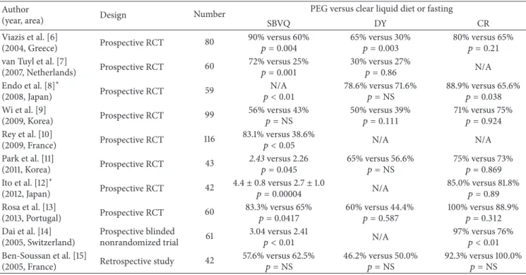
Table 2: Studies comparing SBVQ, DY, and CR between PEG solution versus clear liquid or fasting of small bowel VCE. Author
(year, area) Design Number
PEG versus clear liquid diet or fasting
SBVQ DY CR
Viazis et al. [6]
(2004, Greece) Prospective RCT 80
90% versus 60%
𝑝 = 0.004 65% versus 30%𝑝 = 0.003 80% versus 65%𝑝 = 0.21
van Tuyl et al. [7]
(2007, Netherlands) Prospective RCT 60 72% versus 25% 𝑝 = 0.001 30% versus 27%𝑝 = 0.86 N/A Endo et al. [8]∗ (2008, Japan) Prospective RCT 59 N/A 𝑝 < 0.01 78.6% versus 71.6%𝑝 = NS 88.9% versus 65.6%𝑝 = 0.038 Wi et al. [9] (2009, Korea) Prospective RCT 99 56% versus 43% 𝑝 = NS 50% versus 39%𝑝 = 0.111 71% versus 75%𝑝 = 0.924 Rey et al. [10] (2009, France) Prospective RCT 116 83.1% versus 38.6% 𝑝 < 0.05 N/A N/A Park et al. [11] (2011, Korea) Prospective RCT 43 2.43 versus 2.26 𝑝 = 0.045 65% versus 56.6%𝑝 = NS 75% versus 73%𝑝 = 0.869 Ito et al. [12]∗ (2012, Japan) Prospective RCT 42 4.4± 0.8 versus 2.7 ± 1.0 𝑝 = 0.00004 N/A 85.0% versus 81.8%𝑝 = 0.89 Rosa et al. [13] (2013, Portugal) Prospective RCT 60 83.3% versus 65% 𝑝 = 0.0417 60% versus 44.4%𝑝 = 0.587 100% versus 88.9%𝑝 = 0.312 Dai et al. [14] (2005, Switzerland) Prospective blinded nonrandomized trial 61 3.04 versus 2.41 𝑝 < 0.01 N/A 97% versus 76%𝑝 < 0.01 Ben-Soussan et al. [15]
(2005, France) Retrospective study 42
57.6% versus 62.5%
𝑝 = NS 46.2% versus 50.0%𝑝 = NS 92.3% versus 100.0%𝑝 = NS
PEG: polyethylene glycol, VCE: video capsule endoscopy, RCT: randomized-controlled trial, SBVQ: small bowel visualization quality, DY: diagnostic yield, CR: completion rate, N/A: not applicable, and NS: no significant.∗PEG 500 mL.
According to the Korean Gut Image Study Group guidelines [4], bowel preparation with PEG solution enhances DY and SBVQ, without effect on cecal CR (strong recommendation, moderate quality evidence).
Table 2 shows many studies regarding bowel preparation with comparison of PEG versus clear liquid or fasting for small bowel VCE, including prospective randomized con-trolled trials [6–13], a prospective blinded nonrandomized trial [14], and a retrospective study [15]. Most studies were performed by comparing SBVQ, DY, and cecal CR between 2 L PEG solution and clear diet or fasting groups. Four-liter PEG solution was used in a few studies [10, 14]. In addition, ingestion of a small amount of PEG (500 mL) beginning 30 minutes after swallowing the capsule significantly improves SBVQ and cecal CR, although DY was not affected [8]. Another study regarding a small amount (500 mL) of PEG
solution over 2 hours, beginning 30 minutes after swallowing the capsule, showed increased SBVQ without any difference in cecal CR [12]. Since PEG is completely transparent, a view through PEG was considered better than a view through natural intestinal fluid. However, negative result regarding SBVQ with 2 L PEG was reported in one retrospective study [15].
Two-liter PEG solution bowel preparation is similar to that of 4 liters of PEG in DY, SBVQ, and CR of VCE (weak recommendation, moderate quality evidence). Two studies by Kantianis et al. [16] and Park et al. [11] indicated no significant difference between 2 L and 4 L PEG in regard to small bowel cleansing and CR. Therefore, 2 L PEG should be recommended as preparation for VCE, administered on the day prior to the procedure, as the most commonly used preparation method [17].
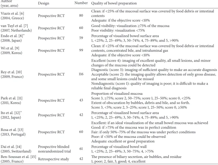
Table 3: Definitions of optimal bowel preparation of VCE among studies with PEG. Author
(year, area) Design Number Quality of bowel preparation
Viazis et al. [6]
(2004, Greece) Prospective RCT 80
Clean: if<25% of the mucosal surface was covered by food debris or intestinal
contents
Adequate: if the objective score<10%
van Tuyl et al. [7]
(2007, Netherlands) Prospective RCT 60
Good visibility: visualization≥75% of the mucosa
Poor visibility: visualization<75%
Endo et al. [8]∗
(2008, Japan) Prospective RCT 59
Percentage of visualized bowel surface area
1,<25%; 2, 25–49%; 3, 50–74%; 4, 75–89%; and 5, >90%
Wi et al. [9]
(2009, Korea) Prospective RCT 99
Clean: if<25% of the mucosal surface was covered by food debris or intestinal
contents, concentrated bile, and intraluminal gas
Adequate: if the objective score<10%
Rey et al. [10]
(2009, France) Prospective RCT 116
Excellent (score 4): imaging of excellent quality, all small lesions, and minor changes of the mucosa could be detected
Diagnostic (score 3): imaging of sufficient quality to make an accurate diagnosis Acceptable (score 2): the imaging quality allows detection of only gross disease, and some small lesions could be missed
Nondiagnostic (score 1): quality of imaging is poor; it is difficult to make a reliable final diagnosis
Park et al. [11]
(2011, Korea) Prospective RCT 43
Proportion of visualized mucosa
Score 3,≥75%; score 2, 50–75%; score 1, 25–50%; score 0, <25%
Extent of obscuration by bubbles, debris and bile, and so forth.
Score 3,<5%; score 2, 5–25%; score 1, 25–50%; score 0, ≥50%
Ito et al. [12]∗
(2012, Japan) Prospective RCT 42
Percentage of visualized bowel surface area
1,<25%; 2, 25–49%; 3, 50–74%; 4, 75–89%; and 5, >90%
Rosa et al. [13]
(2013, Portugal) Prospective RCT 60
Excellent: if an ideal visualization of the small bowel mucosa was achieved
Good: if>75% of the mucosa was in perfect condition
Fair: if only 50%–75% of the mucosa was under perfect conditions
Poor: if<50% of the mucosa could be observed
Adequate: excellent or good preparation Dai et al. [14]
(2005, Switzerland)
Prospective blinded
nonrandomized trial 61
Percentage of visualized bowel wall
1,<25%; 2, 25–49%; 3, 50–75%; 4, >75%
Ben-Soussan et al. [15]
(2005, France) Retrospective study 42
The presence of biliary secretion, air bubbles, and residue 1, poor; 2, fair, 3, good; 4, excellent
PEG: polyethylene glycol, VCE: video capsule endoscopy, and RCT: randomized-controlled trial.∗PEG 500 mL.
In colonoscopy, bowel preparation status is classified as excellent, good, fair, poor, or inadequate. Clinically, most gastroenterologists considered excellent and good bowel preparation status as optimal bowel preparation. However, there was no consensus of optimal bowel preparation for VCE, as each study with PEG suggested various definitions for bowel preparation quality (Table 3). A recent study con-sidered excellent or good preparation (>75% small bowel visualization) as adequate bowel preparation [13]. Therefore, standardized definition of optimal bowel preparation for VCE is necessary.
To date, there has been no consensus regarding optimal timing of bowel preparation for VCE. To evaluate optimal timing of VCE bowel preparation, a single-center random-ized controlled trial was conducted by Black et al. [18]. How-ever, there was no significant difference between the quality and timing (day before VCE versus 4 hours prior to VCE) of small bowel preparation. Intestinal lavage administered one day prior was similar to same-day preparation with regard to overall preparation quality, SBTT, frequency of identified
mucosal abnormalities, general DY, and CR. One of the issues for bowel preparation of VCE is that the distal segment of the small intestine should be improved. The main limitation of this study is that the number of patients (𝑛 = 34) is not sufficient for generalizing to actual practice. Therefore, multicenter large randomized controlled trial is required to clarify optimal timing of bowel preparation for VCE.
According to the 2012 consensus guidelines for bowel preparation [19], purgative is absolutely contraindicated in patients with gastrointestinal obstruction, ileus, ulcer, perfo-ration, or inflammatory bowel diseases. In addition, it is also contraindicated in patients with decreased consciousness, swallowing disorders, and hypersensitivity to oral bowel-cleansing agents and in patients having an ileostomy. There-fore, optimal bowel preparation should be made considering individual patient risk factors.
2.2. Sodium Picosulfate. Recently, various types of bowel
preparation such as PEG, PEG plus ascorbic acid, sodium picosulfate, and phosphate (NaP) are available. There has
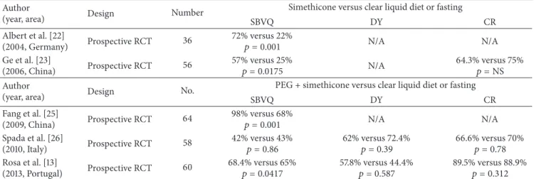
Table 4: Studies comparing SBVQ, DY, and CR between simethicone versus clear liquid or fasting of small bowel VCE. Author
(year, area) Design Number
Simethicone versus clear liquid diet or fasting
SBVQ DY CR Albert et al. [22] (2004, Germany) Prospective RCT 36 72% versus 22% 𝑝 = 0.001 N/A N/A Ge et al. [23] (2006, China) Prospective RCT 56 57% versus 25% 𝑝 = 0.0175 N/A 64.3% versus 75%𝑝 = NS Author
(year, area) Design No.
PEG + simethicone versus clear liquid diet or fasting
SBVQ DY CR Fang et al. [25] (2009, China) Prospective RCT 64 98% versus 68% 𝑝 = 0.001 N/A N/A Spada et al. [26] (2010, Italy) Prospective RCT 58 42% versus 43% 𝑝 = 0.86 62% versus 72.4%𝑝 = 0.39 66.6% versus 70%𝑝 = 0.78 Rosa et al. [13] (2013, Portugal) Prospective RCT 60 68.4% versus 65% 𝑝 = 0.0417 57.8% versus 44.4%𝑝 = 0.587 89.5% versus 88.9%𝑝 = 0.312
PEG: polyethylene glycol, VCE: video capsule endoscopy, RCT: randomized-controlled trial, SBVQ: small bowel visualization quality, DY: diagnostic yield, CR: completion rate, N/A: not applicable, and NS: no significant.
been no published evidence to support the use of sodium picosulfate; however, it is used in some units. Anecdotal evidence suggests that it is not as effective as PEG [3].
2.3. Sodium Phosphate (NaP). NaP is not recommended for
bowel cleansing due to the potential for renal damage and other adverse effects (Grade B) [3]. However, the use of NaP is possible in patients for whom PEG or sodium picosulfate is ineffective or not tolerated (Grade D). According to a previous study conducted using NaP, SBVQ of NaP group is better than overnight fasting (35% versus 4%) [20]. However, recent meta-analysis of NaP-based regimens revealed no significant difference from fasting alone (OR = 1.32, 95% CI
= 0.52–2.96,𝑝 < 0.0001) [21]. Therefore, NaP should not be
used in general.
3. Simethicone
Preparing the small bowel with simethicone has been reported to increase SBVQ by reducing intraluminal air bub-bles [22, 23]. Table 4 demonstrates several studies regarding bowel preparation with simethicone for small bowel VCE [13, 22–26]. Systemic review and meta-analysis of RCTs of simethicone revealed that supplemental use of simethicone prior to VCE enhances SBVQ, especially for patients without purgative, but does not affect the cecal CR [27]. It decreases air bubbles in the colonic lumen but does not improve bowel preparation. Additionally, its effect on DY remains controversial. Bowel preparation by fasting or administration of PEG solution combined with simethicone enhances SBVQ, but it does not affect CR for VCE (strong recommendation, moderate quality evidence) [4].
4. Prokinetics
Prokinetics can be used for shortening of the GTT and may improve cecal CR. To date, various prokinetics including
erythromycin [28–30], mosapride [31], metoclopramide [32– 34], and lubiprostone [35, 36] have been investigated for bowel preparation of VCE. Table 5 exhibits previous studies regarding bowel preparation with various prokinetics for small bowel VCE. Previously, the battery time of VCE was 8 hours and approximate 20% do not reach the colon due to limited recording time [34]. Currently, the battery time of VCE is about 12 hours; therefore the effect of prokinetics on the CR could be minimal.
Lubiprostone, a selective activator of type 2 chloride channels in the apical membrane of the GI epithelium, as a propulsive agent was investigated for decreasing the SBTT by VCE. However, there were opposite results regarding the GTT and SBTT in two studies [35, 36]. Lubiprostone neither decreased the GTT and SBTT nor improved SBVQ for VCE in one double-blind placebo-controlled study [35], while it decreased the SBTT by VCE in another exploratory random-ized, double-blind, controlled study [36]. Bowel preparation with prokinetics does not enhance the SBVQ, DY, or CR of VCE. Therefore, it is not generally recommended (weak recommendation, moderate quality evidence) [4].
5. Miscellaneous
Recently, there have been new studies using substances such as coffee enema or magnesium citrate. Coffee enema is known to induce dilation of bile ducts and excretion of bile through the colon wall. During VCE, excreted bile is one of the causes of poor bowel preparation. Coffee enema for preparation for small bowel VCE was investigated by a pilot study (𝑛 = 34) [37]. Comparison of coffee enema plus 2 L PEG versus 2 L PEG demonstrated greater efficacy of bowel preparations in the mid-to-distal segments of the small bowel in patients who received coffee enema plus 2 L PEG than in those who received PEG only. In one magnesium citrate trial of bowel preparation for VCE [38], there was no significant difference between the group that received the preparation (34 g magnesium) and the control group.
T a ble 5: St udies co m pa ri n g SB V Q ,D Y ,a n d C R b etw een p ro k inet ics ver sus cle ar liq uid o r fast in g o f small b o w el V CE. Au th o r (y ea r, ar ea ) De si gn N u m b er P ro k inet ics P ro k inet ics ver sus p laceb o o r fast in g GT T (min) SB T T (min) SB V Q D Y CR Le u n g et al .[ 28 ] (20 0 5, C hina) Pro sp ec tive no nra n do mized st ud y 38 Er yt hr o m yc in 16 versus 70 𝑝 = 0.005 227 ve rs u s 18 3 𝑝 = 0.18 54 % ve rs u s 6 4 % 𝑝 = 0.74 N/A 96 % ve rs u s 79 % 𝑝=0 .1 3 Ca d d y et al .[ 29 ] (20 0 6, A u stralia) Pro sp ec tive R C T 45 Er yt hr o m yc in 51 versus 38 𝑝 = 0.42 304 ve rs u s 30 2 𝑝 = 0.96 1.9 ver sus 2.2 𝑝 = 0.24 N/A 68 % ve rs u s 78 % 𝑝=0 .4 5 Ni v et al .[ 30 ] (20 0 8, Israel) Retr os p ecti ve bl in d stu dy 10 0 E ry th ro m ycin 21 ve rs u s 28 𝑝 = 0.07 27 9 ve rs u s 27 0 𝑝 = 0.83 2.8 ver sus 2.8 𝑝 = 0.73 48 % ve rs u s 36 % 𝑝= N/A 90 % ve rs u s 84 % 𝑝=0 .3 7 We i et al .[ 31 ] (20 0 7, C hina) Pro sp ec tive R C T 6 0 M o sa p ride 14 ve rs u s 34 𝑝 = 0.035 24 8 ve rs u s 28 1 𝑝 = 0.3492 N/A 73 % ve rs u s 50 % 𝑝 = 0.110 93 % ve rs u s 6 7% 𝑝 = 0.021 Se lb y [3 2] (20 0 5, A u stralia) Pro sp ec tive R C T 15 0 M et o clo p ra mide 31 ve rs u s 4 8 𝑝 = 0.025 23 1 ve rs u s 256 𝑝 = 0.35 10 0% versus 69% 𝑝 = 0.998 51% ver sus 57% 𝑝= N/A 97% ver sus 76% 𝑃 < 0.001 Po st ga te et al .[ 33 ] (20 0 9, UK) Pro sp ec tive R C T 74 M et o cl o p ra mide 17 ve rsus 17 𝑝 = 0.62 26 0 ver sus 27 8 𝑝 = 0.91 38 ve rs u s 37 𝑝 = 0.18 26% ver sus 35% 𝑝=0 .4 5 85 % ve rsus 89 % 𝑝=0 .7 4 Alm eida et al .[3 4] (2010, P o rt ugal) Pro sp ec tive R C T 95 M et o cl o p ra mide 26 ve rs us 28 𝑝 = 0.511 221 ve rs u s 256 𝑝 = 0.083 55 % ve rsus 54 % 𝑝 = 0.545 68 % ve rs u s 65 % 𝑝 = 0.443 81 % versus 77% 𝑝 = 0.422 H o o k s III et al .[ 35 ] (20 0 9, N et h erla nds) Pro sp ec tive R C T 4 0 L u bipro st o n e 126 ver su s 43 𝑝 = 0.0095 188 versus 219 𝑝 = 0.130 NS N/A N /A Mat suu ra et al .[ 36 ] (201 4, Ja pa n) Pro sp ec tive R C T 6 L ub ip rosto ne 58 ve rs u s 23 𝑝 = 0.846 111 ve rs us 17 9 𝑝 = 0.042 3.7 6 ve rs us 2.88 𝑝 < 0.001 N/A N /A V C E: video ca p su le en dosco p y, GT T :ga st ric tra n si t tim e, SB T T :sm all b o w el tra n si t tim e, SB V Q :s m all bo w el vi sualiza tio n q u ali ty ,D Y :d ia gn os tic yi eld ,CR: co m p letio n ra te ,R CT :r an do mized co n tr o lled tr ial ,N /A: no t ap p lic able, and N S: no sig n ifi ca n t.
6. Conclusion
Bowel preparation is generally recommended for small bowel VCE. Currently, a combination of 2 L PEG and simethicone appears to be the optimal bowel preparation before VCE. After reviewing current articles regarding bowel prepara-tion for VCE, we suggest using purgatives such as PEG as first line and sodium picosulfate as second line with antifoaming agent. However, sodium phosphate should not be used except for the patients whom PEG or sodium pico-sulfate is not effective and intolerable. However, prokinetics (erythromycin, metoclopramide, or lubiprostone) are not generally recommended. For each of these agents, including purgative (PEG, sodium picosulfate, and sodium phosphate), consensus is needed regarding optimal timing of bowel preparation. Therefore, best bowel preparation is determined considering individual patient status.
Conflict of Interests
The authors declare that there is no conflict of interests regarding the publication of this paper.
Authors’ Contribution
Hyun Joo Song wrote the paper; Jeong Seop Moon and Ki-Nam Shim revised the paper.
References
[1] T. Rokkas, K. Papaxoinis, K. Triantafyllou, D. Pistiolas, and S. D. Ladas, “Does purgative preparation influence the diagnostic yield of small bowel video capsule endoscopy?: a meta-analysis,” American Journal of Gastroenterology, vol. 104, no. 1, pp. 219– 227, 2009.
[2] S. D. Ladas, K. Triantafyllou, C. Spada et al., “European Society of Gastrointestinal Endoscopy (ESGE): recommendations (2009) on clinical use of video capsule endoscopy to investigate small-bowel, esophageal and colonic diseases,” Endoscopy, vol. 42, no. 3, pp. 220–227, 2010.
[3] E. Mathus-Vliegen, M. Pellis´e, D. Heresbach et al., “Consensus guidelines for the use of bowel preparation prior to colonic diagnostic procedures: colonoscopy and small bowel video cap-sule endoscopy,” Current Medical Research and Opinion, vol. 29, no. 8, pp. 931–945, 2013.
[4] H. J. Song, J. S. Moon, J. H. Do et al., “Guidelines for bowel preparation before video capsule endoscopy,” Clinical Endos-copy, vol. 46, no. 2, pp. 147–154, 2013.
[5] D. Atkins, D. Best, P. A. Briss et al., “Grading quality of evidence and strength of recommendations,” British Medical Journal, vol. 328, no. 7454, article 1490, 2004.
[6] N. Viazis, S. Sgouros, K. Papaxoinis et al., “Bowel preparation increases the diagnostic yield of capsule endoscopy: a prospec-tive, randomized, controlled study,” Gastrointestinal Endoscopy, vol. 60, no. 4, pp. 534–538, 2004.
[7] S. A. C. van Tuyl, H. den Ouden, M. F. J. Stolk, and E. J. Kuipers, “Optimal preparation for video capsule endoscopy: a prospec-tive, randomized, single-blind study,” Endoscopy, vol. 39, no. 12, pp. 1037–1040, 2007.
[8] H. Endo, Y. Kondo, M. Inamori et al., “Ingesting 500 ml of poly-ethylene glycol solution during capsule endoscopy improves the image quality and completion rate to the cecum,” Digestive Diseases and Sciences, vol. 53, no. 12, pp. 3201–3205, 2008. [9] J.-H. Wi, J.-S. Moon, M.-G. Choi et al., “Bowel preparation
for capsule endoscopy: a prospective randomized multicenter study,” Gut and Liver, vol. 3, no. 3, pp. 180–185, 2009.
[10] J.-F. Rey, A. Repici, K. Kuznetsov, V. Boyko, and L. Aabakken, “Optimal preparation for small bowel examinations with video capsule endoscopy,” Digestive and Liver Disease, vol. 41, no. 7, pp. 486–493, 2009.
[11] S. C. Park, B. Keum, Y. S. Seo et al., “Effect of bowel preparation with polyethylene glycol on quality of capsule endoscopy,” Digestive Diseases and Sciences, vol. 56, no. 6, pp. 1769–1775, 2011.
[12] T. Ito, K. Ohata, A. Ono et al., “Prospective controlled study on the effects of polyethylene glycol in capsule endoscopy,” World Journal of Gastroenterology, vol. 18, no. 15, pp. 1789–1792, 2012. [13] B. J. Rosa, M. Barbosa, J. Magalhaes, A. Rebelo, M. J. Moreira,
and J. Cotter, “Oral purgative and simethicone before small bowel capsule endoscopy,” World Journal of Gastrointestinal Endoscopy, vol. 5, no. 2, pp. 67–73, 2013.
[14] N. Dai, C. Gubler, P. Hengstler, C. Meyenberger, and P. Bauer-feind, “Improved capsule endoscopy after bowel preparation,” Gastrointestinal Endoscopy, vol. 61, no. 1, pp. 28–31, 2005. [15] E. Ben-Soussan, G. Savoye, M. Antonietti, S. Ramirez, P.
Ducrott´e, and E. Lerebours, “Is a 2-liter PEG preparation useful before capsule endoscopy?” Journal of Clinical Gastroenterology, vol. 39, no. 5, pp. 381–384, 2005.
[16] A. Kantianis, S. Karagiannis, C. Liatsos et al., “Comparison of two schemes of small bowel preparation for capsule endoscopy with polyethylene glycol: a prospective, randomized single-blind study,” European Journal of Gastroenterology and Hepatol-ogy, vol. 21, no. 10, pp. 1140–1144, 2009.
[17] A. Koulaouzidis, E. Rondonotti, and A. Karargyris, “Small-bowel capsule endoscopy: a ten-point contemporary review,” World Journal of Gastroenterology, vol. 19, no. 24, pp. 3726–3746, 2013.
[18] K. R. Black, W. Truss, C. I. Joiner, S. Peter, and F. H. Weber Jr., “A single-center randomized controlled trial evaluating timing of preparation for capsule enteroscopy,” Clinical Endoscopy, vol. 48, no. 3, pp. 234–238, 2015.
[19] A. Connor, D. Tolan, S. Hughes, N. Carr, and C. Tomson, “Con-sensus guidelines for the safe prescription and administration of oral bowel-cleansing agents,” Gut, vol. 61, no. 11, pp. 1525–1532, 2012.
[20] Y. Niv, G. Niv, K. Wiser, and D. C. Demarco, “Capsule endos-copy—comparison of two strategies of bowel preparation,” Alimentary Pharmacology and Therapeutics, vol. 22, no. 10, pp. 957–962, 2005.
[21] J. Belsey, C. Crosta, O. Epstein et al., “Meta-analysis: Efficacy of small bowel preparation for small bowel video capsule endos-copy,” Current Medical Research and Opinion, vol. 28, no. 12, pp. 1883–1890, 2012.
[22] J. Albert, C.-M. G¨obel, J. Leßke, E. Lotterer, H. Nietsch, and W. E. Fleig, “Simethicone for small bowel preparation for capsule endoscopy: a systematic, single-blinded, controlled study,” Gas-trointestinal Endoscopy, vol. 59, no. 4, pp. 487–491, 2004. [23] Z.-Z. Ge, H.-Y. Chen, Y.-J. Goa, Y.-B. Hu, and S.-D. Xiao, “The
role of simeticone in small-bowel preparation for capsule endoscopy,” Endoscopy, vol. 38, no. 8, pp. 836–840, 2006.
[24] W. Wei, Z.-Z. Ge, H. Lu, Y.-J. Gao, Y.-B. Hu, and S.-D. Xiao, “Purgative bowel cleansing combined with simethicone improves capsule endoscopy imaging,” The American Journal of Gastroenterology, vol. 103, no. 1, pp. 77–82, 2008.
[25] Y.-H. Fang, C.-X. Chen, and B.-L. Zhang, “Effect of small bowel preparation with simethicone on capsule endoscopy,” Journal of Zhejiang University. Science B, vol. 10, no. 1, pp. 46–51, 2009. [26] C. Spada, M. E. Riccioni, P. Familiari et al., “Polyethylene
glycol plus simethicone in small-bowel preparation for capsule endoscopy,” Digestive and Liver Disease, vol. 42, no. 5, pp. 365– 370, 2010.
[27] L. Wu, Y. Cao, C. Liao, J. Huang, and F. Gao, “Systematic review and meta-analysis of randomized controlled trials of Sime-thicone for gastrointestinal endoscopic visibility,” Scandinavian Journal of Gastroenterology, vol. 46, no. 2, pp. 227–235, 2011. [28] W. K. Leung, F. K. L. Chan, S. S. L. Fung, M.-Y. Wong, and J. J. Y.
Sung, “Effect of oral erythromycin on gastric and small bowel transit time of capsule endoscopy,” World Journal of Gastroen-terology, vol. 11, no. 31, pp. 4865–4868, 2005.
[29] G. R. Caddy, L. Moran, A. K. H. Chong, A. M. Miller, A. C. Taylor, and P. V. Desmond, “The effect of erythromycin on video capsule endoscopy intestinal-transit time,” Gastrointesti-nal Endoscopy, vol. 63, no. 2, pp. 262–266, 2006.
[30] E. Niv, I. Bogner, O. Barkey et al., “Effect of erythromycin on image quality and transit time of capsule endoscopy: a two-center study,” World Journal of Gastroenterology, vol. 14, no. 16, pp. 2561–2565, 2008.
[31] W. Wei, Z.-Z. Ge, H. Lu, Y.-J. Gao, Y.-B. Hu, and S.-D. Xiao, “Effect of mosapride on gastrointestinal transit time and diag-nostic yield of capsule endoscopy,” Journal of Gastroenterology and Hepatology, vol. 22, no. 10, pp. 1605–1608, 2007.
[32] W. Selby, “Complete small-bowel transit in patients undergoing capsule endoscopy: determining factors and improvement with metoclopramide,” Gastrointestinal Endoscopy, vol. 61, no. 1, pp. 80–85, 2005.
[33] A. Postgate, P. Tekkis, N. Patterson, A. Fitzpatrick, P. Bassett, and C. Fraser, “Are bowel purgatives and prokinetics useful for small-bowel capsule endoscopy? A prospective randomized controlled study,” Gastrointestinal Endoscopy, vol. 69, no. 6, pp. 1120–1128, 2009.
[34] N. Almeida, P. Figueiredo, P. Freire et al., “The effect of metoclopramide in capsule enteroscopy,” Digestive Diseases and Sciences, vol. 55, no. 1, pp. 153–157, 2010.
[35] S. B. Hooks III, T. J. Rutland, and J. A. Di Palma, “Lubiprostone neither decreases gastric and small-bowel transit time nor improves visualization of small bowel for capsule endoscopy: a double-blind, placebo-controlled study,” Gastrointestinal Endoscopy, vol. 70, no. 5, pp. 942–946, 2009.
[36] M. Matsuura, M. Inamori, H. Endo et al., “Lubiprostone decreases the small bowel transit time by capsule endoscopy: an exploratory, randomised, double-blind, placebo-controlled 3-way crossover study,” Gastroenterology Research and Practice, vol. 2014, Article ID 879595, 6 pages, 2014.
[37] E. S. Kim, H. J. Chun, B. Keum et al., “Coffee enema for preparation for small bowel video capsule endoscopy: a pilot study,” Clinical Nutrition Research, vol. 3, no. 2, pp. 134–141, 2014. [38] K. Ninomiya, K. Yao, T. Matsui et al., “Effectiveness of magne-sium citrate as preparation for capsule endoscopy: a random-ized, prospective, open-label, inter-group trial,” Digestion, vol. 86, no. 1, pp. 27–33, 2012.
Submit your manuscripts at
http://www.hindawi.com
Stem Cells
International
Hindawi Publishing Corporationhttp://www.hindawi.com Volume 2014
Hindawi Publishing Corporation
http://www.hindawi.com Volume 2014
INFLAMMATION
Hindawi Publishing Corporation
http://www.hindawi.com Volume 2014
Behavioural
Neurology
Endocrinology
International Journal of Hindawi Publishing Corporationhttp://www.hindawi.com Volume 2014
Hindawi Publishing Corporation
http://www.hindawi.com Volume 2014
Disease Markers
Hindawi Publishing Corporation
http://www.hindawi.com Volume 2014
BioMed
Research International
Oncology
Journal ofHindawi Publishing Corporation
http://www.hindawi.com Volume 2014
Hindawi Publishing Corporation
http://www.hindawi.com Volume 2014
Oxidative Medicine and Cellular Longevity
Hindawi Publishing Corporation
http://www.hindawi.com Volume 2014
PPAR Research
The Scientific
World Journal
Hindawi Publishing Corporation
http://www.hindawi.com Volume 2014
Immunology Research
Hindawi Publishing Corporation
http://www.hindawi.com Volume 2014
Journal of
Obesity
Journal ofHindawi Publishing Corporation
http://www.hindawi.com Volume 2014
Hindawi Publishing Corporation
http://www.hindawi.com Volume 2014
Computational and Mathematical Methods in Medicine
Ophthalmology
Journal ofHindawi Publishing Corporation
http://www.hindawi.com Volume 2014
Diabetes Research
Journal ofHindawi Publishing Corporation
http://www.hindawi.com Volume 2014
Hindawi Publishing Corporation
http://www.hindawi.com Volume 2014
Research and Treatment
AIDS
Hindawi Publishing Corporation
http://www.hindawi.com Volume 2014
Gastroenterology Research and Practice
Hindawi Publishing Corporation
http://www.hindawi.com Volume 2014