저작자표시-비영리-변경금지 2.0 대한민국 이용자는 아래의 조건을 따르는 경우에 한하여 자유롭게
l 이 저작물을 복제, 배포, 전송, 전시, 공연 및 방송할 수 있습니다. 다음과 같은 조건을 따라야 합니다:
l 귀하는, 이 저작물의 재이용이나 배포의 경우, 이 저작물에 적용된 이용허락조건 을 명확하게 나타내어야 합니다.
l 저작권자로부터 별도의 허가를 받으면 이러한 조건들은 적용되지 않습니다.
저작권법에 따른 이용자의 권리는 위의 내용에 의하여 영향을 받지 않습니다. 이것은 이용허락규약(Legal Code)을 이해하기 쉽게 요약한 것입니다.
Disclaimer
저작자표시. 귀하는 원저작자를 표시하여야 합니다.
비영리. 귀하는 이 저작물을 영리 목적으로 이용할 수 없습니다.
변경금지. 귀하는 이 저작물을 개작, 변형 또는 가공할 수 없습니다.
Master of Medicine
The association between free testosterone level and cognition in elderly men and women
: Korean Frailty and Aging Cohort Study
The Graduate School of the University of Ulsan
Department of Medicine Shin Who Park
[UCI]I804:48009-200000366946 [UCI]I804:48009-200000366946
The association between free testosterone level and cognition in elderly men and women
: Korean Frailty and Aging Cohort Study
Supervisor: Kyoung Hyo Choi
A Dissertation
Submitted to
the Graduate School of the University of Ulsan In partial Fulfillment of the Requirements
for the Degree of Master of Medicine
by
Shin Who Park
Department of Medicine
Ulsan, Korea
February 2021
The association between free testosterone level and cognition in elderly men and women
: Korean Frailty and Aging Cohort Study
This certifies that the dissertation of Shin Who Park is approved.
Committee Chair: Dr. Won Kim __________
Committee Member: Dr. Kyoung Hyo Choi __________
Committee Member: Dr. Seung Hak Lee __________
Department of Medicine
Ulsan, Korea
February 2021
`i ABSTRACT
ObjectiveTo investigate the association between serum free testosterone level and cognitive
function in Korean community-dwelling elderly men and women.
Method This is a cross-sectional study using the Korean Frailty and Aging Cohort Study
database. A total of 2,851 patients were included in this study. Cognitive function was
assessed by the Frontal Assessment Battery and the Korean version of the Consortium to
Establish a Registry for Alzheimer’s Disease Assessment Packet which includes Mini-Mental
State Examination, Trail Making Test, Digit Span, and Word List Memory, Recall and
Recognition. Univariate and multivariate logistic regression analyses were performed to
investigate the association between free testosterone level and cognitive function.
Results Global cognition (OR, 1.362; 95% CI, 1.030-1.801, p-value for trend, 0.002),
psychomotor speed (OR, 1.338; 95% CI, 1.002-1.787, p-value for trend, 0.046), executive
function (OR, 1.390; 95% CI, 1.059-1.825, p-value for trend, 0.040) and verbal memory (OR,
1.518; 95% CI, 1.153-1.999, p-value for trend, 0.012) showed significant association and
trend with free testosterone in women. On the other hand, I did not find significant
association or trend in men’s cognition.
Conclusion There is a dose response relationship between FT and cognition function.
Further investigations are needed to confirm the present findings.
Keywordscognition, cognitive dysfunction, testosterone, aged
`iii Contents
Abstract··· i
Contents ··· iii
Lists of tables ··· iv
Lists of figures ··· v
Introduction··· 1
Methods ··· 3
Results ··· 9
Discussion··· 18
Conclusion ··· 21
References ··· 22
Abstract (Korean)··· 27
Lists of tables
Table 1. Characteristics of the study population ··· 10
Table 2. Odds ratio of low serum free testosterone level for low cognitive function
in elderly men ··· 13
Table 3. Odds ratio of low serum free testosterone level for low cognitive function
in elderly women··· 14
`v Lists of figures
Figure 1. Flow chart of participant selection ··· 4
Figure 2. Odds ratio of each quartile of serum free testosterone level for decreased cognitive
function in elderly men ··· 16
Figure 3. Odds ratio of each quartile of serum free testosterone level for decreased cognitive
function in elderly women ··· 17
INTRODUCTION
Testosterone has been thought to play a neuroprotective role in the brain1-3). Testosterone can pass through the blood–brain barrier and affect neuronal cells4). And it act via androgen receptors, which are present in neurons throughout the central nervous system5, 6). Eventually it has ability of neuroprotective antioxidant and anti-apoptotic potential7-9). In situations of testosterone deprivation, cognitive decline was aggravated via increasing oxidative stress, glial activity and apoptosis10).
Serum testosterone levels decrease with age. Previous studies have shown a greater decrease in serum free testosterone (FT) levels compared to total testosterone (TT) levels11, 12). Serum FT is known as a biologically more active fraction of circulating testosterone13). It is thought that the decline of FT in the elderly may lead to a decrease in its neuroprotective function, leading to a decrease in cognitive function. In basic studies, there have been many positive evidences for the neuroprotective function of testosterone affecting the central nervous system. On the other hand, the evidences from observational and interventional studies in human were inconsistent and unclear14-23). Previous studies investigating testosterone levels in association with cognitive function have found a wide variety in the direction of results, ranging from positive, negative or no associations. In order to understand the relationship between human cognitive function and testosterone, it is necessary to consider the causes of inconsistent results in existing studies.
Testosterone is known to be an important hormone in women as well as men. With physiological actions mediated directly or via aromatization to estradiol throughout the body, testosterone is an essential hormone for women and affects women‘s central nervous system and cognitive function24). In women, relatively small differences in testosterone levels (in nmol/ L) could potentially lead to large differences in endogenous estradiol levels (in pmol/L)25). Despite the crucial role of testosterone and the high circulating concentrations of this hormone relative to estradiol in women, studies of its action and the effects of
2
testosterone deficiency and replacement in women are scarce26). It is thought that more study on association between women's cognition and testosterone is needed.
Most of the previous observational studies of cognitive function and testosterone had limitation that they conducted studies in small groups and researches on Asians are rare. In particular, there have been no large-scale cross sectional studies of Asian men and women. In addition, the wide age distribution of existing study made it difficult to show the characteristics of a particular age group, and few studies have measured the multi-domain of cognitive function. Furthermore, most previous studies have not considered the effect of physical function despite the fact that physical function is associated with cognitive function in elderly27-29).
In this study, I conducted the cross-sectional study of the association between levels of endogenous FT and cognitive function in Korean men and women aged 70-84 using the Korean Frailty and Aging Cohort Study (KFACS) database, which is a nationwide multicenter large-scale cohort study. And I sought to determine which specific cognitive domains are associated with testosterone.
METHODS
1. Data Sources and Study Population
This is a cross-sectional study using the Korean Frailty and Aging Cohort Study (KFACS) database. The KFACS is a nationwide multicenter large-scale cohort study conducted in 10 centers in urban and rural regions throughout South Korea. The baseline survey was conducted in 2016-2017, and recruited sex- and age-stratified community-dwelling people aged 70-84 years. The purpose of KFACS was to identify risk factors and prevention of frailty for community-dwelling older adults. Details of the design of the KFACS have been presented elsewhere30). A total of 3,014 community-dwelling elderly completed baseline survey. Of them, 2,851 participants were identified after the exclusion of 147 people who had previous medical history of cerebrovascular diseases and 16 people who did not answer their cerebrovascular medical history. Finally, eligible participants (n=2710) were selected after the exclusion of those with missing data (n=141) (Figure 1).
4 Fig. 1. Flow chart of participant selection.
KFACS, Korean Frailty and Aging Cohort Study; SPPB, Short Physical Performance Battery
2. Free Testosterone Measurement
Blood samples were obtained in the morning after 8 hours of fasting to minimize circadian variation. And sera were stored at -80 until the time of analysis. Serum total testosterone, ℃ free testosterone (FT) and sex hormone-binding globulin levels were assayed with
radioimmunoassay kits. Free testosterone was estimated using mass action equations as described by Vermeulen et al.31).
3. Cognitive Function Assessment
Cognitive function was assessed by the Korean version of the Consortium to Establish a Registry for Alzheimer’s Disease Assessment Packet (CERAD-K)32, 33) and Frontal Assessment Battery (FAB)34).
In this study, Mini-Mental State Examination in the CERAD-K (MMSE-KC) was used for global cognitive function assessment. The MMSE-KC differs from the original MMSE in 3 items: orientation, attention and calculation, and language. Considering the high illiteracy rate among elderly Korean people, ‘reading and writing’ was replaced with ‘judgment’ and the ‘100-7 calculation’ was replaced with ‘speaking backward’35). The highest score is 30 and a higher score indicates a higher level of cognitive function.
Word List Memory is a free-recall memory test that assesses the learning ability for new verbal information. Three trials of a 10-item word list are presented. Immediately following each trial, the subject is asked to recall as many items as possible. The maximum number of correct responses is 30 for the 3 trials. Word List Recall assesses the ability to delayed recall the 10 words given in the Word List Memory task after a delay of a few minutes. Word List Recognition evaluates the recognition of the target words presented in the Word List Memory task when presented among the 10 distractor words. Final score is calculated as the
6
total number of correct answers for both the 10 target words and the 10 distractor words minus 10. A score of zero is given if the result is less than zero, the maximum score of this task is 1033).
Psychomotor speed and attention was assessed by Trail Making Test part A (TMT). The subject was asked to draw a line as rapidly as possible joining consecutive numbers (1-25). A maximum time of 360 sec is allowed36, 37).
Digit Span (DS) test was used to test verbal episodic memory and working memory.
Respondents were asked to recall numbers forward (range 3-9) and backward (range 2-8).
The sum of points (28 points) that gave the correct answer by doing it twice in the same number of forwards and backwards (14 points each)38).
FAB was conducted to assess the executive function affecting both cognitive and motor behavior. The maximum possible sum of the scores is 1834). Well-trained clinical research coordinators measured the cognitive function studies.
The lowest quartile group of the value measured by TMT, DS, Word List, and FAB, and less than 24 points in MMSE-KC were considered clinically relevant indication of cognitive impairment.
4. Physical Performance: Short Physical Performance Battery (SPPB)
SPPB was conducted to measure the physical performance. It consisted of the chair stand test (4 points), balance test (4 points), and a 4 m gait speed test (4 points), with a total score of 12 points. In the chair stand test the participants were initially seated. On verbal command, they stood up then sat down five times. The time in seconds to complete the task was recorded using a stopwatch. Balance was measured in three tests, following an explanation.
In the side-by-side stand test, feet were positioned together and balance was maintained for 10 s. In the semi-tandem stand test, each participant stood with a toe of the dominant foot touching the middle of the opposite foot for 10 s. In the tandem stand test, each participant
stood with the toe of the dominant foot touching the heel of opposite foot for 10 s. In the walking speed test, the four-m gait test was used. The average time of two trials of the walking speed test was recorded39). Less than 9 points of SPPB considered a poor physical function group40).
5. Covariates
Covariates were selected based on the possible mechanism of the effect on cognitive function. Demographic variables included age, sex, and duration of education. Age was categorized into three groups: 70–74, 75–79, and 80–84 years. Duration of education was categorized as junior high school graduate or below (0-9 years) and high school admission or above (>9years). Health behaviors included smoking status, alcohol consumption. Smoking status was divided into “current smoker” or “non- or ex-smoker”. Alcohol consumption was divided into “drinking twice a month or more” or “once a month or less.” Comorbidities included depressive disorder history diagnosed by physicians.
6. Statistical analysis
Statistical analysis was performed using SPSS 21.0 (IBM Corporation, Chicago, IL, USA).
The subjects’ characteristics for men and women were compared by student’s t-test for continuous variables and chi-squared tests for categorical variables. Logistic regression analysis was performed to investigate the association between serum FT level and each cognitive function domain, and p-value for trend. All logistic regression analyses were performed separately for each sex. Participants were placed into four different groups according to quartiles of FT level: G1, highest; G2, high; G3, low; G4, lowest. Different confounder adjustment models were constructed as follows: unadjusted univariate; model 1, adjusted by age group; model 2, adjusted by age group and education duration; and model 3, further adjusted by current smoker status, alcohol intake, depressive disorder history, and
8
SPPB. Odds ratios (ORs) were accordingly calculated with corresponding 95 % confidence intervals (CI) and p-values of < 0.05 were considered to be statistically significant.
RESULTS
1. Clinical characteristics of the subjects
The characteristics of the study subjects according to the sex are shown in Table 1. Of the 2,710 eligible subjects, 1,282 were men and 1,428 were women. The mean MMSE-KC score was 26.2±3.0 in men, and 25.1±3.4 in women. 14.7% of men (n=189) and 26.8% of women (n=382) scored less than 24. Serum free testosterone levels showed significant differences between sexes. Mean values of free testosterone levels were 9.42±3.37 pg/mL in men and 0.91±0.89 pg/mL in women. The mean education year was 10.53±4.68 in men, and 6.80±4.65 in women.
10 Table 1. Characteristics of the study population
Characteristics Men (N=1,282) Women (N=1,428) p-value
Age (years) 76.3 ± 3.9 75.7 ± 3.9 <0.001
70-74 478 (37.3) 617 (43.2)
75-79 491 (38.3) 510 (35.7)
80-84 313 (24.4) 301 (21.1)
Education (years) 10.5 ± 4.7 6.8 ± 4.7 <0.001
≤Junior high school 586 (45.7) 1066 (74.6)
≥High school 696 (54.3) 362 (25.4)
SPPB (point) 11.2 ± 1.2 10.6 ± 1.5 <0.001
<9 55 (4.3) 141 (9.9)
≥9 1227 (95.7) 1287 (90.1)
Serum FT (pg/mL) 9.42 ± 3.37 0.91 ± 0.89 <0.001
G1 13.60 ± 2.29 1.79 ± 1.34
G2 10.29 ± 0.57 0.99 ± 0.11
G3 8.35 ± 0.55 0.66 ± 0.09
G4 5.38 ± 1.80 0.21 ± 0.18
Current smoker 149 (11.6) 14 (1.0) <0.001
Depression 21 (1.6) 50 (3.5) 0.002
Alcohol consumption
≥2/month 646 (50.4) 167 (11.7) <0.001
MMSE-KC(point) 26.2 ± 3.0 25.1 ± 3.4 <0.001
<24 189 (14.7) 382 (26.8)
≥24 1093 (85.3) 1046 (73.2)
TMT(sec) 65.51 ± 40.50 97.96 ± 71.94 <0.001
FAB (point) 14.2 ± 2.7 12.9 ± 3.0 <0.001
DS (point) 11.6 ± 3.7 9.8 ± 3.8 <0.001
Word List (point)
Memory 16.5 ± 4.0 17.1 ± 4.4 0.001
Recall 5.5 ± 2.0 5.6 ± 2.1 0.691
Recognition 8.6 ± 1.8 8.6 ± 1.8 0.896
Values are presented as the mean ± standard deviation or as a number (%).
p-values were obtained using the student t-test or chi-square test.
SPPB, Short Physical Performance Battery; FT, free testosterone; G, quartile group;
MMSE-KC, Mini-Mental Status Examination in the Korean version of the CERAD Assessment Packet; TMT, Trail Making Test; FAB, Frontal Assessment Battery; DS, Digit Span.
12
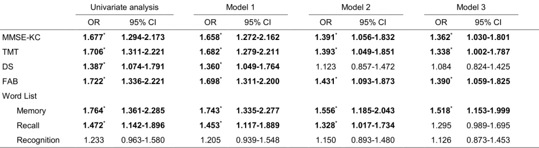
2. Association between FT level and cognitive function
In men, there was no significant association between FT level and low cognitive function (Table 2). On the other hand, in women, low FT level showed association with decreased cognitive function in most of domains. In univariate analysis, low FT level was associated with MMSE-KC (OR, 1.677; 95% CI, 1.294-2.173), TMT (OR, 1.706; 95% CI, 1.311-2.221), DS (OR, 1.387; 95% CI, 1.074-1.791), FAB (OR, 1.722; 95% CI, 1.336-2.221), Word List Memory (OR, 1.764; 95% CI, 1.361-2.285), and Word List Recall (OR, 1.472; 95% CI, 1.142-1.896). After adjusting for all potential confounding factors including age, duration of education, alcohol consumption, smoking, depression and SPPB (Model 3), these associations were constantly observed in MMSE-KC (OR, 1.362; 95% CI, 1.030-1.801), TMT (OR, 1.338; 95% CI, 1.002-1.787), FAB (OR, 1.390; 95% CI, 1.059-1.825), and Word List Memory (OR, 1.518; 95% CI, 1.153-1.999) (Table 3).
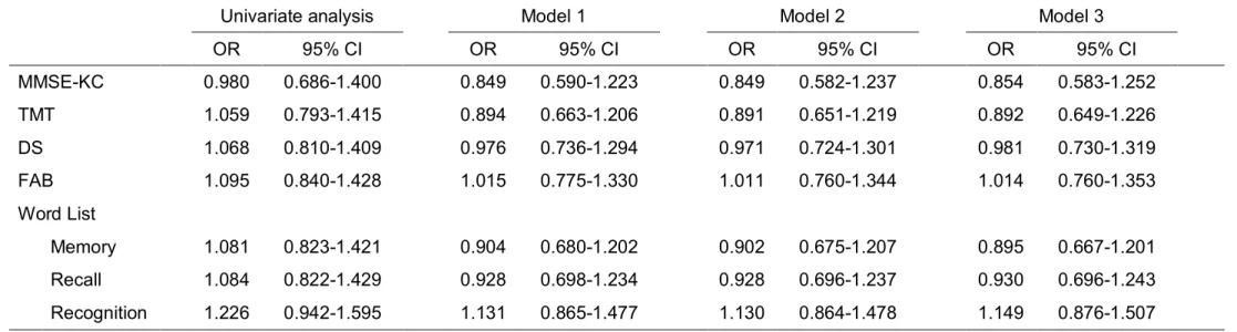
Table 2. Odds ratio of low serum free testosterone level for low cognitive function in elderly men
Univariate analysis Model 1 Model 2 Model 3
OR 95% CI OR 95% CI OR 95% CI OR 95% CI
MMSE-KC 0.980 0.686-1.400 0.849 0.590-1.223 0.849 0.582-1.237 0.854 0.583-1.252
TMT 1.059 0.793-1.415 0.894 0.663-1.206 0.891 0.651-1.219 0.892 0.649-1.226
DS 1.068 0.810-1.409 0.976 0.736-1.294 0.971 0.724-1.301 0.981 0.730-1.319
FAB 1.095 0.840-1.428 1.015 0.775-1.330 1.011 0.760-1.344 1.014 0.760-1.353
Word List
Memory 1.081 0.823-1.421 0.904 0.680-1.202 0.902 0.675-1.207 0.895 0.667-1.201
Recall 1.084 0.822-1.429 0.928 0.698-1.234 0.928 0.696-1.237 0.930 0.696-1.243
Recognition 1.226 0.942-1.595 1.131 0.865-1.477 1.130 0.864-1.478 1.149 0.876-1.507
The odds ratio of developing cognitive function decrease in the lowest free testosterone quartile group relative to the rest was analyzed by logistic regression analysis. Low cognitive function was defined as the lowest quartile in each cognitive domain or less than 24 points in MMSE-KC.
*p<0.05
Model 1 was adjusted for age group.
Model 2 was adjusted for age group and education duration.
Model 3 was adjusted for age group, education duration, alcohol consumption, smoking, depression and SPPB.
OR, odds ratio; CI, confidence interval; MMSE-KC, Mini-Mental Status Examination in the Korean version of the CERAD Assessment Packet; TMT, Trail Making Test; FAB, Frontal Assessment Battery; DS, Digit Span; SPPB, Short Physical Performance Battery.
14
Table 3. Odds ratio of low serum free testosterone level for low cognitive function in elderly women
Univariate analysis Model 1 Model 2 Model 3
OR 95% CI OR 95% CI OR 95% CI OR 95% CI
MMSE-KC 1.677* 1.294-2.173 1.658* 1.272-2.162 1.391* 1.056-1.832 1.362* 1.030-1.801 TMT 1.706* 1.311-2.221 1.682* 1.279-2.211 1.393* 1.049-1.851 1.338* 1.002-1.787
DS 1.387* 1.074-1.791 1.360* 1.049-1.764 1.123 0.857-1.472 1.084 0.824-1.425
FAB 1.722* 1.336-2.221 1.698* 1.311-2.200 1.431* 1.093-1.873 1.390* 1.059-1.825 Word List
Memory 1.764* 1.361-2.285 1.743* 1.335-2.277 1.556* 1.185-2.043 1.518* 1.153-1.999 Recall 1.472* 1.142-1.896 1.453* 1.117-1.889 1.328* 1.017-1.734 1.295 0.989-1.695 Recognition 1.233 0.963-1.580 1.205 0.939-1.548 1.150 0.893-1.480 1.126 0.873-1.453
The odds ratio of developing cognitive function decrease in the lowest free testosterone quartile group relative to the rest was analyzed by logistic regression analysis. Low cognitive function was defined as the lowest quartile in each cognitive domain or less than 24 points in MMSE-KC.
*p<0.05
Model 1 was adjusted for age group.
Model 2 was adjusted for age group and education duration.
Model 3 was adjusted for age group, education duration, alcohol consumption, smoking, depression and SPPB.
OR, odds ratio; CI, confidence interval; MMSE-KC, Mini-Mental Status Examination in the Korean version of the CERAD Assessment Packet; TMT, Trail Making Test; FAB, Frontal Assessment Battery; DS, Digit Span; SPPB, Short Physical Performance Battery.
3. Association between FT quartile and the risk of low cognitive function
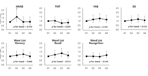
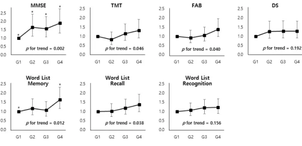
In men, the analysis based on four categories revealed no significant FT level dependent association and no significant trend to cognitive function after adjusting for all potential confounding factors (Figure. 2). Contrarily, in women, the odds ratios of G2 (OR, 1.654;
95% CI, 1.125-2.431), G3 (OR, 1.569; 95% CI, 1.072-2.296), and G4 (OR, 1.898; 95% CI, 1.311-2.750) increased significantly compared to the highest FT level quartile group (G1) in MMSE. And in Word List Memory, the odds ratio of G4 increased significantly (OR, 1.631;
95% CI, 1.148-2.319) compared to the G1 (Figure. 3). The trend between the lower FT and the higher odds ratio of cognitive decline was significant in MMSE (p-value for trend, 0.002), TMT (p-value for trend, 0.046), FAB (p-value for trend, 0.040), Word List Memory (p-value for trend, 0.012), and Word List Recall (p-value for trend, 0.038) (Figure. 3). However, this trend was not observed in elderly men.
16
Fig. 2. Odds ratio of each quartile of serum free testosterone level for decreased cognitive function in elderly men.
The odds ratios of developing cognitive dysfunction in the respective quartile group relative to the 1st quartile group (G1) and p-value for trend were analyzed by logistic regression analysis. The quartile groups were numbered in descending order.
This model was adjusted for age group, education duration, alcohol consumption, smoking, depression and SPPB.
* p <0.05; Gn, nth quartile group of free testosterone level; MMSE, Mini-Mental Status Examination in the Korean version of the CERAD Assessment Packet; TMT, Trail Making Test; DS, Digit Span; FAB, Frontal Assessment Battery.
Fig. 3. Odds ratio of each quartile of serum free testosterone level for decreased cognitive function in elderly women.
The odds ratios of developing cognitive dysfunction in the respective quartile group relative to the 1st quartile group (G1) and p-value for trend were analyzed by logistic regression analysis. The quartile groups were numbered in descending order.
This model was adjusted for age group, education duration, alcohol consumption, smoking, depression and SPPB.
* p <0.05; Gn, nth quartile group of free testosterone level; MMSE, Mini-Mental Status Examination in the Korean version of the CERAD Assessment Packet; TMT, Trail Making Test; DS, Digit Span; FAB, Frontal Assessment Battery.
18 DISCUSSION
I conducted the cross-sectional study of the association between levels of endogenous FT and cognitive function in Korean men and women aged 70-84. The results of the present study suggest sex differential associations of FT levels with cognitive function. In older men, there was no significant association between level of FT and cognitive function and there was no significant trend according to the FT quartiles either. This result is consistent with the findings of a previous study conducted by Zhao et al., which studied in Asian men and showed no association between cognition and testosterone19). On the other hand, many previous observational studies showed controversial results suggesting positive association14,
17, 18, 56) and negative association20, 21) in older men. In addition, previous interventional studies57-59)and meta-analysis studies60-62)have shown inconsistent results.
In older women, on the other hand, there were significant positive associations between FT levels and multi domains of cognitive function. Low FT level was associated with decreased cognitive function in the domains of global cognition, psychomotor speed, attention, executive function and verbal memory. And the association between FT quartiles and the risk of low cognitive function revealed significant FT level dependent association in global cognition (MMSE) and verbal memory (Word List Memory). In global cognition, the risk of impairment was significantly increased in high, low, and lowest FT quartiles compared to the highest FT quartile. In verbal memory, the risk of impairment was significantly increased in the lowest FT quartile compared to the highest FT quartile. Furthermore, significant trends showed not only in global cognition and verbal memory, but also in psychomotor speed (TMT), executive function (FAB), and delayed verbal memory (Word List Recall), suggesting that the lower the FT level, the greater the risk of cognitive decline. These associations and trends were particularly prominent in the global cognition, which revealed the strongest association with FT level among other domains. In previous studies on women,
cognitive function was associated with several domains such as verbal fluency23), verbal learning and memory22, 43), visuospatial ability51, 52, 53), mathematical ability15). Considering the various results of previous studies, present study is considered to be consistent with them and represent comprehensively. However, as in men's studies, previous studies in women have not reached consensus.
Testosterone acts on the entire central nervous system via androgen receptors present in neurons5, 6, 54). Testosterone is known to have neuroprotective functions such as neuro- protective antioxidant, anti-apoptotic potential7-9), and increase neuronal viability55). Roselli et al. reported the sex differences in neural responsiveness of the brain to androgen66, 67). In the same vein, findings of this study can be explained by sex specific influence on cognition.
Apart from study of aforementioned studies, the regulation and expression of androgen receptors in both sexes’ neural tissue demonstrated dose-response relationship with testosterone in a study by Lu et al.68). Considering the significant difference in serum FT levels between men and women (9.42±3.37 pg/mL vs. 0.91±0.89 pg/mL, respectively;
p<0.001), I could hypothesize that there is a dose response relationship between testosterone and cognitive function, and neuroprotective function of the FT in the brain decreases when the level of FT is very low.
In previous studies, various methods of measuring testosterone have been used. This might be a potential reason for the discrepancies among the previous studies. Many of them have measured TT22, 23)instead of FT. FT is bioactive form of testosterone and is considered to be the clinically important marker31). In elderly men, for example, FT level drops sharply due to increased sex hormone-binding globulin, but TT does not69). Furthermore, in some other studies, salivary hormonal samples15, 70) or genetically predicted testosterone level19) were used instead of measuring serum testosterone. This also could account for the inconsistent results of previous studies. Given the very low levels of FT in women and elderly men, it is important to maintain consistency of measurement methods. This is why serum FT was
20 measured in this study.
The results of present study were obtained by adjusting various variables that affect cognitive function, such as age, duration of education, alcohol consumption, smoking, depression and SPPB. Variables adjusted for previous studies varied in a wide range and it seemed lacking consensus on confounding factors on cognitive function. In this study, I tried to include various factors that influence cognitive function. Present study is valuable as the first study to adjust SPPB as a confounding factor to investigate the association between FT and cognitive function. Poor physical function linked to cognitive impairment in older individuals27-29, 46-50). Therefore, SPPB, a typical indicator of the elderly's physical function, was selected as a variable39, 40). None of the previous studies of the testosterone and cognition considered the effect of physical performance on cognitive function. Some studies adjusted body mass index (BMI) as a confounder16, 21, 41), the relationship between BMI and cognitive function is unclear42-45), however.
The major strength of this study is that it is the first study to analyze nationwide multicenter large-scale Asian cohort study to investigate the association between testosterone and cognitive function. Also, this study has the advantage of making various efforts to overcome the limitations of previous studies. I measured serum FT and investigated the association with multi domains of cognition. Additionally, a substantial range of covariates affecting cognitive function, including SPPB, were analyzed.
Study Limitations
Several limitations of my study deserve mention. Its cross-sectional design did not allow evidence of any cause–effect relationship between FT and cognitive function. I plan to conduct a follow-up longitudinal study in the near future. Since this study included only Asian older people, there may be limitations in its application to other ethnic groups.
CONCLUSIONS
FT levels and global cognition, psychomotor speed, executive function and verbal memory had significant association in elderly women. And significant trends also existed, suggesting that the lower the FT level, the greater the risk of cognitive function decrease. On the other hand, in elderly men, there was no association between FT and cognition. Presumably, there is a dose response relationship between FT and multi domains of cognition. Future longitudinal and interventional studies are needed to confirm the present findings.
22 REFERENCES
1. Siddiqui AN, Siddiqui N, Khan RA, Kalam A, Jabir NR, Kamal MA, et al. Neur oprotective Role of Steroidal Sex Hormones: An Overview. CNS Neurosci Ther. 201 6;22(5):342-50.
2.McEwen BS, Milner TA. Understanding the broad influence of sex hormones and s ex differences in the brain. Journal of neuroscience research. 2017;95(1-2):24-39.
3.Zarate S, Stevnsner T, Gredilla R. Role of Estrogen and Other Sex Hormones in B rain Aging. Neuroprotection and DNA Repair. Front Aging Neurosci. 2017;9:430.
4.Iqbal MJ, Dalton M, Sawers RS. Binding of testosterone and oestradiol to sex hor mone binding globulin, human serum albumin and other plasma proteins: evidence for non-specific binding of oestradiol to sex hormone binding globulin. Clin Sci (Lond).
1983;64(3):307-14.
5.Belle MD, Lea RW. Androgen receptor immunolocalization in brains of courting an d brooding male and female ring doves (Streptopelia risoria). Gen Comp Endocrinol.
2001;124(2):173-87.
6.Białek M, Zaremba P, Borowicz KK, Czuczwar SJ. Neuroprotective role of testoster one in the nervous system. Pol J Pharmacol. 2004;56(5):509-18.
7.Chisu V, Manca P, Lepore G, Gadau S, Zedda M, Farina V. Testosterone induces neuroprotection from oxidative stress. Effects on catalase activity and 3-nitro-L-tyrosin e incorporation into alpha-tubulin in a mouse neuroblastoma cell line. Arch Ital Biol.
2006;144(2):63-73.
8.Nguyen TV, Jayaraman A, Quaglino A, Pike CJ. Androgens selectively protect agai nst apoptosis in hippocampal neurones. J Neuroendocrinol. 2010;22(9):1013-22.
9.Tehranipour M, Moghimi A. Neuroprotective effects of testosterone on regenerating spinal cord motoneurons in rats. J Mot Behav. 2010;42(3):151-5.
10.Chunchai T, Apaijai N, Keawtep P, Mantor D, Arinno A, Pratchayasakul W, et al.
Testosterone deprivation intensifies cognitive decline in obese male rats via glial hyp eractivity, increased oxidative stress, and apoptosis in both hippocampus and cortex.
Acta physiologica (Oxford, England). 2019;226(1):e13229.
11.Gray A, Feldman HA, McKinlay JB, Longcope C. Age, disease, and changing sex hormone levels in middle-aged men: results of the Massachusetts Male Aging Study.
J Clin Endocrinol Metab. 1991;73(5):1016-25.
12.Feldman HA, Longcope C, Derby CA, Johannes CB, Araujo AB, Coviello AD, et al. Age trends in the level of serum testosterone and other hormones in middle-aged men: longitudinal results from the Massachusetts male aging study. J Clin Endocrino l Metab. 2002;87(2):589-98.
13.Ly LP, Sartorius G, Hull L, Leung A, Swerdloff RS, Wang C, et al. Accuracy of calculated free testosterone formulae in men. Clin Endocrinol (Oxf). 2010;73(3):382- 8.
14.Thilers PP, Macdonald SW, Herlitz A. The association between endogenous free te stosterone and cognitive performance: a population-based study in 35 to 90 year-old men and women. Psychoneuroendocrinology. 2006;31(5):565-76.
15.Gouchie C, Kimura D. The relationship between testosterone levels and cognitive ability patterns. Psychoneuroendocrinology. 1991;16(4):323-34.
16.Hogervorst E, De Jager C, Budge M, Smith AD. Serum levels of estradiol and te stosterone and performance in different cognitive domains in healthy elderly men and
women. Psychoneuroendocrinology. 2004;29(3):405-21.
17.Yaffe K, Lui LY, Zmuda J, Cauley J. Sex hormones and cognitive function in ol der men. J Am Geriatr Soc. 2002;50(4):707-12.
18.Boss L, Kang DH, Bergstrom N, Leasure JL. Endogenous sex hormones and cogn itive function in the elderly. Aging clinical and experimental research. 2015;27(4):515 -21.
19.Zhao JV, Lam TH, Jiang C, Cherny SS, Liu B, Cheng KK, et al. A Mendelian r andomization study of testosterone and cognition in men. Sci Rep. 2016;6:21306.
20.Yonker JE, Eriksson E, Nilsson LG, Herlitz A. Negative association of testosteron e on spatial visualization in 35 to 80 year old men. Cortex; a journal devoted to the
study of the nervous system and behavior. 2006;42(3):376-86.
21.Martin DM, Wittert G, Burns NR, Haren MT, Sugarman R. Testosterone and cogn itive function in ageing men: data from the Florey Adelaide Male Ageing Study (FA MAS). Maturitas. 2007;57(2):182-94.
22.Wolf OT, Kirschbaum C. Endogenous estradiol and testosterone levels are associat ed with cognitive performance in older women and men. Horm Behav. 2002;41(3):25 9-66.
23.Drake EB, Henderson VW, Stanczyk FZ, McCleary CA, Brown WS, Smith CA, e t al. Associations between circulating sex steroid hormones and cognition in normal e lderly women. Neurology. 2000;54(3):599-603.
24.Davis SR, Wahlin-Jacobsen S. Testosterone in women—the clinical significance. T he Lancet Diabetes & Endocrinology. 2015;3(12):980-92.
25.Hogervorst E, Matthews FE, Brayne C. Are optimal levels of testosterone associat ed with better cognitive function in healthy older women and men? Biochim Biophys
Acta. 2010;1800(10):1145-52.
26.Davis SR, Wahlin-Jacobsen S. Testosterone in women--the clinical significance. Th e lancet Diabetes & endocrinology. 2015;3(12):980-92.
27.Fitzpatrick AL, Buchanan CK, Nahin RL, Dekosky ST, Atkinson HH, Carlson MC, et al. Associations of gait speed and other measures of physical function with cogni tion in a healthy cohort of elderly persons. J Gerontol A Biol Sci Med Sci. 2007;62 (11):1244-51.
28.Auyeung TW, Kwok T, Lee J, Leung PC, Leung J, Woo J. Functional decline in cognitive impairment--the relationship between physical and cognitive function. Neuro epidemiology. 2008;31(3):167-73.
29.Cohen JA, Verghese J, Zwerling JL. Cognition and gait in older people. Maturitas.
2016;93:73-7.
30.Won CW, Lee Y, Choi J, Kim KW, Park Y, Park H, et al. Starting Construction of Frailty Cohort for Elderly and Intervention Study. Annals of Geriatric Medicine a nd Research. 2016;20(3):114-7.
31.Vermeulen A, Verdonck L, Kaufman JM. A critical evaluation of simple methods for the estimation of free testosterone in serum. J Clin Endocrinol Metab. 1999;84(1 0):3666-72.
32.Lee JH, Lee KU, Lee DY, Kim KW, Jhoo JH, Kim JH, et al. Development of th
24
e Korean version of the Consortium to Establish a Registry for Alzheimer's Disease Assessment Packet (CERAD-K): clinical and neuropsychological assessment batteries.
J Gerontol B Psychol Sci Soc Sci. 2002;57(1):P47-53.
33.Lee DY, Lee KU, Lee JH, Kim KW, Jhoo JH, Kim SY, et al. A normative stud y of the CERAD neuropsychological assessment battery in the Korean elderly. J Int Neuropsychol Soc. 2004;10(1):72-81.
34.Kim TH, Huh Y, Choe JY, Jeong JW, Park JH, Lee SB, et al. Korean version of frontal assessment battery: psychometric properties and normative data. Dement Geria tr Cogn Disord. 2010;29(4):363-70.
35.Ki S, Yun J, Kim J, Lee Y. Association Between Dental Implants and Cognitive Function in Community-dwelling Older Adults in Korea. J Prev Med Public Health.
2019;52(5):333-43.
36.Bowie CR, Harvey PD. Administration and interpretation of the Trail Making Test.
Nat Protoc. 2006;1(5):2277-81.
37.Seo EH, Lee DY, Kim KW, Lee JH, Jhoo JH, Youn JC, et al. A normative stud y of the Trail Making Test in Korean elders. Int J Geriatr Psychiatry. 2006;21(9):844 -52.
38.Wechsler D. Wechsler Adult Intelligence Scale–Fourth Edition. San Antonio, TX:
Pearson Assessment; 2008.
39.Yoon DH, Hwang SS, Lee DW, Lee CG, Song W. Physical Frailty and Cognitive Functioning in Korea Rural Community-Dwelling Older Adults. J Clin Med. 2018;7 (11).
40.Chen LK, Liu LK, Woo J, Assantachai P, Auyeung TW, Bahyah KS, et al. Sarco penia in Asia: consensus report of the Asian Working Group for Sarcopenia. J Am Med Dir Assoc. 2014;15(2):95-101.
41.Hogervorst E, Bandelow S, Combrinck M, Smith AD. Low free testosterone is an independent risk factor for Alzheimer's disease. Experimental gerontology. 2004;39(1 1-12):1633-9.
42.Kirton JW, Dotson VM. The interactive effects of age, education, and BMI on co gnitive functioning. Neuropsychology, development, and cognition Section B, Aging, n europsychology and cognition. 2016;23(2):253-62.
43.Ng TP, Feng L, Niti M, Yap KB. Albumin, haemoglobin, BMI and cognitive perf ormance in older adults. Age and ageing. 2008;37(4):423-9.
44.Kuo HK, Jones RN, Milberg WP, Tennstedt S, Talbot L, Morris JN, et al. Cognit ive function in normal-weight, overweight, and obese older adults: an analysis of the Advanced Cognitive Training for Independent and Vital Elderly cohort. J Am Geriat r Soc. 2006;54(1):97-103.
45.Jeong SK, Nam HS, Son MH, Son EJ, Cho KH. Interactive effect of obesity inde xes on cognition. Dement Geriatr Cogn Disord. 2005;19(2-3):91-6.
46.Legdeur N, Badissi M, Yaqub M, Beker N, Sudre CH, Ten Kate M, et al. What determines cognitive functioning in the oldest-old? The EMIF-AD 90+ Study. J Gero ntol B Psychol Sci Soc Sci. 2020. doi: 10.1093/geronb/gbaa152.
47.Kuo HK, Leveille SG, Yu YH, Milberg WP. Cognitive function, habitual gait spe ed, and late-life disability in the National Health and Nutrition Examination Survey (NHANES) 1999-2002. Gerontology. 2007;53(2):102-10.
48.Li CY, Wu SC. Effects of cognitive impairment and loss of physical capacities on survival of the elderly. Neuroepidemiology. 1999;18(6):322-6.
49.Bassett SS, Folstein MF. Cognitive impairment and functional disability in the abs ence of psychiatric diagnosis. Psychological medicine. 1991;21(1):77-84.
50.Warren EJ, Grek A, Conn D, Herrmann N, Icyk E, Kohl J, et al. A correlation b etween cognitive performance and daily functioning in elderly people. Journal of geri atric psychiatry and neurology. 1989;2(2):96-100.
51.Pintzka CW, Evensmoen HR, Lehn H, Håberg AK. Changes in spatial cognition a nd brain activity after a single dose of testosterone in healthy women. Behav Brain Res. 2016;298(Pt B):78-90.
52.Postma A, Meyer G, Tuiten A, van Honk J, Kessels RP, Thijssen J. Effects of te stosterone administration on selective aspects of object-location memory in healthy yo ung women. Psychoneuroendocrinology. 2000;25(6):563-75.
53.Aleman A, Bronk E, Kessels RP, Koppeschaar HP, van Honk J. A single adminis tration of testosterone improves visuospatial ability in young women. Psychoneuroendo crinology. 2004;29(5):612-7.
54.Rosario ER, Pike CJ. Androgen regulation of beta-amyloid protein and the risk of Alzheimer's disease. Brain Res Rev. 2008;57(2):444-53.
55.Nguyen TV, Yao M, Pike CJ. Androgens activate mitogen-activated protein kinase signaling: role in neuroprotection. J Neurochem. 2005;94(6):1639-51.
56.Muller M, Aleman A, Grobbee DE, de Haan EH, van der Schouw YT. Endogeno us sex hormone levels and cognitive function in aging men: is there an optimal leve l? Neurology. 2005;64(5):866-71.
57.Resnick SM, Matsumoto AM, Stephens-Shields AJ, Ellenberg SS, Gill TM, Shuma ker SA, et al. Testosterone Treatment and Cognitive Function in Older Men With Lo w Testosterone and Age-Associated Memory Impairment. JAMA. 2017;317(7):717-27.
58.Wahjoepramono EJ, Asih PR, Aniwiyanti V, Taddei K, Dhaliwal SS, Fuller SJ, et al. The Effects of Testosterone Supplementation on Cognitive Functioning in Older Men. CNS Neurol Disord Drug Targets. 2016;15(3):337-43.
59.Maki PM, Ernst M, London ED, Mordecai KL, Perschler P, Durso SC, et al. Intr amuscular testosterone treatment in elderly men: evidence of memory decline and alte red brain function. J Clin Endocrinol Metab. 2007;92(11):4107-14.
60.Buskbjerg CR, Gravholt CH, Dalby HR, Amidi A, Zachariae R. Testosterone Supp lementation and Cognitive Functioning in Men-A Systematic Review and Meta-Analys is. J Endocr Soc. 2019;3(8):1465-84.
61.Tan S, Sohrabi HR, Weinborn M, Tegg M, Bucks RS, Taddei K, et al. Effects of Testosterone Supplementation on Separate Cognitive Domains in Cognitively Healthy Older Men: A Meta-analysis of Current Randomized Clinical Trials. The American j ournal of geriatric psychiatry : official journal of the American Association for Geriat ric Psychiatry. 2019;27(11):1232-46.
62.Corona G, Torres LO, Maggi M. Testosterone Therapy: What We Have Learned F rom Trials. The journal of sexual medicine. 2020;17(3):447-60.
63.Ota H, Akishita M, Akiyoshi T, Kahyo T, Setou M, Ogawa S, et al. Testosterone deficiency accelerates neuronal and vascular aging of SAMP8 mice: protective role of eNOS and SIRT1. PLoS One. 2012;7(1):e29598.
26
64.Hawley WR, Grissom EM, Martin RC, Halmos MB, Bart CL, Dohanich GP. Test osterone modulates spatial recognition memory in male rats. Horm Behav. 2013;63(4):
559-65.
65.Handelsman DJ. Global trends in testosterone prescribing, 2000-2011: expanding th e spectrum of prescription drug misuse. The Medical journal of Australia. 2013;199 (8):548-51.
66.Roselli CE. Sex differences in androgen receptors and aromatase activity in microd issected regions of the rat brain. Endocrinology. 1991;128(3):1310-6.
67.Roselli CE, Klosterman SA, Fasasi TA. Sex differences in androgen responsiveness in the rat brain: regional differences in the induction of aromatase activity. Neuroen docrinology. 1996;64(2):139-45.
68.Lu SF, McKenna SE, Cologer-Clifford A, Nau EA, Simon NG. Androgen receptor in mouse brain: sex differences and similarities in autoregulation. Endocrinology. 199 8;139(4):1594-601.
69.Basaria S, Dobs AS. Risks Versus Benefits of Testosterone Therapy in Elderly Me n. Drugs & Aging. 1999;15(2):131-42.
70.Hooven CK, Chabris CF, Ellison PT, Kosslyn SM. The relationship of male testos terone to components of mental rotation. Neuropsychologia. 2004;42(6):782-90.
노인에서 혈중 유리 테스토스테론과 인지기능 간의 연관성
목적 한국 노인들의 유리 테스토스테론 혈중 농도와 인지 기능의 연관성을 조사 하고자 하였다.
방법 본 연구는한국 노인 노쇠 코호트 자료를 이용한 단면적 연구로 총 2851명 의 환자가 포함되었다. 연구 대상자들의 유리 테스토스테론 혈중 농도를 측정하 고 인지기능을 평가하였다. 인지기능은 신경심리평가 (Korean version of the C onsortium to Establish a Registry for Alzheimer’s Disease Assessment Pack et; CERAD-K)와 전두엽 기능검사 (Frontal Assessment Battery)를 사용하여 평 가하였다. 유리 테스토스테론 혈중 농도와 인지 기능의 연관성을 조사하기 위해
단변량 및 다변량 로지스틱 회귀 분석을 시행하였다.
결과 여성에서 전반적 인지기능 (OR, 1.362; 95% CI, 1.030-1.801, p-value for trend, 0.002), 정신운동 속도(OR, 1.338; 95% CI, 1.002-1.787, p-value for t rend, 0.046), 집행기능(OR, 1.390; 95% CI, 1.059-1.825, p-value for trend, 0.040), 언어적 기억력(OR, 1.518; 95% CI, 1.153-1.999, p-value for trend, 0.
012) 영역에서 유리 테스토스테론 농도와 유의한 연관성이 있었다. 반면에 남성 에서는 이러한 연관성이 관찰되지 않았다.
결론 유리 테스토스테론 혈중 농도와 인지기능 사이에는 용량 반응 관계가 있다 고 생각된다. 보다 확실한 결론을 위해서는 향후 추가적인 연구가 필요하다.