INTRODUCTION
Executive function (EF) refers to “higher-level” cognitive functions involved in the control and regulation of “lower-lev- el” cognitive processes and goal-directed, future-oriented be- haviors.1 EFs include planning, abstract reasoning, processing
speed, working memory, cognitive flexibility, set-shifting, in- hibitory control, and generative fluency.1 Executive dysfunc- tion is a crucial feature of several neurodegenerative disorders such as fronto-temporal dementia (FTD), Parkinson’s disease (PD), and progressive supranuclear palsy (PSP).2
For many years, Alzheimer’s disease (AD) had been known to have memory dysfunction as an initial and prominent symptom, with the executive function being relatively spared in the early stage of the disease.3 In recent years, however, re- searchers reported that even mild AD patients exhibit impair- ments in a variety of EF tests, and over a 2-year period, mild
Disproportionate Decline of Executive Functions in Early Mild Cognitive Impairment, Late Mild Cognitive
Impairment, and Mild Alzheimer’s Disease
Sangsoon Kim,1 Yeonwook Kang,2,3 Kyung-Ho Yu,3 Byung-Chul Lee3
1Department of Psychiatry, Gachon University Gil Medical Center, Incheon, Korea
2Department of Psychology, Hallym University, Chuncheon, Korea
3Department of Neurology, Hallym University Sacred Heart Hospital, Anyang, Korea
Background and Purpose Many literatures indicate that executive dysfunction exists in mild cognitive impairment (MCI) as well as Al- zheimer’s disease (AD). However, there are few studies that found how early the deficits of the executive function (EF) exist in MCI. The pres- ent study investigated the presence of executive dysfunctions in the earliest stage of MCI, and the sub-domains of EF which are dispropor- tionately impaired earlier than others.
Methods The participants were 41 normal elderly (NE), 86 with amnestic multi-domain MCI, and 41 with mild AD. The MCI group was further sub-divided into two groups: Early MCI (EMCI, n=45) and late MCI (n=41), based on the Clinical Dementia Rating-Sum of Boxes.
All participants were given neuropsychological tests to assess the sub-domains of EF, such as verbal fluency, psychomotor speed, inhibitory control, and mental set-shifting.
Results Impairment of semantic fluency was observed in EMCI, with gradual worsening as cases approached mild AD. Phonemic fluency and psychomotor speed were also impaired at the early stage of MCI relative to the NE, but maintained at the same level up to mild AD.
EMCI exhibited the same degree of performance with NE for inhibitory control and mental set-shifting; however, they progressively wors- ened from EMCI to mild AD.
Conclusions These results suggest that impairments of EF exist even in the earliest stage of the MCI, with a disproportionate decline in the sub-domains of EF.
Key Words mild cognitive impairment, Alzheimer’s disease, executive function, verbal fluency, psychomotor speed, inhibitory control, mental set-shifting.
Received: November 21, 2016 Revised: December 19, 2016 Accepted: December 19, 2016
Correspondence: Yeonwook Kang, PhD, Department of Psychology, Hallym University, 1 Hallimdaehak-gil, Chuncheon 24252, Korea Tel: +82-33-248-1724, Fax: +82-33-256-3424, E-mail: [email protected]
cc This is an Open Access article distributed under the terms of the Cre- ative Commons Attribution Non-Commercial License (http://creative- commons.org/licenses/by-nc/3.0) which permits unrestricted non-com- mercial use, distribution, and reproduction in any medium, provided the ori- ginal work is properly cited.
DND
ORIGINAL ARTICLE
Sangsoon Kim et al.
Disproportionate Decline of EFs in MCI and mild AD
AD with dysexecutive clinical phenotype progresses more rapidly than AD with amnestic phenotype.4 Previous studies have also shown that deficits in EF are common even in am- nestic mild cognitive impairment (aMCI).5-9 A growing body of evidence has shown that aMCI combined other cognitive deficits leads to greater risk for conversion into AD, than in the case of memory dysfunction alone.10 Rozzini et al.11 found that aMCI patients with worsening of EF and functional status but not of memory, were more likely to progress to AD at a 1-year follow up.
Some researchers demonstrated that the Clinical Dementia Rating-Sum of Boxes (CDR-SB) is a more detailed quantitative general index for staging of severity of dementia than the CDR-Global Score (CDR-GS), and is more accurate for track- ing changes across time.12,13 O’Bryant et al.14 suggested that CDR-GS of 0.5 does not represent a homogeneous group, and CDR-SB score of 2.5 may have potential for discriminating be- tween MCI (questionable impairment) and very early AD with CDR-GS of 0.5. Moreover, the Alzheimer’s Disease Neu- roimaging Initiative (ADNI Go and ADNI 2) sub-categorized MCI into early MCI (EMCI) and late MCI (LMCI), to predict conversion into AD and to develop intervention strategies for the earliest stages of the disease.15
Although the aforementioned literatures consistently indi- cate the relationship between executive dysfunction and aMCI as well as AD, there is a lack of research that indicates how ear- ly the deficits of EF exist in aMCI. The present study investi- gated whether executive dysfunctions occur in the earlier stage of aMCI (EMCI), and which sub-domains of EF are dispro- portionately impaired earlier than others.
METHODS
Participants
Forty-one community-dwelling elders [normal elderly (NE)], without subjective memory complaints, voluntarily participat- ed in the study from July 2015 to September 2015. All were screened based on Christensen’s health screening criteria16 and a total score on the Korean-Mini Mental State Examination (K-MMSE)17 higher than the 16th percentile. The patients with cognitive impairments were selected from the patients who visited the Department of Neurology at Hallym Universi- ty Sacred Heart Hospital from July 2013 to April 2015. Patients who had a stroke, significant ischemic changes on brain MRI, movement problems, or salient personality changes, were ex- cluded. Eighty-six patients with amnestic multi-domain MCI (aMCI) met Petersen’s criteria for MCI,18 and 41 patients with mild AD, met the clinical criteria for probable AD proposed by the NINCDS-ADRDA.19 All patients underwent brain
MRI. Additionally, based on the CDR-SB, 86 aMCI patients with a CDR-GS of 0.5 were classified into EMCI (n=45, CDR- SB: 0.5−2.0) and LMCI (n=41, CDR-SB: 2.5−4.0) subgroups.
Materials and procedure
All participants were administered the Seoul Neuropsycho- logical Screening Battery, 2nd Edition (SNSB-II)20 by trained neuropsychologists, which included measures of attention, language, visuospatial, memory, and frontal/executive func- tions, as well as the K-MMSE and CDR. We adopted the sub- tests of frontal/executive function in the SNSB-II for the pres- ent study: Go-No go Test, Korean-Color Word Stroop Test (K- CWST), Controlled Oral Word Association Test (COWAT:
Semantic and phonemic fluency), Digit Symbol Coding (DSC), and Korean-Trail Making Test-Elderly’s version (K- TMT-E). The score on each test was converted to a standardized z-score, except for the Go-No go test and K-CWST: Word, where raw scores were used.
Statistical analysis
An ANOVA and chi-square test were used to detect group differences for demographic variables such as age, education, and sex. We performed a multivariate analysis of variance (MANOVA) on the z-scores of each test measure. In addition, we did post-hoc analyses with Bonferroni correction for multi- ple comparisons to analyze group differences. All p-values were two-tailed, and statistical significance was accepted at p<0.05.
RESULTS
Characteristics of demographic variable, K-MMSE and CDR-SB
Demographic variables, K-MMSE score and CDR-SB of the NE, aMCI, and mild AD are presented in Table 1. There were no significant group differences among the NE, aMCI, and mild AD with regards to age, sex, and education. However, significant group differences were observed in the total score of K-MMSE (F(3, 164)=52.73, p<0.001) and CDR-SB (F(2, 124)=54.56, p<0.001). Results of the post-hoc analysis indicated mild AD with the lowest score on the K-MMSE, and the highest score on the CDR-SB. Post-hoc analysis also revealed significant dif- ferences between the EMCI and LMCI; the LMCI exhibited poorer performance on the K-MMSE and had a greater score on the CDR-SB than the EMCI, respectively. The NE showed significantly higher score on K-MMSE than the EMCI.
Profile of executive functions
The EF performances of the four groups included in the
DND
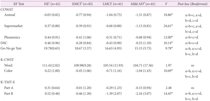
study are shown in Table 2. MANOVA revealed a significant difference among groups on EF tests (λ=0.41, F(3, 164)=6.04, p<0.001). Significant group differences were observed in COW- AT: Animal (F(3, 164)=18.86, p<0.001), COWAT: Supermarket (F(3, 164)=24.61, p<0.001), COWAT: Phonemics (F(3, 164)=12.00, p<0.001), DSC (F(3, 164)=10.14, p<0.001), Go-No go Test (F(3, 164)=9.78, p<0.001), K-CWST: Color (F(3, 164)=10.69, p<0.001), and K-TMT-E: Part B (F(3, 164)=14.45, p<0.001), but not for K- CWST: Word and K-TMT-E: Part A.
Results of the post-hoc analysis for the COWAT: Animal and Supermarket showed that EMCI was more impaired than the NE, although there were no differences between the EMCI and LMCI. Also, mild AD exhibited worse performance than the EMCI, whereas there were no significant differences be- tween the LMCI and mild AD. With regard to COWAT: Pho- nemics and DSC, all patient groups revealed significant im- pairments compared to the NE, but all patient groups were impaired similarly. For the Go-No go Test, K-CWST: Color and K-TMT-E: Part B, there was no difference between the NE and EMCI; however, the LMCI exhibited worse performance than the NE. Although the LMCI revealed no significant dif- ference compare to the EMCI and mild AD, the latter showed poorer performance than the NE and EMCI. There were no significant differences for all groups in the K-CWST: Word and K-TMT-E: Part A.
DISCUSSION
The present study examined whether impairment of EF ex- ists in MCI as well as mild AD, and studied which sub-do- mains of EF are differentially affected at the earlier stages. The results indicated significant declines of EF in either EMCI or LMCI for 7 out of the 9 measures in 5 EF tests, except in K- CWST: Word and K-TMT-E: Part A.
With regard to the COWAT: Animal and Supermarket, the results indicated that semantic fluency was impaired at the early stage of MCI (EMCI), which gradually worsens up to mild AD. Semantic fluency has been recognized to be depen- dent not only upon the integrity of semantic memory, but also
on executive functioning; semantic fluency is more impaired following focal temporal and frontal lobe damage.21 Previous studies suggest that semantic memory impairment is often present in aMCI as well as in dementia of the Alzheimer’s type.
It is said to be associated with injury of the temporal lobe struc- ture.21 In line with the findings of recent literature, the results of the present study indicated that semantic fluency deficits exist even in the earliest stage of the MCI, with progression through AD, suggesting that semantic fluency is a sensitive measure to detect the early stage of aMCI, and could be a good index to monitor the progression to AD.
Results for post-hoc analysis for the COWAT: Phonemics and DSC showed that phonemic fluency and psychomotor speed were impaired at the early stage of aMCI relative to NE, but were maintained at the same level to mild AD, unlike se- mantic fluency. Phonemic fluency measures the generating of as many words as possible based on orthographic criteria. It requires effortful self-initiated retrieval processing.22 In addi- tion, phonemic fluency imposes greater demands on executive skills, than semantic fluency, and many authors have reported that phonemic fluency is more sensitive to frontal, as opposed to non-frontal, lesions.22 The results of the present study indi- cated that the effortful self-initiated retrieval processing for words based on lexical representation was already impaired even in the early stage of aMCI. DSC is associated with visuo- spatial perception, processing speed, sustained attention, vi- suomotor coordination, and incidental memory. It is especially recognized for use in assessing the psychomotor speed.23 Sev- eral authors have emphasized that the marked characteristic of normal aging is the slowing of processing speed,24,25 which it- self is a very sensitive measure of brain abnormality.26 Unlike the DSC, however, we could not find any differences on K- TMT: Part A which is a known measure of processing speed among all groups. This result indicated that DSC is a more sensitive test to detect the impairments of psychomotor speed than TMT: Part A. It also showed that psychomotor slowing was already present at the early stage of aMCI. Recent studies reported that psychomotor slowing is a crucial determinant of performance on verbal fluency tests in normal elderly as well Table 1. Demographic characteristics of the participants, K-MMSE, and CDR-SB
NEa (n=41) EMCIb (n=45) LMCIc (n=41) Mild ADd (n=41) F or χ2 Post-hoc (Bonferroni)
Age 72.73 (6.19) 70.39 (9.81) 73.95 (7.25) 73.88 (8.21) 2.32 ns
Education 9.88 (3.64) 10.52 (4.20) 8.38 (4.80) 8.88 (4.51) 1.88 ns
Sex (M:F) 10:31 20:25 11:30 11:30 χ2=5.31 ns
K-MMSE 28.78 (1.39) 26.58 (2.05) 23.86 (2.67) 22.22 (3.66) 52.73* a>b>c>d
CDR-SB - 1.24 (0.48) 2.91 (0.73) 4.84 (0.71) 54.56* b<c<d
*p<0.001.
AD: Alzheimer’s disease, CDR-SB: Clinical Dementia Rating-Sum of Boxes, EMCI: early MCI, K-MMSE: Korean-Mini Mental State Examination, LMCI: late MCI, MCI: mild cognitive impairment, NE: normal elderly.
Sangsoon Kim et al.
Disproportionate Decline of EFs in MCI and mild AD
as dementia of the Alzheimer’s type.27 Our results showed the same pattern of decline both in the COWAT: Phonemics and in the DSC, supporting the common role of psychomotor speed for both tests.
With regard to the Go-No go Test, K-CWST: Color naming, and K-TMT-E: Part B, we found that EMCI performed simi- larly to NE, though inhibitory control and mental set-shifting progressively worsened from MCI (EMCI and LMCI) to mild AD. Go-No go Test and K-CWST (Stroop test) assess the abili-
ty to inhibit the automatized or previously learned responses, and select the appropriate responses.5,6 Some authors have re- ported that inhibitory control, which is measured by the Hay- ling and Stroop tests, was the most frequently and severely im- paired in aMCI compared to normal elderly controls, relative to other EF tests.5 In contrast, other researchers have shown that tests for inhibition of prepotent responses (Go-No go Test and Stroop tests) failed to uncover significant group differenc- es between normal control and aMCI.6 In our results, we ob- Table 2. Performance on the executive function tests in NE, EMCI, LMCI, and mild AD
EF Test NEa (n=41) EMCIb (n=45) LMCIc (n=41) Mild ADd (n=41) F Post-hoc (Bonferroni) COWAT
Animal -0.05 (0.82) -0.77 (0.94) -1.04 (0.75) -1.31 (0.87) 18.86* a>b=c, a>d,
b>d, c=d
Supermarket 0.37 (0.80) -0.39 (0.91) -0.68 (0.80) -1.13 (0.81) 24.61* a>b=c, a>d,
b>d, c=d
Phonemics 0.44 (0.91) -0.41 (1.06) -0.31 (0.71) -0.68 (0.94) 12.00* a>b=c=d
DSC 0.46 (0.96) -0.28 (0.84) -0.43 (0.90) -0.53 (1.10) 10.14* a>b=c=d
Go-No go Test 19.78(0.65) 18.67 (3.37) 16.63 (4.93) 15.15 (5.75) 9.78* a=b, a>c=d,
b=c, b>d K-CWST
Word 111.41(2.82) 109.98(9.28) 105.54 (11.93) 104.71 (17.36) 1.97 ns
Color 0.22 (1.00) -0.45 (1.06) -0.71 (1.16) -1.04 (1.43) 10.69* a=b, a>c=d,
b=c, b>d K-TMT-E
Part A 0.31 (0.64) -0.01 (1.20) -0.29 (1.23) -0.15 (0.94) 2.48 ns
Part B 0.52 (0.48) -0.46 (1.30) -1.39 (2.07) -2.16 (3.07) 14.45* a=b, a>c=d,
b=c, b>d
*p<0.001.
AD: Alzheimer’s disease, COWAT: Controlled Oral Word Association Test, DSC: Digit Symbol Coding, EF: executive function, EMCI: early MCI, K- CWST: Korean-Color Word Stroop Test, K-TMT-E: Korean-Trail Making Test-Elderly’s version, LMCI: late MCI, MCI: mild cognitive impairment, NE: normal elderly.
Table 3. Performance on the executive function tests in NE, aMCI, and mild AD
EF Test NEa (n=41) aMCIb (n=86) Mild ADc (n=41) F Post-hoc (Bonferroni)
COWAT
Animal -0.05 (0.82) -0.90 (0.86) -1.31 (0.87) 23.71† a>b>c
Supermarket 0.37 (0.80) -0.53 (0.87) -1.13 (0.81) 33.57† a>b>c
Phonemics 0.44 (0.91) -0.36 (0.90) -0.68 (0.94) 16.74† a>b=c
DSC 0.46 (0.96) -0.35 (0.87) -0.53 (1.10) 13.32† a>b=c
Go-No go Test 19.78(0.65) 17.70 (4.28) 15.14 (5.75) 12.60† a>b>c
K-CWST
Word 111.41(2.82) 107.86 (10.79) 104.71(17.36) 3.42* a=b, a>c, b=c
Color 0.22 (1.00) -0.57 (1.11) -1.04 (1.43) 12.21† a>b=c
K-TMT-E
Part A 0.31 (0.64) -0.14 (1.21) -0.15 (0.94) 2.96 ns
Part B 0.52 (0.48) -0.91 (1.76) -2.16 (3.07) 18.62† a>b>c
*p<0.05, †p<0.001.
AD: Alzheimer’s disease, aMCI: amnestic mild cognitive impairment, COWAT: Controlled Oral Word Association Test, DSC: Digit Symbol Coding, EF: executive function, K-CWST: Korean-Color Word Stroop Test, K-TMT-E: Korean-Trail Making Test-Elderly’s version, NE: normal elderly.
DND
served that EMCI performed similarly to NE, but LMCI ex- hibited a poorer performance than NE on the Go-No go Test and K-CWST: Color. K-TMT-E: Part B measures the working memory, divided attention, and processing speed; however, it mainly requires mental set-shifting ability.28 Some authors re- ported that MCI performed similarly to control group on TMT: Part B,7 whereas other authors found that TMT: Part B was impaired in MCI compared to normal control.6 In the present study, we found that EMCI performed similarly to NE;
in contrast, LMCI was more impaired than NE on the K- TMT-E: Part B. Many reasons could explain the inconsistent conclusions of previous studies indicating the use of different tasks, but we believe it is primarily because of different criteria for the recruitment of aMCI patients. In the current study, we subdivided aMCI patients with a CDR-GS of 0.5 into EMCI and LMCI, according to CDR-SB. As a post-hoc analysis, we re-analyzed after merging the two MCI groups (EMCI and LMCI) into one group (aMCI). We found group differences between the aMCI and NE in the Go-No go Test, K-CWST:
Color, and K-TMT-E: Part B (Table 3). Since the EMCI, but not LMCI, still maintained the inhibitory control and mental set-shifting abilities, these results supported that subdividing aMCI into EMCI and LMCI based on CDR-SB would help in understanding the spectrum of impairments of EF in the dis- ease, and to identify the progression of dementia.
To summarize, there were no group differences in simple processing speed among the NE, aMCI, and mild AD. We found that the EMCI performed worse than the NE in verbal fluency and psychomotor speed tests; however, there were no differences between the EMCI and NE in inhibitory control and set-shifting abilities. Also, no differences were observed between the EMCI and LMCI in any of the 9 measures in 5 EF tests; but the LMCI revealed worse performance than the NE in 7 measures. The EMCI exhibited better performance on se- mantic fluency, Go-No go Test, K-CWST: Color, and K-TMT:
Part B than the mild AD; the LMCI did not show any differ- ences with the mild AD on any measures in 5 EF tests.
These results indicated that EF impairments exist even in the earlier stage of the aMCI in several sub-domains, with some sub-domains being similar to NE. This means that the semantic and phonemic fluency and psychomotor speed de- cline earlier, whereas inhibitory control and mental set-shifting abilities decline later. Functional neuroimaging studies showed that patients with left dorsolateral prefrontal cortex (DLPFC) or inferior frontal gyrus lesions are impaired on verbal fluen- cy,29,30 whereas psychomotor speed is associated with superior frontal lobe (middle frontal gyrus).26 It has known that anterior cingulate cortex (ACC) and orbitofrontal cortex (OFC) play an important role in Stroop and Go-No go Test performanc-
es.31-33 Set-shifting or cognitive flexibility are also known to be related with increased activation in the DLPFC and medial pre- frontal cortex (PFC).34 In addition, some researchers reported that the most substantial age-related decline is the volume of prefrontal grey matter including both DLPFC and OFC ar- eas.35,36 Several authors, however, found a relative preservation of the OFC and ACC, although they found the strongest vol- ume reduction within the DLPFC regions.35,37,38 Taking togeth- er these findings from functional and structural neuroimaging with the present study results, we can assume that functions of the PFC which are more related to lateral regions decline at earlier stage of aMCI, whereas the EFs associated with the more orbital or medial part of PFC are impaired at later stage of aMCI.
Our results indicated that executive dysfunction is an im- portant feature of aMCI. Thus, it should be noted that if a clin- ical setting cannot afford to administer a comprehensive neu- ropsychological assessment for aMCI, we suggest that at least two kinds of EF tests should be administered for staging aMCI. The results of the present study indicated that the COWAT and DSC are more sensitive for the detection of an early-stage of aMCI, whereas the Go-No go Test, K-CWST:
Color, and K-TMT: Part B are useful for the detection of late- stage aMCI.
There are several limitations to the current study. First, this study was retrospective with a relatively small sample size. Sec- ond, we could not control the drug effects on each patient group. Third, we did not have biomarker evidences such as hy- pometabolism, β-amyloid deposition, and cerebrospinal fluid (CSF) proteins which would prove that our aMCI is the pro- dromal stage of AD, although we excluded patients who had a stroke, significant ischemic changes on brain MRI, movement problems, or salient personality changes. Fourth, all the EF tests included in our study were traditional “paper-and-pencil”
tests used in clinical settings. We suggest this study should be replicated using more sensitive experimental tasks that mea- sure the EF in detail. Finally, this study was a cross-sectional study; thus, future research should be conducted as a longitu- dinal base.
Conflicts of Interest
The authors have no financial conflicts of interest.
REFERENCES
1. Alvarez JA, Emory E. Executive function and the frontal lobes: a me- ta-analytic review. Neuropsychol Rev 2006;16:17-42.
2. Miller BL, Cummings JL. The Human Frontal Lobes: Functions and Disorders. 2nd. New York: Guilford Press, 2007
3. Broks P, Lines C, Atchison L, Challenor J, Traub M, Foster C, et al.
Neuropsychological investigation of anterior and posterior cortical
Sangsoon Kim et al.
Disproportionate Decline of EFs in MCI and mild AD
function in early-stage probable Alzheimer’s disease. Behav Neurol 1996;9:135-148.
4. Dickerson BC, Wolk DA; Alzheimer’s Disease Neuroimaging Initia- tive. Dysexecutive versus amnesic phenotypes of very mild Alzheim- er’s disease are associated with distinct clinical, genetic and cortical thinning characteristics. J Neurol Neurosurg Psychiatry 2011;82:45- 5. Johns EK, Phillips NA, Belleville S, Goupil D, Babins L, Kelner N, et 51.
al. The profile of executive functioning in amnestic mild cognitive im- pairment: disproportionate deficits in inhibitory control. J Int Neuro- psychol Soc 2012;18:541-555.
6. Zhang Y, Han B, Verhaeghen P, Nilsson LG. Executive functioning in older adults with mild cognitive impairment: MCI has effects on plan- ning, but not on inhibition. Neuropsychol Dev Cogn B Aging Neuro- psychol Cogn 2007;14:557-570.
7. Traykov L, Raoux N, Latour F, Gallo L, Hanon O, Baudic S, et al. Ex- ecutive functions deficit in mild cognitive impairment. Cogn Behav Neurol 2007;20:219-224.
8. Zheng D, Dong X, Sun H, Xu Y, Ma Y, Wang X. The overall impair- ment of core executive function components in patients with amnes- tic mild cognitive impairment: a cross-sectional study. BMC Neurol 2012;12:138.
9. Chen NC, Chang CC, Lin KN, Huang CW, Chang WN, Chang YT, et al. Patterns of executive dysfunction in amnestic mild cognitive im- pairment. Int Psychogeriatr 2013;25:1181-1189.
10. Summers MJ, Saunders NL. Neuropsychological measures predict de- cline to Alzheimer’s dementia from mild cognitive impairment. Neu- ropsychology 2012;26:498-508.
11. Rozzini L, Chilovi BV, Conti M, Bertoletti E, Delrio I, Trabucchi M, et al. Conversion of amnestic Mild Cognitive Impairment to dementia of Alzheimer type is independent to memory deterioration. Int J Geriatr Psychiatry 2007;22:1217-1222.
12. Lynch CA, Walsh C, Blanco A, Moran M, Coen RF, Walsh JB, et al.
The clinical dementia rating sum of box score in mild dementia. De- ment Geriatr Cogn Disord 2006;21:40-43.
13. O’Bryant SE, Waring SC, Cullum CM, Hall J, Lacritz L, Massman PJ, et al. Staging dementia using clinical dementia rating scale sum of boxes scores: a Texas Alzheimer’s research consortium study. Arch Neurol 2008;65:1091-1095.
14. O’Bryant SE, Lacritz LH, Hall J, Waring SC, Chan W, Khodr ZG, et al. Validation of the new interpretive guidelines for the clinical demen- tia rating scale sum of boxes score in the national Alzheimer’s coordi- nating center database. Arch Neurol 2010;67:746-749.
15. Aisen PS, Petersen RC, Donohue MC, Gamst A, Raman R, Thomas RG, et al. Clinical core of the Alzheimer’s disease neuroimaging initia- tive: progress and plans. Alzheimers Dement 2010;6:239-246.
16. Christensen KJ, Multhaup KS, Nordstrom S, Voss K. A cognitive bat- tery for dementia: development and measurement characteristics. J Consult Clin Psychol 1991;3:168-174.
17. Kang YW. A normative study of the Korean-Mini Mental State Exami- nation (K-MMSE) in the elderly. Korean J Psychol 2006;25:1-12.
18. Petersen RC. Clinical practice. Mild cognitive impairment. N Engl J Med 2011;364:2227-2234.
19. McKhann G, Drachman D, Folstein M, Katzman R, Price D, Stadlan EM. Clinical diagnosis of Alzheimer’s disease: report of the NINCDS- ADRDA Work Group under the auspices of Department of Health and Human Services Task Force on Alzheimer’s Disease. Neurology 1984;34: 939-944.
20. Kang Y, Jahng SM, Na DL. Seoul Neuropsychological Screening Battery, 2nd ed (SNSB-II). Seoul: Human Brain Research & Consult- ing Co., 2012.
21. Henry JD, Crawford JR, Phillips LH. Verbal fluency performance in dementia of the Alzheimer’s type: a meta-analysis. Neuropsychologia 2004;42:1212-1222.
22. Henry JD, Crawford JR. A meta-analytic review of verbal fluency per- formance following focal cortical lesions. Neuropsychology 2004;18:
284-295.
23. Fabrigoule C, Rouch I, Taberly A, Letenneur L, Commenges D, Mazaux JM, et al. Cognitive process in preclinical phase of demen- tia. Brain 1998;121:135-141.
24. Stephens R. Age-related decline in Digit-Symbol performance: eye- movement and video analysis. Arch Clin Neuropsychol 2006;21:101- 25. Salthouse TA. The processing-speed theory of adult age differences in 107.
cognition. Psychol Rev 1996;103:403-428.
26. Turken A, Whitfield-Gabrieli S, Bammer R, Baldo JV, Dronkers NF, Gabrieli JD. Cognitive processing speed and the structure of white matter pathways: convergent evidence from normal variation and le- sion studies. Neuroimage 2008;42:1032-1044.
27. Rodríguez-Aranda C, Waterloo K, Sparr S, Sundet K. Age-related psy- chomotor slowing as an important component of verbal fluency: evi- dence from healthy individuals and Alzheimer’s patients. J Neurol 2006;253:1414-1427.
28. Muir RT, Lam B, Honjo K, Harry RD, McNeely AA, Gao FQ, et al.
Trail Making Test elucidates neural substrates of specific poststroke executive dysfunctions. Stroke 2015;46:2755-2761.
29. Paulesu E, Goldacre B, Scifo P, Cappa SF, Gilardi MC, Castiglioni I, et al. Functional heterogeneity of left inferior frontal cortex as revealed by fMRI. Neuroreport 1997;8:2011-2017.
30. Phelps EA, Hyder F, Blamire AM, Shulman RG. FMRI of the prefron- tal cortex during overt verbal fluency. Neuroreport 1997;8:561-565.
31. Botvinick MM, Cohen JD, Carter CS. Conflict monitoring and ante- rior cingulate cortex: an update. Trends Cogn Sci 2004;8:539-546.
32. Menon V, Adleman NE, White CD, Glover GH, Reiss AL. Error-relat- ed brain activation during a Go/NoGo response inhibition task. Hum Brain Mapp 2001;12:131-143.
33. Rolls ET, Hornak J, Wade D, McGrath J. Emotion-related learning in patients with social and emotional changes associated with frontal lobe damage. J Neurol Neurosurg Psychiatry 1994;57:1518-1524.
34. Zakzanis KK, Mraz R, Graham SJ. An fMRI study of the Trail Mak- ing Test. Neuropsychologia 2005;43:1878-1886.
35. Raz N, Gunning FM, Head D, Dupuis JH, McQuain J, Briggs SD, et al. Selective aging of the human cerebral cortex observed in vivo: dif- ferential vulnerability of the prefrontal gray matter. Cereb Cortex 1997;
7:268-282.
36. Tisserand DJ, Pruessner JC, Sanz Arigita EJ, van Boxtel MP, Evans AC, Jolles J, et al. Regional frontal cortical volumes decrease differen- tially in aging: an MRI study to compare volumetric approaches and voxel-based morphometry. Neuroimage 2002;17:657-669.
37. Salat DH, Kaye JA, Janowsky JS. Selective preservation and degener- ation within the prefrontal cortex in aging and Alzheimer disease. Arch Neurol 2001;58:1403-1408.
38. Tisserand DJ, van Boxtel M, Gronenschild E, Jolles J. Age-related vol- ume reductions of prefrontal regions in healthy individuals are differ- ential. Brain Cogn 2001;47:182-185.