저작자표시-비영리-변경금지 2.0 대한민국 이용자는 아래의 조건을 따르는 경우에 한하여 자유롭게
l 이 저작물을 복제, 배포, 전송, 전시, 공연 및 방송할 수 있습니다. 다음과 같은 조건을 따라야 합니다:
l 귀하는, 이 저작물의 재이용이나 배포의 경우, 이 저작물에 적용된 이용허락조건 을 명확하게 나타내어야 합니다.
l 저작권자로부터 별도의 허가를 받으면 이러한 조건들은 적용되지 않습니다.
저작권법에 따른 이용자의 권리는 위의 내용에 의하여 영향을 받지 않습니다. 이것은 이용허락규약(Legal Code)을 이해하기 쉽게 요약한 것입니다.
Disclaimer
저작자표시. 귀하는 원저작자를 표시하여야 합니다.
비영리. 귀하는 이 저작물을 영리 목적으로 이용할 수 없습니다.
변경금지. 귀하는 이 저작물을 개작, 변형 또는 가공할 수 없습니다.
理學博士 學位論文
蔚山大學校 大學院 生命科學科
朴賢貞
염증에 의한 골 소실 기전 연구
Studies of bone loss mechanism by inflammation
[UCI]I804:48009-200000291282 [UCI]I804:48009-200000291282
염증에 의한 골 소실 기전 연구
울 산 대 학 교 대 학 원 생 명 과 학 과
박 현 정 지도교수 최혜선
이 논문을 이학박사 학위 논문으로 제출함
2019년 11월
Doctor of Philosophy
Studies of bone loss mechanism by inflammation
The Graduate School of the University of Ulsan Department of Biological
Sciences
Hyun-Jung Park
Department of Biological Sciences Ulsan, Korea
November 2019
Studies of bone loss mechanism by inflammation
Supervisor : Hye Seon Choi, Ph.D
A Dissertation
submitted to
The Graduate School of the University of Ulsan In partial Fulfillment of the Requirements
for the Degree of
Doctor of Philosophy by
Hyun-Jung Park
This certifies that the dissertation of Hyun-Jung Park is approved
Studies of bone loss mechanism by inflammation
1
국 문 요 약
우리 몸을 구성하고 있는 뼈는 몸을 지탱해주고, 내부 중요 장기를 보호해줄 뿐만 아니라 줄기세포를 만들어내는 가장 중요한 기관 중 하나이다. 뼈는 골을 흡수하는 파골세포와 골 형성에 관여하는 조골세포에 의해 항상성을 유지하고, 일생동안 끊임없이 골재형성 (bone remodeling) 과정을 거치게 된다. 과도한 염증반응이 일어나거나, 폐경기 여성의 경우 골흡수와 골형성의 균형이 무너져 골 흡수 정도가 증가하게 되면서 골다공증이 유발되는 것으로 알려져 있다. 골을 파괴하는 파골세포는 조혈모세포로를 기원으로 하고, 조골세포에서 분비되는 macrophage-colony stimulating factor (M-CSF) 와 receptor activator of nuclear factor-ĸB ligand (RANKL)에 의해 분화되어 여러 개의 단핵세포가 모여 거대한 크기의 다핵세포를 이루는 특징을 가지고 있다. 성숙한 파골세포는 integrin αvβ3에 의해 뼈에 부착된 후 actin ring을 형성하고, proton과 가수분해효소에 의해 뼈의 흡수가 시작된다.
Autophagy는 병리학적, 생태학적 조건 하에서 불필요하거나 기능에 문제가 있는
성분을 제거하는 과정이다. 과도한 autophagy는 골 량을 감소시키는 것으로 보고되고 있다.
본 연구는 염증에 의한 골소실에서 autophagy의 역할에 초점을 맞추었고, LPS로 유도된 골소실에서 autophagy 억제제인 4-PBA와 lycorine의 효과를 확인하여 골다공증의 새로운 치료 및 진단방법에 대한 기본 정보를 제시하였다.
Keywords: osteoclast, autophagy, LPS, inflammatory bone loss, 4-PBA, lycorine
2
목 차
국 문 요 약 ... 1
목 차 ... 2
List of original publications ... 4
List of Figures and Tables ... 5
서 론 ... 6
실험재료 및 방법... 9
1. 시약 및 배지 ... 9
2. 실험동물과 연구설계 ... 10
3. 파골세포 분화 ... 11
4. 세포생존률 측정 (MTT assay) ... 11
5. 파골세포 활성도 측정 (Bone resorption assay) ... 11
6. RNA 추출 및 quantitative polymerase chain reaction (q-PCR) ... 12
7. 산성 소포성 소기관 염색 (acidic vesicular organelles staining) ... 13
8. Cathepsin K 분석 ... 13
9. Electrophoretic mobility shift assays (EMSA) ... 13
10. Western blot ... 14
11. Transfection and siRNA ... 14
12. Intracellular, mitochondrial 활성산소종 (ROS) 측정 ... 14
13. 카르복시메틸화에 의한 산화된 TRPML1 검출 ... 14
14. 통계처리 ... 15
3
실 험 결 과 ... 16
1. LPS에 의한 파골세포 분화와 autophagy의 연관성 ... 16
1.1 LPS에 의한 파골세포 분화 증가 ... 16
1.2 LPS로 유도된 autophagy에 의한 파골세포 형성 증가 ... 16
1.3 LPS로 유도된 동물 실험에서 autophagy 억제제인 3-MA의 효과 ... 17
2. Autophagy를 저해하는 4-PBA가 LPS로 유도된 골 소실에 미치는 영향 ... 24
2.1 LPS로 유도된 골소실에서 4-PBA의 효과 ... 24
2.2 4-PBA에 의한 파골세포 분화 억제 ... 25
2.3 4-PBA에 의한 NF-κB 신호전달 저해 ... 25
2.4 4-PBA에 의한 autophagy 억제 ... 26
3. LPS로 유도된 골 소실에서 mROS/TRPML1/TFEB를 저해하는 lycorine의 효과 ... 37
3.1 LPS로 유도된 골소실에서 lycorine의 효과 ... 37
3.2 lycorine에 의한 파골세포 분화와 활성 억제 ... 37
3.3 lycorine에 의한 autophagy 감소 ... 38
3.4 lycorine에 의한 mitochondria ROS, TRPML1 산화 감소에 따른 TFEB의 핵 이동 억제 ... 38
고 찰 ... 51
참 고 문 헌 ... 53
Abstract ... 58
4
List of original publications
The present thesis is based on the following original articles:
1. Mol Cells. 2017 Nov 30;40(11): 880-887.
Lipopolysaccharide (LPS)-induced autophagy is responsible for enhanced osteoclastogenesis
Ok-Joo Sul,Hyun-Jung Park,Ho-Jung Son, and Hye-Seon Choi
2. Biochem Pharmacol. 2018 May; 151:9-17.
4-Phenylbutyric acid protects against lipopolysaccharide-induced bone loss by modulating autophagy in osteoclasts
Hyun-Jung Park, Ho-Jung Son,Ok-Joo Sul, Jae-Hee Suh, Hye-Seon Choi
3. Oxidative Medicine and Cellular Longevity. Volume 2019, Article ID 8982147, 11 pages Lycorine attenuated autophagy in osteoclasts via an axis of mROS/TRPML1/TFEB to reduce LPS-induces bone loss
Hyun-Jung Park, Malihatosadat Gholam-Zadeh,Jae-Hee Suh, and Hye-Seon Choi
5
List of Figures
Figure 1. LPS increases the number and activity of OCs in vitro ... 18
Figure 2. LPS induces autophagy, increasing OC formation ... 20
Figure 3. Effect of autophagy inhibitor 3-MA on LPS-induced bone loss ... 22
Figure 4. 4-PBA protects mice from LPS-induced bone loss ... 28
Figure 5. 4-PBA inhibits LPS-induced OC differentiation ... 31
Figure 6. 4-PBA impairs LPS-induced NF-κB signaling required for osteoclastogenesis . ... 33
Figure 7. 4-PBA attenuates LPS-induced autophagy in OCs ... 35
Figure 8. Lycorine protects against LPS-induced bone loss in mice ... 40
Figure 9. Lycorine inhibits LPS-induced OC differentiation and OC activity in vitro 44
Figure 10. Lycorine decreases LPS-induced autophagy in OCs ... 46
Figure 11. Lycorine reduces TFEB nuclear translocation by attenuating oxidation of TRPML1 via decreased mitochondrial ROS upon LPS stimulation in OC ... 48
List of Tables
Table 1. Specific primers used in PCR and real-time quantitative PCR (RT-qPCR) analysis ... 12Table 2. Trabecular microarchitecture and biochemical markers of LPS with or without lycorine-treated mice ... 42
6
서 론
우리 몸을 구성하고 있는 골은 뼈를 흡수하고 파괴하는 파골세포 (osteoclast)와 새로운 뼈를 생성하는 조골세포 (osteoblast)에 의해 일생동안 생성과 소멸을 반복하는 매우 역 동적인 조직이다 [Boyle et al., 2003]. 정상적인 상태에서는 파골세포와 조골세포의 균형이 잘 조절되어 건강한 뼈를 유지하게 되지만, 노령화나 폐경기 여성의 급격한 에스트로겐 발현 저하, 감염에 의한 세균의 구성요소나, 만성적인 염증반응으로 인해 파골세포의 수 와 활성이 증가하여 과도한 골 흡수가 유도되면 골다공증이라 불리는 골 질환을 야기하 게 된다 [Raggatt and Partridge, 2010]. 조혈모세포에서 유래된 단핵구/대식세포가 macrophage colony-stimulating factor (M-CSF)와 receptor activator of nuclear factor-κB
ligand (RANKL)의 자극을 받으면 여러 단계를 거쳐 다핵을 가진 파골세포가 형성된다
[Roodman, 2006]. 그람음성 세균의 세포벽 구성성분인 Lipopolysaccharide (LPS)는 염증세 포를 불러들이고, proinflammatory cytokine을 활성화시켜 염증반응을 유발하는 대표적인 물질인데 [Henderson B and Nair SP, 2003], 다양한 연구 결과들을 통해 파골세포의 분화 를 자극한다고도 알려져 있다. 쥐의 복강에 LPS를 투여하게 되면 활성산소종 (ROS)에 의 해 파골세포의 분화가 유도되면서 파골세포 수와 활성이 증가되어 [Sul et al., 2017; Sul et al., 2019; Itoh et al., 2003] 대퇴골 침식이 증가되는데 [Orcel et al., 1993], in vitro에서 LPS 의 효과는 파골세포 형성과 관련하여 두가지 결과가 나오는 것으로 알려져 있다. LPS 자 극 이전에 RANKL의 자극을 받은 세포는 파골세포로의 형성이 크게 증가되지만 [Sul et al., 2017; Park et al., 2015], LPS와 RANKL의 자극이 동시에 주어지게 될 때에는 파골세포 의 형성을 현저하게 억제하는 것으로 보아 [Takami et al., 2002] LPS는 파골세포의 생성을 증가시키기는 하나 전구세포에는 작용하지 않는 것을 알 수 있다.
Autophagy는 영양소가 고갈되었을 때 세포의 생존을 증진시키는 기작으로 처음 밝혀
졌지만 불필요하거나, 기능에 이상이 있는 세포 구성성분들을 제거할 때에도 관여를 한 다. Phagophore를 형성하고, lysosome과의 융합을 통해 내용물들이 분해, 재사용하는 과 정을 거쳐 세포의 항상성이 유지되므로 autophagy 과정에 문제가 있을 경우 여러 질병 의 원인이 되는 것으로 알려져 있다. 사람의 전유전체연관성 데이터(genome-wide association data)에 따르면 autophagy가 골다공증 발병과 관련이 있는 것으로 밝혀져 있 어 [Zhang et al., 2010] , autophagy와 골사간에는 긴밀한 관계가 있음을 알 수 있다. In vitro 상에서 autophagy를 억제하는 PI3K 억제제 중 하나인 nocodazole 처리시 파골세
7
포의 podosome belt 형성에 결함을 유발하여 파골세포의 활성을 저해하고 [Ti et al., 2015]
wortmannin 과 LY301497 역시 파골세포에 의한 골 흡수를 줄여줄 뿐만 아니라 [Sato et
al., 1996] 난소절제 마우스 모델에서 유발되는 골소실을 예방하는 것으로 보아
autophagy 억제는 파골세포의 활성에 영향을 미친다는 것을 알 수 있다. 뿐만 아니라
autophagic cargo 수용체인 SQSTM1 (p62)에 돌연변이가 유발되면 NF-κB의 과도한 활성 으로 파골세포의 숫자와 활성이 증가하는 Pagetic disorder 가 유발되고 [Helfrich and Hocking, 2008; McManus and Roux, 2012], autophagosome 형성과 관련된 ATG5와 ATG7 에 문제가 생길 시 파골세포의 기능에 영향을 미치는 것으로 알려져 있다 [DeSelm et al., 2011]. Myeloid 계열 특이적으로 ATG5나 ATG7을 결핍시키면 파골세포의 ruffled border 가 제기능을 하지 못해 골 흡수 능력이 감소되어 골밀도가 증가되고 [DeSelm et al., 2011], MAILC3A를 knockdown 시키면 actin ring 형성을 저해해 골 흡수가 감소되는 것 으로 나타난다 [Chung et al., 2012]. 또한 [Lin et al., 2013]에 따르면 류마티스 관절염 환 자에게서 파골세포가 증가되는데 autophagy 과정 초기 단계에 phagophore의 신장에 관 여하는 beclin1과 Atg7이 관여하는 것으로 보아 autophagy가 파골세포의 골 흡수에 영 향을 미친다는 것을 알 수 있다.
4-Phenylbutyric acid (4-PBA)는 저분자 물질로 요소순환장애 (urea cycle disorders)가 있 는 환자에게 정맥주사로 250-300mg/kg/d 투여 시 치료 효과를 보이는 FDA 승인 약물 이다 [Maestri et al., 1996]. 4-PBA를 7일간 400 mg/kg/d 농도로 투여하였을 때 혈장에서 일정 농도를 유지하는 것으로 확인되었고 [Lin et al., 2009], 4-PBA는 ammonia scavenger 로서의 역할과 histone deacetylase 억제제, endoplasmic reticulum (ER) stress 억제제 [Iannitti and Palmieri, 2011; Kim et al., 2013], 항암제로서의 역할을 하는 것으로도 알려져 있다 [Gilbert et al., 2001].
Lycorine은 Lycoris radiata (Amaryllidaceae)에서 추출된 pyrrolo phenanthridine ring- type의 알칼로이드로, 항 염증 [Çitoğlu et al., 2012], free radical 제거 [Ghane et al., 2018], 항 바이러스 [Liu et al., 2011], 항암효과 [Cao et al., 2013; Jin and Yao, 2016]를 가지고 있 어 한방에서는 낮은 독성을 가진 한약재로 사용되고 있다 [Zupkó et al., 2009]. 분자생물 학적 측면에서 lycorine은 NF-κB 활성을 억제하고 [Park, 2014], p38을 감소, STAT 활성화 를 통해 LPS로 유도된 염증을 감소시킨다 [Kang et al., 2012]. 뿐만 아니라 lycorine은 HMGB1의 분해를 통해 autophagy를 억제하고 [Roy et al., 2016], MAPK를 억제하여 난소 절제에 의한 골소실을 예방하는 것으로 알려져 있다 [Chen et al., 2015].
본 연구에서는 autophagy가 LPS로 유도된 파골세포 형성과 골 소실에 관여가 되어있
8
다는 것을 확인하고, 4-PBA와 lycorine이 LPS로 유도된 autophagy와 파골세포 활성을 약 화시킬 수 있는지 상세한 분자 메커니즘을 통해 확인해보았다.
9
실험재료 및 방법
1. 시약 및 배지
10% fetal bovine serum (FBS), penicillin, streptomycin, α-Minimum essential medium (α- MEM) 은 Welgene (Gyeongsan, Korea)에서 구입하였으며, Opti-MEM은 Gibco (Grand Island, NY, USA)에서, Recombinant mouse M-CSF and RANKL 은 R&D Systems사 (Minneapolis, MN, USA)에서 구입하였다. 3-methyladenine, LPS, MTT (3-(4,5- dimethylthiazol-2-yl)-2,5-diphenyltetrazolium bromide), Leukocyte acid phosphatase (TRAP) kit, Mayer’s hematoxylin, toluidine blue, Bafilomycin A, diphenyl iodonium (DPI)은 Sigma Aldrich (St. Louis, MO, USA)의 제품을 사용하였다. 4-PBA, HRP-conjugated 2차 항체, small interfering RNA (siRNA) against ATG7 (sc-41448), scrambled siRNA (scRNA, sc-37007), Small interfering RNA (siRNA) against TRPML1 (sc-44520)과 MitoTEMPO는 Santa Cruz Biotechnology (Santa Cruz, CA, USA)에서, lycorine은 Chengdu Biopurity Phytochemicals (Chengdu, Sichuan, China)사에서 구입하였다. Dentine slices와 RatLaps EIA kit는 Immunodiagnostic Systems (Fountain Hills, AZ, USA)사에서 구매하였으며 BAY 11-7082와 JSH23은 Calbiochem (Darmstadt, Germany)의 제품을 사용하였다. Osteocalcin EIA,는 Biomedical Technologies (Stoughton, MA, USA)사에서 Alkaline phosphatase kit는 BioAssay Systems (Hayward, CA, USA )에서, Cathepsin K activity assay kit는 Abcam (Cambridge, UK) 에서 구매하였다. N-(Biotinoyl)-N′-(iodoacetyl) ethylenediamine (BIAM)은 Invitrogen (Carlsbad, CA, USA) 제품을 사용하였다.
MCP-1 (coating and detection)에 대한 항체는 R&D System (Minneapolis, MN, USA)사 에서, LC3B (#2775), lamin B1 (#13435)는 Cell Signaling Technology (Danvers, MA, USA), p62 (H00008878-M01)는 Abnova (Taipei, Taiwan), TFEB (A303-673A)는 Bethyl Laboratories, Inc. (Montgomery, TX, USA), TRPML1 (ACC-081)은 from Alomone Labs (Jerusalem, Israel) 사에서, β-actin (A5441)은 from Sigma Chemical (St. Louis, MO, USA)에서 구입하였다. NE- PER nuclear and cytoplasmic extraction reagents 와 LightShift Chemiluminescent EMSA kit 는 Pierce (Waltham, MA, USA) 제품을 사용하였고, 2′,7′-dichlorofluorescein diacetate (H2DCFDA)는 Thermo Fisher Scientific (Waltham, MA, USA)에서 구매하였다.
M-MLV reverse transcriptase와 SYBR Green Real-Time PCR Master Mixes 은Promega (Madison, WI, USA)에서, QIAzol reagent는QIAGEN (Hilden, Germany), Lipofectamine 3000
10
과 MitoSOX Red는 Invitrogen (Carlsbad, CA, USA)에서 구입하여 사용하였다.
2. 실험동물과 연구설계
실험동물은 10주령의 C57BL/6J, 암컷 mouse를 사용하여 울산대학교 Immunomodulation Research Center (IRC) 동물사육실에서 사육하였다. 모든 실험 동물은 울산대학교 IRC의 동물실험윤리위원회 (IACUC)의 규정에 따라 수행하였으며, 각 실험의 승인 ID는 다음과 같다.
# UOU-2014-010, 실험동물은 무작위로 3그룹 : PBS를 투여한 대조군 (n=5), LPS (5mg/kg)를 투여한 그룹 (n=6), LPS와 3-MA 투여 (n=3)으로 나누어 진행하였다. 총 3주 간 복강 내 주사를 시행하였다.
# UOU-2014-014, 실험동물은 무작위로 다음과 같이 나누었다 : PBS를 투여한 대조군 (vehicle) (n=5), PBS와 4-PBA를 투여한 그룹 (n=6), LPS를 투여한 그룹 (n=6), LPS와 4- PBA를 투여한 그룹 (n=6) 총 4그룹으로 나누었고 LPS는 5mg/kg의 양으로 복강내 주사 를 실시하였다. LPS와 PBS는 일주일에 한번씩 복강 내 주사를 시행하였고 [Gao et al., 2007], 4-PBA는 240mg/kg의 양으로 HCl과 NaOH로 pH 7.4를 맞춘 후 매일 복강 내 주 사를 시행하였다. LPS와 PBS, 4-PBA는 각 마리당 200μl씩, 3주간 투여하였다.
# HSC-15-11, PBS 투여 (n=5), PBS와 lycorine 투여 (n=5), LPS (n=5), LPS와 lycorine 투 여 (n=6) 총 4그룹으로 나누어 실험을 진행하였다. LPS는 5mg/kg의 농도로 200μl의 인 산완충용액 (PBS)에 녹여 일주일에 한번씩 복강 내 주사를 하였고, lycorine 6mg/kg의 농 도로 맞춘 후 1N HCl과 1N NaOH로 pH 7.2를 맞춘 후 이틀에 한번 복강 내 주사를 시행 하였고 총 3주간의 실험 기간을 거쳤다.
시료 투여가 끝난 mouse는 이산화탄소 질식방법을 사용해 희생하여 오른쪽 대퇴골을 적출한 후 10% PFA (paraformaldehyde)에 고정하였다. 이후 고해상도 Micro CT (μCT) SkyScan 1176 System (Bruker-Micro CT, Kontich, Belgium)을 이용하여 골밀도 (bone mineral density, BMD)를 측정하였다. 픽셀크기 6.9μm, 임계값 77-255 mg/cc에서 촬영을 수행하였고, 해면골 (trabecular bone)의 분석 범위는 성장판이 끝나는 지점의 0.1mm 아 래에서부터 1.6mm 두께에 해당하는 곳을 분석하였다. 총 75~125개의 단층 슬라이드를 사용하여 CTan 소프트웨어를 사용해 bone volume fraction (BV/TV), trabecular thickness (Tb.Th), trabecular space (Tb.Sp.), trabecular number (Tb.N), bone mineral density (BMD)를 측정하였다. 이후 CTvol 소프트웨어 (ver 1.11 ; skyscan)를 사용해 3차원 이미지를 구현하
11 였다.
생체내 골 흡수 표지자인 CTX-1 (collagen-type I fragments)을 측정하기 위해 혈청을 사용하였고 RatLaps EIA assay kit를 사용하여 제조사의 지시에 따라 측정하였다. 혈청 내 osteocalcin과 ALP (alkaline phosphatase)를 측정하기 위해 osteocalcin EIA kit와 colorimetric kinetic determination kit를 사용하였고, MCP-1의 경우 sandwich ELISA 방법 을 사용하여 진행하였다.
3. 파골세포 분화
4-5주령된 C57BL/6J 쥐의 대퇴골과 경골로부터 골수세포를 채취하고 10% FBS가 함유 된 α-MEM 배지에 M-CSF를 넣고 37℃ 배양기에서 3일간 배양하였다. 3일 후 부착된 대 식세포를 사용하여 실험하였다. 파골세포로의 분화를 위해 M-CSF (30ng/ml)와 RANKL (40ng/ml)을 40시간 동안 처리하고, 이후 생성된 파골전구세포에 M-CSF (30ng/ml)와 LPS (50ng/ml)를 처리하고, 3-MA와 4-PBA, lycorine을 농도별로 처리하여 48시간 동안 배양하 였다. 만들어진 파골세포는 3.7% PFA로 고정시킨 후 TRAP 용액으로 염색하여 핵이 3개 이상인 파골세포의 개수를 측정하였다 [Van et al., 2013]. 또한 파골세포의 면적과 직경을 측정하고, 한세포당 가지고 있는 핵의 개수를 구하였다 [Okayasu et al., 2012].
4. 세포생존률 측정 (MTT assay)
세포생존은 MTT 분석법으로 측정하였다. 세포를 세척한 후 MTT (3-(4,5- dimethylthiazol-2-yl)-2,5-diphenyltetrazolium bromide) (0.5 mg/ml)를 첨가하고 3시간 동안 37℃에서 배양하였다. DMSO에 의해 MTT가 formazan으로 분해되는 양을 ELISA reader를 이용하여 540nm에서 흡광도를 측정하였다.
5. 파골세포 활성도 측정 (Bone resorption assay)
파골세포의 활성을 검사하기 위해 상아질 조각위에 파골세포를 키우면 흡수공 (resorption pits)이 형성되어, 골이 흡수되는 정도를 측정할 수 있다 [Jimi et al., 1999].
성숙한 파골세포를 만들기 위해 M-CSF와 RANKL로 분화를 유도하고, 3-4일 후 성숙한
12
파골세포를 떼어내어 같은 수만큼의 세포를 상아질 조각위에 넣어준다. M-CSF와 LPS를 처리하고 3-MA, 4-PBA, lycorine의 유무에 따라 4일간 배양을 한 뒤 상아질 조각위에 붙어있는 파골세포를 3.7% PFA로 고정 후 TRAP 염색을 시행한다. 이후 붙어있는 세포를 떼어내기 위해 1M NH4OH를 처리하고 초음파진동을 시행했다. TDW로 세척한 후 상아질 조각을 Mayer’s hematoxylin과 0.5% sodium borate가 들어있는 1% toluidine blue 용액을 사용하여 염색한 후 현미경으로 형성된 흡수공을 관찰하였다. 흡수공 면적은 Image J 소프트웨어를 사용하여 분석하였다.
6. RNA 추출 및 quantitative polymerase chain reaction (q-PCR)
총 RNA는 TRIzol 시약을 사용해 세포로부터 추출하였고, 1st strand cDNA는 random primer 및 M-MLV 역전사 효소로 역전사시켰다. qPCR은 SYBR Green Real-Time PCR Master Mixes와 적절한 primer를 사용하였고, StepOnePlus TM Real Time System을 사용하여 분석하였다. 18S rRNA (RPS) 유전자는 상대유전자와 병행하여 증폭시켰다.
RPS와 비교한 상대적인 copy수는 2-△△Ct를 이용하여 계산하였다. 실험에서 사용한
primer 들은 Table 1과 같다.
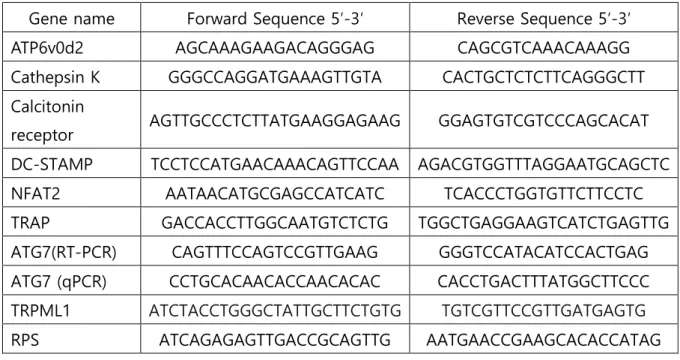
Table 1. Specific primers used in PCR and real-time quantitative PCR (RT-qPCR) analysis.
Gene name Forward Sequence 5′-3′ Reverse Sequence 5′-3′
ATP6v0d2 AGCAAAGAAGACAGGGAG CAGCGTCAAACAAAGG
Cathepsin K GGGCCAGGATGAAAGTTGTA CACTGCTCTCTTCAGGGCTT
Calcitonin
receptor AGTTGCCCTCTTATGAAGGAGAAG GGAGTGTCGTCCCAGCACAT
DC-STAMP TCCTCCATGAACAAACAGTTCCAA AGACGTGGTTTAGGAATGCAGCTC
NFAT2 AATAACATGCGAGCCATCATC TCACCCTGGTGTTCTTCCTC
TRAP GACCACCTTGGCAATGTCTCTG TGGCTGAGGAAGTCATCTGAGTTG
ATG7(RT-PCR) CAGTTTCCAGTCCGTTGAAG GGGTCCATACATCCACTGAG
ATG7 (qPCR) CCTGCACAACACCAACACAC CACCTGACTTTATGGCTTCCC
TRPML1 ATCTACCTGGGCTATTGCTTCTGTG TGTCGTTCCGTTGATGAGTG
RPS ATCAGAGAGTTGACCGCAGTTG AATGAACCGAAGCACACCATAG
13
7. 산성 소포성 소기관 염색 (acidic vesicular organelles staining)
자가포식작용을 특정 짓기 위해 산성 소포성 소기관 (자가포식소체와 자가용해소체)을 acridine orange (AO)를 사용하여 염색한 후 flow cytometry로 정량하였다 [Bil et al., 2010].
AO는 형광을 띄는 약염기 물질로, 산성화된 공간에 축적되어 밝은 적색으로 형광을 낸다. AO로 염색된 세포에서 세포질은 밝은 녹색으로, 핵은 흐릿한 적색으로 보이고 적색형광의 강도는 산성 소포성 소기관이 가지고 있는 산성 정도에 비례한다. AO 1μg/ml의 양으로 세포를 20분간 염색하고, 2번의 씻음 과정을 거쳐 flow cytometer로 분석한다.
8. Cathepsin K 분석
BMM (bone marrow derived macrophage) (2.5x10⁶ cells/well)을 M-CSF와 RANKL로
40시간동안 배양시켜 파골세포 전구체로 분화시킨 후, 48시간동안 M-CSF와 LPS, 4-
PBA를 넣어 배양 후 상층액을 얻어 분석에 사용한다. 상층액을 이용해 형광 기반
분석인 Cathepsin K activity assay kit를 사용하여 분비된 cathepsin K의 활성을 측정하였다.
9. Electrophoretic mobility shift assays (EMSA)
Biotin이 부착된 NF-κB, NF-Y double-stranded oligonucleotides를 Bioneer Co.(Korea)에서 합성하였다 ; NF-κB, 5′-agttgaggggactttcccaggc-3′ ; NF-Y, 5′-gaccgtacgtgattggttaatctctt-3′.
파골전구세포에 LPS (100ng/ml)로 자극시킨 후 제조사의 지시에 따라 NE-PER nuclear and cytoplasmic extraction reagents를 사용하여 세포의 핵을 추출하였다. 부착반응을 위해 1x binding buffer (50 ng/ml poly(dI-dC), 0.05% Nonidet P-40, 5mM MgCl2, 10mM EDTA, and 2.5% glycerol)에 biotin이 부착된 표적 DNA와 핵 추출물 3μg을 넣어 20분간 실온에 방치하였다. 반응이 끝난 샘플은 0.5x Tris borate / EDTA buffer를 사용, 6%
polyacrylamide gel 상에서 전기영동 하였다. 전기영동이 끝난 gel을 charged nylon membrane (Hybond-N+)으로 transfer 한 후 membrane을 crosslinking 하여 고정시켰다.
이후 HRP가 붙은 streptavidin을 기질로 사용하여 분석하였다.
14 10. Western blot
배양된 세포를 차가운 PBS로 씻어준 후 채취하여 추출완충액 (50mM Tris-HCl, pH 8.0, 150mM NaCl, 1mM EDTA, 0.5% Nonidet P-40, 0.01% protease inhibitor mixture)에 용해시킨다. 세포로부터 추출한 단백질은 SDS-PAGE를 통하여 gel에 크기별로 나열해 준 후 nitrocellulose membrane으로 옮겨준다. membrane은 1시간동안 0.05% Tween 20이 포함된 Tris-buffered saline에 탈지유를 녹인 용액으로 blocking 시켜주고, LC3, p62, TRPML1, TFEB, β-actin, lamin B1에 대한 항체를 넣어 4℃에서 overnight incubation 시켜준다. 이후에 Membrane을 씻어준 뒤, HRP가 붙어있는 2차 항체로 1시간 동안 incubation 시키고 chemiluminescence substrate를 이용하여 현상한다.
11. Transfection and siRNA
M-CSF와 RANKL로 40시간동안 배양시킨 BMM 세포에 ATG7과 TRPML1의 siRNA, 대조군으로 사용한 scrambled siRNA를 lipofectamine 3000과 섞어 혼합물로 만든 후 transfection 하였다. 각각의 세포는 Opti-MEM에서 3.75μl 용량의 lipofectamine 3000과 사전 배양 후 siRNA와 처리하였고, transfection 후 M-CSF와 LPS를 처리하여 일정 시간이 경과한 후 결과 분석을 하였다.
12. Intracellular, mitochondrial 활성산소종 (ROS) 측정
Intracellular ROS는 2’,2’-dichlorofluorescein diacetate (H2DCFDA)를 사용하여, 미토콘드리아에서 생성된 ROS는 mitoSOX Red mitochondrial superoxide indicator를 사용하여 측정하였다. M-CSF와 RANKL로 40시간동안 배양한 파골전구세포에 M-CSF와 LPS로 자극을 준 다음 세포를 떼어내었다. 이후 H2DCFDA와 mitoSOX Red를 각각 처리하여 37℃에서 30분간 배양하고 flow cytometer로 분석한다.
13. 카르복시메틸화에 의한 산화된 TRPML1 검출
M-CSF와 RANKL의 자극을 받은 파골전구세포에 M-CSF와 LPS로 분화를 유도한 후
lycorine (1.6μM) 유.무 조건에 따라 이틀간 배양 후, 배양액을 제거하고 세포를 떼어내어
15
액체질소에 동결시킨다. 산화된 단백질 검출을 위해서는 N-(biotinoyl)-N′-(iodoacetyl) ethylenediamine (BIAM)이 포함된 추출완충액 (50mM Tris-HCl, pH 7.5, 150mM NaCl, 0.5%
Triton X-100, 10μg/ml aprotinin, and 10μg/ml leupeptin)의 산소가 제거되어야 하기에
20분간 낮은 유량으로 질소 가스에 노출시켜 산소를 제거해준다. 동결된 세포에
추출완충액을 넣어 용해시키고 면역침강법 (immunoprecipitation, IP)을 사용, 추출된 단백질에 1차 항체로 TRPML1을 넣어준다. 이후 BIAM이 붙어있는 면역복합체는 HRP가 붙어있는 2차 항체로 배양 후 현상한다.
14. 통계처리
모든 실험은 세번 이상 반복하였다. 실험 결과는 mean ± SD로 표시하였다. 두 그룹 사이의 차이는 t-test로 검증하였으며, 세군 이상의 비교에서는 one-way ANOVA를 수행한 후 Bonferroni posttest를 통해 유의성을 검증하였다. 또한 서로 다른 두개의 실험인자 간의 영향을 확인하기 위해서 Two-way ANOVA를 수행하였다. 모든 경우 p < 0.05에서 통계적으로 유의한 것으로 보인다고 판정하였다.
16
실 험 결 과
1. LPS에 의한 파골세포 분화와 autophagy의 연관성
1.1 LPS에 의한 파골세포 분화 증가
파골전구세포 (pre-OC)에 대한 LPS의 직접적인 영향을 확인하기 위해 다른 세포의 영향을 배제한 조건에서, LPS가 파골세포의 형성과 뼈흡수를 증가시키는지를 알아보았다.
이전 보고된 바에 따르면 LPS와 RANKL의 자극을 동시에 주게 되면 파골세포 분화를 오히려 억제한다고 하여 [Takami et al., 2002], RANKL로 전처리 하여 키운 파골전구세포에 LPS를 처리하였다. LPS 처리 48시간 후 tartrate-resistant acid phosphatase (TRAP) 양성 다핵세포 개수를 확인한 결과, LPS 농도 의존적으로 증가됨을 알 수 있었고, 파골세포의 최대 직경 및 면적, 융합지수의 경우 RANLKL에 의해 형성된 파골세포 보다는 그 정도가 낮지만, LPS에 의해 증가되는 것을 알 수 있었다 (Fig 1A). 다음으로 LPS가 골 흡수를 증가시키는지 알아보기 위해 성숙한 파골세포 (mature OC)를 상아질조각 (dentin disc)위에 배양하였고, 대조군과 비교 시 흡수공 형성이 증가되는 것으로 보아 (Fig 1B) LPS는 파골세포의 활성을 증가시킴을 알 수 있었다.
1.2 LPS로 유도된 autophagy에 의한 파골세포 형성 증가
Autophagy는 파골세포의 수와 기능을 증가시키는 것으로 보고 되어있고, [DeSelm et al., 2011; Xiu et al., 2014] TNF-α 발현이 높은 류마티스 관절염에서 골 소실이 유도될 때 autophagy가 관여한다고 알려져 있다 [Cejka et al., 2010]. 이를 바탕으로 LPS가
autophagy를 유도함으로써 파골세포의 형성을 증가시킨다는 가설을 세웠고,
autophagy를 억제하였을 때 파골세포 관련 유전자들의 발현을 확인해보았다. Autophagy
억제제인 3-MA는 파골세포의 생존력에 영향을 미치지 않는 농도로 사용하였고 (Fig 2A), Fig 2B에서 확인할 수 있듯이 3-MA 처리시 LPS에 의해 증가된 TRAP, cathepsin K, calcitonin receptor, DC-STAMP, ATP6v0d2의 유전자 발현을 감소시키는 것을 알 수 있었다. 또한 3-MA는 LPS에 의해 유도되는 흡수공 형성도 저해시키는 것으로 나타났다 (Fig 1B). 그 다음 LPS가 파골세포에 미치는 영향이 autophagy에 의한 것임을 확인하기 위해, pH에 따라 염색이 되는 acridine orange를 사용하여 자가용해소체
17
(autolysosome)를 염색하고 FACs로 측정하였다 (Fig 2C). LPS 처리 48시간 후 자가용해소체가 관찰되었고, 3-MA 처리 시 자가용해소체가 감소됨을 알 수 있었다.
자가포식소체 (autophagosome)과 자가용해소체의 융합을 막는 bafilomycin A1 처리시 유사한 패턴을 보이는 것으로 보아 LPS는 autophagy를 유도함으로써 파골세포 형성을 증가시킴을 알 수 있었다.
1.3 LPS로 유도된 동물 실험에서 autophagy 억제제인 3-MA의 효과
Autophagy 억제제인 3-MA 처리시 LPS로 유도된 파골세포 형성이 저해됨을 in vitro 상에서 확인하였기 때문에, 생체 내 파골세포 형성에 있어서 3-MA의 효과를 확인해보았다. 염증성 골소실에서 autophagy의 영향을 확인하기 위해 쥐에게 LPS를 주사하였고, 대퇴골을 추출해 μCT를 사용하여 골밀도를 측정하였다. LPS를 투여한 군과
PBS를 주사한 대조군 사이의 몸무게나 크기 변화는 관찰되지 않았지만 LPS를 주사한
그룹에서 해면골의 소실이 현저하게 증가됨을 알 수 있었다. 뿐만 아니라 bone volume (BV/TV), trabecular thickness (Tb.Th), bone mineral density (BMD) 값이 대조군과 비교하여 감소되었고 trabecular space (Tb.Sp)는 증가되었는데, 3-MA를 투여한 경우 LPS에 의한 골소실이 의미있게 억제됨을 알 수 있었다 (Fig 3A). 또한 조직학적 결과를 통해 파골세포의 숫자를 확인해보았을 때, 3-MA가 파골세포의 수를 줄여주는 것으로 보아 (Fig 3B) autophagy 억제는 염증성 골소실을 완화시킬 수 있음을 확인하였다.
18
19
Figure 1. LPS increases the number and activity of OCs in vitro
(A) BMMs were prepared and incubated with M-CSF (M, 30 ng/ml) and RANKL (R, 40 ng/ml) for 40 h, washed thoroughly, and incubated further for 48 h with LPS at the indicated concentration or RANKL in the presence of M-CSF. Cells were fixed and TRAP-positive MNCs per well were counted. Thereafter, more than 70 TRAP-positive MNCs in each culture were randomly selected, the area, and the maximum diameter of the formed OCs were measured. The fusion index was presented as the average number of nuclei per OCs formed in the cultures, which indicted the number of cells that participates in fusion. The representative photos are shown. Scale bar; 200 μm. (B) Mature OCs were incubated on whole dentine slices with M-CSF and LPS with or without 3-methyladenine (3-MA, 3 mM) for 3 days. Slices were stained with Mayer’s hematoxylin. Representative photos of resorption pits formed by LPS-induced OCs are shown. Scale bar: 50 μm. Resorption pit areas were measured. Results are expressed as means ± SEM of 3–6 cultures per variable.
**P < 0.01; ***P < 0.001 compared with vehicle (V)-treated cells. #P < 0.05; ##P < 0.01;
###P < 0.001 compared with LPS-treated cells. Similar results were obtained in three independent experiments
20
21
Figure 2. LPS induces autophagy, increasing OC formation
BMMs were prepared and incubated with M-CSF (M, 30 ng/ml) and RANKL (R, 40 ng/ml) for 40 h, washed thoroughly, and incubated further with LPS (50 ng/ml) and M-CSF under the indicated conditions. (A) Cell viability was measured by MTT assay. Pre OCs treated with LPS were incubated with or without 3-MA (3 mM). After 48 h, cells were washed and incubated with MTT (3-(4,5-Dimethylthiazol-2-yl)-2,5-Diphenyltetrazolium Bromide) for 3 h and lysed in 50% dimethylformamide. Absorbance was determined at 595 nm with a microplate reader. (B) RNA from cells stimulated with LPS with or without 3-MA (3 mM) for 24 h was analyzed by qPCR. (C) LPS-induced AVO formation was analyzed by flow cytometry. FL-1H indicated green color intensity (cytoplasm and nucleus), while FL3-H showed red color intensity (AVO). As a positive control, LPS was added with bafilomycin A1. ***P < 0.001 compared with vehicle (V)-treated cells. ##P < 0.01; ###P < 0.001 compared with LPS-treated cells. Similar results were obtained in three independent experiments.
22
23
Figure 3. Effect of autophagy inhibitor 3-MA on LPS-induced bone loss
(A) Mice were sacrificed 3 weeks after the first LPS injection. Representative μCT images of distal mouse femora 1.0 mm from the growth plate of mice treated with PBS (n = 5), LPS (5 mg/kg/week) (n = 6) and LPS +3-MA (5 mg/kg/d) (n = 3). (B) To examine TRAP-positive OCs in vivo, mouse femora were excised, cleaned with a soft tissue, and decalcified in EDTA.
Representative histological sections of the distal femoral metaphysis of mice from each of the 3 groups were stained for TRAP to identify OCs (indicated by arrows) in order to calculate OC.N/BS (OC number divided by total bone surface) Scale bar: 50 μm in the representative photos. Inset showed higher magnification of the image. *p < 0.05;
**p < 0.01; ***p < 0.001 compared with PBS-treated mice. #p < 0.05; ##p < 0.01;
###p < 0.001 compared with LPS-treated mice.
24
2. Autophagy를 저해하는 4-PBA가 LPS로 유도된 골 소실에 미치는 영향
2.1 LPS로 유도된 골소실에서 4-PBA의 효과
LPS로 유도된 염증성 골소실에서 4-PBA의 효과를 확인하기 위해 쥐에게 LPS와 다양한 농도의 4-PBA 를 3 주간 주사한 후, 대조군과 LPS 를 주사한 군에서의 몸무게나 크기 변화에는 변화가 없음을 확인하였다. 이후 대퇴골을 추출해 x-ray 촬영을 진행하였고 4- PBA 를 120mg/kg, 500mg/kg 로 주사한 군보다, 240mg/kg 으로 주사한 군에서 LPS 로 유도된 골 소실이 가장 많이 회복되는 것을 확인하였다 (Fig 4A). 또한 μCT 를 사용하여 해면골의 골밀도를 측정한 결과 LPS 의 투여에 의해 현저한 골소실이 유도되었으며 4- PBA를 투여한 군에서는 LPS에 의한 골 소실이 회복되는 것으로 나타났다. Bone volume (BV/TV)과 trabecular thickness (Tb.Th)의 경우 LPS에 의해 유의성 있게 낮아지고, 4-PBA 투여시 일정수준의 증가를 보였다. Trabecular space (Tb.Sp)는 LPS 에 의해 증가했고, 4- PBA 에 의해 감소됨을 보였지만 trabecular number (Tb.N)의 변화는 관찰할 수 없었다.
또한 4-PBA 를 단독으로 처리한 경우 PBS 를 투여한 대조군과 비교하여 차이가 없음을 알 수 있었다 (Fig 4B). 그 다음 대퇴골을 이용해 골 조직형태학적 분석을 실시한 결과, LPS 를 투여한 경우 파골세포의 면적 (OC.S/BS)이 증가되는데, 4-PBA 투여시 증가된 파골세포 면적이 감소됨을 확인하였다. 하지만 파골세포의 숫자 (OC.N/BS)의 경우 LPS 투여에 의해 증가하지만, 4-PBA를 통한 감소는 관찰할 수 없었다 (Fig 4C). 결과적으로 4- PBA 는 LPS 로 유도된 파골세포의 숫자보다는 면적을 줄여주는 역할을 한다는 것을 확인할 수 있었다. 이러한 결과를 바탕으로 in vivo 골 분해 마커인 CTX-1 (C-telopeptide of type I collagen)의 농도를 혈청 내에서 확인하였고, LPS 투여시 증가된 수치가 4-PBA에 의해 줄어드는 것을 확인하였다. 하지만 in vivo 골 형성 마커인 ALP(alkaline phosphatase)와 osteocalcin 은 LPS 와 4-PBA 투여시 변화가 없는 것으로 보아 4-PBA 는 파골세포에만 영향을 미친다는 것을 알 수 있었다. 또한 4-PBA는 LPS에 의해 증가되는 혈청 MCP-1 의 발현도 줄여 줌으로써 LPS 에 의한 전신염증 반응도 줄여준다는 것을 확인하였다 (Fig 4D).
25 2.2 4-PBA에 의한 파골세포 분화 억제
In vitro 상에서 4-PBA가 LPS로 유도된 파골세포 형성을 억제시키는지 확인해보기 위해
RANKL 을 처리하여 파골전구세포를 만든 다음, LPS 를 넣어주어 성숙한 파골세포로의
분화를 유도하였다. LPS 처리 48 시간 후 파골세포가 형성되는 것을 TRAP 염색을 통해 확인하였고, LPS 에 의해 파골세포 수가 증가함을 확인할 수 있었다. 하지만 4-PBA 를 처리한 경우 LPS 처리군과 비교하여 파골세포의 개수에는 차이를 보이지 않았지만, 파골세포의 면적과 최대 직경, 융합지수에서는 유의미한 감소를 보이는 것으로 나타났다 (Fig 5A). 4-PBA는 파골세포의 생존력에 영향을 미치지 않는 농도를 사용하였다 (Fig 5B).
이러한 결과를 바탕으로 파골세포 관련 유전자들의 발현을 확인해보았고, LPS 처리시 DC-STAMP, ATP6v0d2, calcitonin receptor 발현이 증가되는 것이 4-PBA 에 의해 현저히 줄어드는 것을 알 수 있었다. 하지만 TRAP 과 NFATc1 유전자 발현은 차이를 보이지 않았으며 cathepsin K의 경우도 마찬가지 결과를 얻었지만 (Fig 5C), 분비되는 cathepsin K 의 양을 확인 한 결과 4-PBA 처리시 현저한 감소를 확인할 수 있었다 (Fig 5D). 그 다음 4-PBA 가 골 흡수 능력을 감소시키는지 확인해보았고 LPS 처리군과 비교하였을 때 골 흡수된 면적이 현저히 줄어듦으로 보아 4-PBA 는 파골세포 형성보다는 활성을 억제하는 역할을 한다는 것을 알 수 있었다.
2.3 4-PBA에 의한 NF-κB 신호전달 저해
지금까지 결과를 통해 4-PBA 가 파골세포 활성을 저해한다는 것을 확인하였으므로, 어떠한 기작을 통해 파골세포 활성을 억제하는지 확인하였다. NF-κB 는 파골세포 분화에 필수적인 신호전달 기작 중 하나로 NF-κB1 과 NF-κB2 유전자가 double knockout 된 쥐의 경우 자세한 기작은 확인되지 않았지만 파골세포의 활성이 감소되어 골화석증이 형성되는 것으로 알려져 있다 [Hou GQ et al., 2013]. 따라서 4-PBA 가 LPS 로 유도되는
NF-κB활성에 관여하는지를 확인해보기 위해 EMSA를 시행하였다. RANKL로 전처리를 한
파골전구세포에 LPS 를 처리하여 파골세포로 분화를 유도한 후 NF-κB 가 DNA 에 결합하는 정도를 확인하였다 (lane 2). 4-PBA 처리시 농도 의존적으로 (0.5mM, 1mM) NF-κB 가 DNA 에 결합함이 줄어들었다 (lane 3 과 4). NF-Y 는 결합정도를 측정하는 대조군으로 사용하였다 (Fig 6A).
4-PBA 가 NF-κB 활성을 저해하는 것인지 한번 더 확인해보기 위해 p65 가 핵으로
이동하는 것을 억제하는 JSH23과 IκBα 인산화를 막는 BAY 11-7082를 세포에 처리하였다.
26
두 억제제 모두 파골세포의 면적, 최대직경, 융합지수를 현저히 줄여주는 것으로 확인되었는데, 4-PBA 와 억제제를 같이 처리한 경우 더 이상의 감소를 보이지 않는 것으로 보아 (Fig 6B), 4-PBA 가 파골세포에 미치는 영향이 NF-κB 억제로 인한 현상임을 알 수 있다.
2.4 4-PBA에 의한 autophagy 억제
염증반응으로 인한 골흡수 증가로 골 손실이 유도되는 류마티스 관절염의 경우 autophagy가 강화되어 있다고 알려져 있다 [Lin et al., 2013]. 파골세포에서 NF-κB신호는 autophagy 활성에 관여한다고 보고되어 있으므로 [McManus and Roux, 2012] 4-PBA가
NF-κB 활성을 차단함으로써 autophagy 에 영향을 미친다는 가설을 세웠다. 따라서 4-
PBA 에 의해 autophagy 가 줄어드는지 알아보기 위해 자가포식소체 (autophagosome)이 형성되는 정도를 확인해보았다. 배양한 세포로부터 단백질을 추출하여 LC3 (microtubule- associated protein light chain 3) 항체를 사용, western blot 을 시행한 결과 LPS 처리군에서 LC3II (lapidated form of LC3)의 발현이 증가되는 것으로 보아 자가포식 기전 (autophagic flux)이 활성화됨을 확인할 수 있었다. Bafilomycin A1 처리시 LC3II의 축적이 유도되게 되는데, 4-PBA 는 LC3II 의 축적을 억제 시킴을 알 수 있었다. p62/STSQM1 은
autophagy 가 진행됨에 따라 분해되는 단백질로써 자가포식 기전의 또 다른 마커로
사용된다 [McManus and Roux, 2012]. LPS 처리 시 autophagy 과정이 진행됨에 따라 p62가 분해되는 것으로 나타났는데, 4-PBA 처리 시 반대의 결과를 보였다 (Fig 7A). 또한 자가용해소체가 형성되는 정도를 확인해보기 위해 pH 에 민감한 형광염료인 acridine orange를 사용하여 FACs로 분석하였다. Fig 7B에서 확인할 수 있듯이 LPS처리 48시간 이후 자가용해소체 생성 비율이 61.6% 로 나타났는데, 4-PBA 처리시 53.1%로 줄어 든 것으로 나타났고 대조군으로는 bafilomycin A1 을 사용해 자가용해소체의 생성이 줄어드는 것을 확인하였다. 따라서 4-PBA 는 LPS 로 유도된 autophagy 를 억제한다는 것을 알 수 있었다. 다음으로 파골세포에 대한 4-PBA 의 효과가 autophagy 와 관련 있는지 검증해보기 위해 ATG7 유전자를 silencing 시켜보았다. ATG7 silencing 에 의해 파골세포의 면적과 융합지수가 감소되는데 4-PBA 처리 시 더 이상의 감소 효과를 보이지 않는 것을 알 수 있었다 (Fig 7C). 또한 NF-κB 가 autophagy 에 영향을 미치는 것인지를 확인하기 위해 NF-κB 활성을 차단한 후 autophagy에 대한 영향을 알아보았다.
BAY 11-7082 및 JSH23을 사용하여 NF-κB신호를 억제하였을 때 LPS로 유도되는 LC3II
27
발현이 감소되는데 4-PBA 와 NF-κB 억제제 동시 처리 시 감소효과가 없어지는 것으로 나타났다 (Fig 7D).
28
29
30
Figure 4. 4-PBA protects mice from LPS-induced bone loss
(A) Representative images of the distal femurs from X-ray radiograms of mice. (B) Representative μCT images of distal mouse femora 1.0 mm from the growth plate of mice treated with PBS (n = 5), LPS (5 mg/kg/week) (n = 6), LPS + 4-PBA (240 mg/kg/d) (n = 6), or 4-PBA only (240 mg/kg/d) (n = 6). Mice were treated for 3 weeks beginning at the age of 10 weeks. (C) To examine TRAP-positive OCs in vivo, mouse femora were excised, cleaned with a soft tissue, and decalcified in EDTA. Representative histological sections of the distal femoral metaphysis of mice from each of the 4 groups were stained for TRAP to identify OCs (indicated by arrows) in order to calculate OC.N/BS (OC number divided by total bone surface) and OC.S/BS (OC surface area divided by total bone surface area). Scale bar: 50 μm in the representative photos. Inset showed higher magnification of the image. (D) Biochemical markers of LPS with or without 4-PBA-treated mice. *p < 0.05; **p < 0.01;
***p < 0.001 compared with PBS-treated mice. #p < 0.05; ##p < 0.01; ###p < 0.001
compared with LPS-treated mice. Statistical differences were calculated using two-way ANOVA followed by Bonferroni’s post-testes.
31
32
Figure 5. 4-PBA inhibits LPS-induced OC differentiation
BMMs (104 cells/well) were prepared and incubated with RANKL (40 ng/ml) in the presence of M-CSF (30 ng/ml) for 40 h, washed, and then incubated further for 24 h (C) or 48 h (A, B, D) with LPS (50 ng/ml) ± 4-PBA (0.5 mM, 1 mM) at the indicated concentration in the presence of M-CSF (30 ng/ml). LPS and 4-PBA were dissolved in PBS as a vehicle. (A) Cells were fixed after 48 h, and more than 70 TRAP-positive MNCs in each culture were randomly selected. The area which is surrounded by bold line and maximum diameter which is indicated by double arrow of the formed OCs were measured. The fusion index is presented as the average number of nuclei per TRAP-positive MNC formed in the culture. The Representative photos are shown. Scale bar: 200 μm in the representative OC photos. (B) Cell viability was measured by MTT assay. ns, no significant difference compared with V- treated cells. Similar results were obtained in 3 independent experiments. (C) RNA from cells stimulated with LPS in the presence or absence of 4-PBA (1 mM) was analyzed by qPCR. The expression level before RANKL pre-treatment was set to 1. (D) Secreted cathepsin K activity was determined in conditioned medium from cells treated under the indicated conditions. (E) Mature OCs were incubated further on whole dentine slices with M-CSF and LPS in the presence or absence of 4-PBA (1 mM) for 4 d. After TRAP staining, the cells were removed, and the slices were stained with toluidine blue. Representative photos of TRAP-positive OCs and resorption pits are shown. Scale bar: 50 μm in the representative photos. Total pit area/number of TRAP-positive OCs was calculated. *p < 0.05;
**p <0.01; ***p < 0.001 compared with vehicle (V)-treated pre-OCs. #p < 0.05; ##p < 0.01;
###p < 0.001 compared with LPS-treated cells. Similar results were obtained from three
independent experiments.
33
34
Figure 6. 4-PBA impairs LPS-induced NF-κB signaling required for osteoclastogenesis (A) BMMs (8 × 10⁵ cells) were prepared and incubated with RANKL (40 ng/ml) in the presence of M-CSF (30 ng/ml) for 40 h, washed, and then stimulated with vehicle (V) (lane 1) or LPS (100 ng/ml) (lane 2) with 4-PBA (0.5 mM: lane 3, 1 mM: lane 4) in the presence of M-CSF (30 ng/ml) for 1 h. A 100-fold excess of unlabeled probe (lane 5) was used as a negative control. NF-Y DNA binding activity was measured as an internal control. (B) BMMs pretreated with RANKL (40 ng/ml) and M-CSF (30 ng/ml) for 40 h were treated with BAY 11-7082 (3 μM) or JSH-23 (10 μM) and then stimulated with LPS (50 ng/ml) and M-CSF (30 ng/ml) in the presence or absence of 4-PBA (1 mM) for 48 h. Cells were fixed after 48 h, and more than 70 TRAP-positive MNCs in each culture were randomly selected. The area, maximum diameter of the formed OCs and fusion index were measured as described in Fig 5A. ***p < 0.001 compared with RANKL-pretreated cells treated with vehicle (V).
##p <0.01; ###p < 0.001 compared with LPS-treated cells. Similar results were obtained in
three independent experiments.
35
36
Figure 7. 4-PBA attenuates LPS-induced autophagy in OCs
BMMs were prepared and incubated with RANKL (40 ng/ml) in the presence of M-CSF (30 ng/ml) for 40 h, washed, and then incubated further for the indicated period with or without 4-PBA (1 mM) in the presence of LPS (50 ng/ml) and M-CSF (30 ng/ml). (A) The levels of LC3II and p62 were analyzed after 24 and 48 h of LPS stimulation, with or without 4-PBA. Bafilomycin A1 (30 nM) was added 4 h before harvesting to stimulate the accumulation of LC3II. The quantified levels of LC3II and p62 are shown normalized to ß- actin. (B) LPS-induced AVO formation after 48 h was analyzed by flow cytometry. Positive control cells were incubated without FBS for 20 h. Values represent the percentage of AVO- containing cells of the representative experiments shown. (C) BMMs were incubated with RANKL (40 ng/ml) in the presence of M-CSF (30 ng/ml) for 40 h, washed, transfected with 50 nM of scRNA or siATG7, and incubated further for the indicated periods with LPS (50 ng/ml) and M-CSF (30 ng/ml). siRNA-mediated silencing of ATG7 was confirmed by RT- PCR and qPCR. Cells were fixed after 48 h, and more than 70 TRAP-positive MNCs in each culture were randomly selected. The area and fusion activity of the formed OCs were measured. Representative photos are shown. Scale bar: 200 μm in the representative OC photos. (D) The formation of LC3II was analyzed at 48 h after stimulation with LPS, with or without 4-PBA, in the presence of BAY 11-7032 (3 μM) and JSH-23 (10 μM). **p < 0.01;
***p < 0.001 compared with RANKL-pretreated cells treated with vehicle. #p < 0.05;
##p < 0.01; ###p < 0.001 compared with LPS-treated cells. +p < 0.05; +++p <0.001 between
the two indicated groups. Similar results were obtained in three independent experiments.
37
3. LPS로 유도된 골 소실에서 mROS/TRPML1/TFEB를 저해하는 lycorine의 효과
3.1 LPS로 유도된 골소실에서 lycorine의 효과
우리는 이전 연구결과들을 통해 LPS가 생체 내 활성산소를 증가시킴으로써 파골세포의 분화 및 활성이 높아지고, autophagy가 유도되어 골소실이 유도된다는 것을 확인하였다.
Lycorine은 항 염증 [Çitoğlu et al., 2012], 활성산소 제거 [Ghane et al., 2018], autophagy 억제제 [Roy et al., 2016] 로서의 역할을 하는 것으로 알려져 있기 때문에 lycorine이 마우스에서 LPS로 유도된 골 소실을 약화시킬 수 있을 것이라는 가설을 세웠다. 먼저 LPS에 의한 lycorine의 영향을 조사하기 위해 10주령의 마우스에 3주간 LPS를 주사하여 염증성 골 소실을 유도하였고, lycorine을 주사한 군과 대조군으로 PBS를 주사한 군의 대퇴골을 추출하여 x-ray 촬영을 진행하였다. lycorine 2.5mg/kg와 6mg/kg에 비해 4mg/kg에서 골 소실이 가장 많이 저해됨을 알 수 있었다 (Fig 8A). LPS 주사 시 대조군으로 PBS를 주사한 군과 비교하였을 때 현저한 골 소실이 유도되었는데, bone mineral density (BMD), bone volume (BV/TV), trabecular thickness (Tb.Th), trabecular space (Tb.Sp) 값을 통해 확인할 수 있었다 (Fig 8B). 하지만 LPS와 함께 lycorine을 투여한 경우
LPS로 유도된 골소실과 염증반응이 현저히 저해되는 것으로 나타났고 lycorine 만
투여한 경우 PBS를 투여한 군과 비교하여 유의미한 차이를 보이지 않는 것을 알 수 있었다 (Fig 8B, Table 2). 또한 골조직에 TRAP염색을 시행하여 파골세포의 면적과 수를 확인해 보았을 때, lycorine 투여 시 LPS에 의해 증가된 파골세포의 크기와 개수가 현저히 줄어드는 것을 확인할 수 있었다. 이러한 결과를 바탕으로 골 흡수 마커인 CTX- 1의 농도를 혈청내에서 측정해보았고, LPS와 lycorine을 같이 투여한 군에서 농도가 유의미하게 감소되는 것을 알 수 있었다. 하지만 골 형성 마커인 ALP와 osteocalcin은 lycorine 투여에 영향을 받지 않았고 LPS 주사로 인해 혈청 MCP-1의 농도가 높아진 것이 lycorine에 의해 줄어드는 것으로 보아 (Table 2), LPS 주사 로 유도되는 전신 염증반응에 의한 골소실이 lycorine에 의해 감소됨을 확인하였다.
3.2 lycorine에 의한 파골세포 분화와 활성 억제
In vivo 상에서 lycorine 이 LPS 에 의한 골소실을 보호하는 역할을 보였기 때문에, in vitro 상에서 lycorine 의 효과를 확인해보았다. RANKL 로 파골전구세포를 만들고, LPS 를 처리한 후 48 시간이 지나 성숙한 파골세포로의 분화가 유도되는 것을 TRAP 염색을 통해 확인하였다. 또한 파골세포의 면적, 최대 직경을 측정하고 하나의 파골세포가
38
가지고 있는 핵의 개수를 세어 융합지수를 확인해 본 결과 LPS에 의해 증가된 결과값이 lycorine 에 의해 감소됨을 알 수 있었다 (Fig 9A). lycorine 은 세포의 생존율에 영향을 미치지 않는 농도를 사용하였다 (Fig 9B). 이전 연구 결과에 따르면 LPS 처리시 파골세포 관련 유전자들의 발현이 증가하였기 때문에 [Sul et al., 2017] lycorine 처리 시 파골세포 관련 유전자들의 발현에 변화가 있는지 살펴보았다. lycorine 을 처리하면 LPS 에 의해 증가되었던 TRAP, calcitonin receptor, cathepsin K, DC-STAMP, ATP6v0d2 가 현저하게 줄어드는 것으로 나타났다 (Fig 9C). 또한 in vitro 상에서 골흡수 능력을 측정하기 위해 성숙한 파골세포로 분화를 유도한 뒤, 상아질 조각 위에 파골세포를 배양하였다. 이후 LPS와 lycorine을 처리해 준 뒤 흡수공이 형성되는 정도를 확인하였고, 흡수공의 면적이 LPS 단독 처리군과 비교하여 현저히 감소되는 것으로 보아 (Fig 9D), lycorine 은 파골세포의 활성을 억제시킴을 알 수 있었다.
3.3 lycorine에 의한 autophagy 감소
과도한 autophagy는 류마티스 관절염에 의한 염증성 골소실을 유도하는 것으로 알려져 있다 [Lin et al., 2013]. 이전 연구결과를 통해 LPS가 autophagy를 유도하여 파골세포의 분화와 활성에 영향을 미친다는 것을 확인하였기 때문에, lycorine 이 LPS 에 의한 파골세포의 분화와 활성을 저해하는데 autophagy 가 관여할 것이라 생각하였다. 따라서
autophagy 가 관련되어 있는지 알아보기 위해 자가포식소체가 형성되는 정도를 LC3 와
p62/STSQM1 단백질의 발현으로 검증해보았다. LPS 에 의해 LC3II 발현은 증가되고
p62/STSQM1 의 분해가 유도되는데 lycorine 처리시 LC3II 축적이 저하되고
p62/STSQM1의 분해 또한 현저하게 줄어드는 것으로 나타났다 (Fig 10).
3.4 lycorine에 의한 mitochondria ROS, TRPML1 산화 감소에 따른 TFEB의 핵 이동 억제
mitochondria ROS (mROS)는 lysosome의 칼슘 채널인 TRPML1을 산화시켜 autophagy 에 기여하는 것으로 보고되고 있어 [Zhang X et al., 2016] ROS가 autophagy 유도에 중요 한 역할을 한다는 것을 알 수 있다. 선행 연구결과에서 LPS는 cytosolic ROS (cROS)를 증 가시켜 autophagy를 유도한다는 것을 보였기 때문에 [Sul et al., 2017], lycorine이 ROS를 감소시켜 LPS에 의한 autophagy를 줄여준다는 가설을 세웠다. 예상대로 LPS처리 후 16 시간, 24시간이 지나 각각 발현이 증가되는 mROS와 cROS가 lycorine에 의해 현저하게
39
줄어드는 것을 확인하였다 (Fig 11A). NADPH oxidase 억제제인 DPI와 mROS 제거제인 Mito-TEMPO는 LPS에 의해 유도되는 mROS를 감소시키고 (Fig 11B), LC3II 발현과 p62의 분해를 감소시켜 LPS에 의해 유도되는 autophagic flux를 줄여주는 것뿐만 아니라 (Fig 11C), 파골세포의 수와 면적 또한 감소시키는 것을 알 수 있다. 하지만lycorine을 DPI 또 는 Mito-TEMPO와 함께 첨가하였을 시 더 이상의 mROS, autophagy flux, 파골세포 분화 의 감소는 발견되지 않는 것으로 보아 (Fig 11B-D) lycorine의 억제 효과는 LPS에 의해 유 도된 ROS를 감소시킴으로써 매개되는 현상임을 확인하였다. 또한 mROS 는lysosome의 양이온 채널인 mucolipin subfamily (TRPML1)을 산화시켜 transcription factor EB (TFEB)가 핵으로 이동하는데 도움을 주는 것으로 알려져 있어서 [Zhang et al., 2016], lycorine이 TRPML1의 산화를 억제하는지 확인해보았다. Fig 11E에서 볼 수 있듯이 LPS는 TRPML1의 산화를 촉진하기 때문에 reduced form 발현은 줄어들어 있지만, lycorine 처리시 Mito- TEMPO에서 보이는 결과와 비슷하게 reduced form의 TRPML1 발현이 증가되는 것으로 나타났다. 따라서 lycorine에 의해 조절되는 TRPML1의 기능을 확인하기 위해 TRPML1 유전자를 knockdown 시켰다. TRPML1이 사라지게 되면 LPS에 의해 증가되는 파골세포의 수나, 면적, 핵 융합지수가 현저히 감소되지만, TRPML1이 감소되어 있는 상태에서 lycorine을 처리해주면 더 이상의 감소가 나타나지 않는 것 (Fig 11F)으로 보아 lycorine은 TRPML1을 통해 억제효과를 나타냄을 알 수 있었다. 다음으로 lycorine이 LPS에 의한 TFEB의 핵 이동을 저해하는지 확인해보았고, Fig 11G에서 보이는 것과 같이 LPS 처리 시 TFEB가 세포질에서 핵으로 이동을 많이 하게 되는데, lycorine은 TFEB의 핵 이동을 현저 히 줄여주는 것으로 나타났다. Mito-TEMPO는 대조군으로 사용하였다.
40
41
Figure 8. Lycorine protects against LPS-induced bone loss in mice
(A) Representative images and relative intensities of the distal femurs from X-ray radiograms of mice treated with PBS (n = 5), LPS (5 mg/kg/week) (n = 5), or LPS+lycorine (2.5 mg/kg/d, n = 5; 4 mg/kg/d, n = 5; 6 mg/kg/d, n = 6) were measured using the ImageJ program. ∗p < 0.05, ∗∗p < 0.01, and ∗∗∗p < 0.001 compared with PBS-treated mice. #p
< 0.05 and ###p < 0.001 compared with LPS-treated mice. Similar results were obtained in three independent experiments. (B) Representative μCT images of distal femora 1.0 mm from the growth plate of mice treated with PBS (n = 5), LPS (5 mg/kg/week) (n = 5), LPS+lycorine (6 mg/kg/d) (n = 6), or lycorine only (6 mg/kg/d) (n = 5). Mice were treated for 3 weeks beginning at the age of 10 weeks. (C) To examine TRAP-positive OCs in vivo, mouse femora were excised, cleaned with a soft tissue, and decalcified in EDTA.
Representative histological sections of the distal femoral metaphysis of mice from each of the 4 groups were stained for TRAP to identify OCs (indicated by arrows head) to calculate OC.N/BS (OC number divided by total bone surface) and OC.S/BS (OC surface area divided by total bone surface area). Scale bar: 20 μm in the representative photos. ∗p < 0.05 and
∗∗p < 0.01 compared with PBS-treated mice. #p < 0.05 and ###p < 0.001 compared with LPS-treated mice. Differences between groups were analyzed by two-way ANOVA, followed by Bonferroni posttests to compare the effect of lycorine (OC.S/BS and OC.N/BS; p < 0.01) and the effect of LPS (OC.N/BS; p < 0.01) and interaction (OC.S/BS; p < 0.05). Similar results were obtained in three independent experiments.
42
Vehicle (PBS) LPS
PBS Lycorine PBS Lycorine
BMD [mg/cm3]
213.0±7.026 208.3±3.173 147.2±8.770 a'' 182.7±5.250 b' BV/TV [%]
16.85±0.754 17.88±0.264 12.07±0.578 a'' 15.28±0.447 b'' Tb.Th [μm]
74.38±1.392 76.01±1.516 57.46±2.518 a'' 68.71±1.503 b'' Tb.Sp [μm]
349.7±10.67 355.6±7.538 474.6±30.33 a'' 408.0±8.441 b CTX [ng/ml]
26.16±0.974 24.29±3.068 51.37±2.848 a'' 24.20±1.839 b'' ALP [U/L]
47.21±1.239 41.07±0.478 43.29±5.014 33.46±2.062 OCN [ng/ml]
23.54±1.907 26.56±0.529 26.11±0.561 24.85±1.142 MCP-1 [pg/ml]
150.1±9.156 153.3±11.43 278.8±23.47 a' 170.1±34.22 b'
43
Table 2. Trabecular microarchitecture and biochemical markers of LPS with or without lycorine-treated mice
PBS (n = 5); LPS (dissolved in PBS, 5 mg/kg) (n = 5); LPS+lycorine (dissolved in PBS, 6 mg/kg) (n = 6); lycorine (n = 5). Data are represented as mean ± SD. Differences between groups were analyzed by two-way ANOVA, followed by Bonferroni posttests to compare the effect of lycorine (BMD, p < 0.05; Tb.Th, p < 0.01; BV/TV and CTX-1, p < 0.001) and the effect of LPS (MCP-1, p < 0.05; BMD, Tb.Th, Tb.Sp, and CTX-1, p < 0.001) and interaction (BV/TV, Tb.Th, Tb.Sp, and MCP-1, p < 0.05; BMD, p < 0.01; CTX-1, p < 0.001). a′ p < 0.01 and a′′ p < 0.001 compared with vehicle-injected mice. b p < 0.05, b′ p < 0.01, and b′′ p <
0.001 compared with LPS-injected mice.
44
45
Figure 9. Lycorine inhibits LPS-induced OC differentiation and OC activity in vitro BMMs (104 cells/well) were prepared and incubated with RANKL (40 ng/ml) in the presence of M-CSF (30 ng/ml) for 40 h, washed, and then incubated further for 48 h (A, B, D) or 24 h (C) with LPS (50 ng/ml)±lycorine (1.6 μM, 3.2 μM) at the indicated concentration in the presence of M-CSF (30 ng/ml). LPS and lycorine were dissolved in PBS as a vehicle. Cells were fixed, and more than 70 TRAP-positive MNCs in each culture were randomly selected.
The area, surrounded by a dotted line, and the maximum diameter, indicated by a double arrow of the formed OCs, were measured. The fusion index was presented as the average number of nuclei per TRAP-positive MNC formed in the culture. Representative photos are shown. Scale bar: 200 μm in the representative OC photos (A). Cell viability was measured by MTT assay. No significant difference was found compared with PBS-treated cells (B).
RNA from cells stimulated with LPS in the presence or absence of lycorine (1.6 μM) was analyzed by qPCR. The expression level before RANKL pretreatment was set to 1 (C). Mature OCs were incubated further on whole dentine slices with M-CSF and LPS in the presence or absence of lycorine (1.6 μM) for 4 d. After TRAP staining, the cells were removed, and the slices were stained with toluidine blue. Representative photos of TRAP-positive OCs and resorption pits are shown. Scale bar: 50 μm in the representative photos. Total pit area/number of TRAP-positive OCs was calculated (D). ∗p < 0.05, ∗∗p < 0.01, and ∗∗∗p <
0.001 compared with PBS-treated pre-OCs. #p < 0.05, ##p < 0.01, and ###p < 0.001 compared with LPS-treated cells. Similar results were obtained from three independent experiments.
46
47
Figure 10. Lycorine decreases LPS-induced autophagy in OCs
BMMs were prepared and incubated with RANKL (40 ng/ml) in the presence of M-CSF (30 ng/ml) for 40 h, washed, and then incubated further for the indicated period with or without lycorine (1.6 μM) in the presence of LPS (50 ng/ml) and M-CSF (30 ng/ml). The levels of LC3II and p62 were analyzed after 48 h of LPS stimulation with or without lycorine.
Bafilomycin A1 (25 nM) was added 4 h before harvesting to stimulate the accumulation of LC3II. Quantification of LC3II and p62 normalized to β-actin was plotted. ∗p < 0.05 and
∗∗p < 0.01 compared with RANKL-pretreated cells treated with PBS (V). # p < 0.05 and
###p < 0.001 compared with LPS-treated cells. Similar results were obtained in three independent experiments.
48
49