Copyright © 2014 The Korean Society for Bone and Mineral Research
This is an Open Access article distributed under the terms of the Creative Commons Attribution Non-Commercial Li- cense (http://creativecommons.org/licenses/by-nc/3.0/) which permits unrestricted non-commercial use, distribu- tion, and reproduction in any medium, provided the original work is properly cited.
Methanol Extract of Croton Pycnanthus Benth.
Inhibits Osteoclast Differentiation by Suppressing the MAPK and NF-κB Signaling Pathways
Jiyeon Lee, Hong-Hee Kim
Department of Cell and Developmental Biology, BK21 Program and Dental Research Institute, Seoul National University, Seoul, Korea
Background: Osteoclasts are differentiated from monocytes/macrophage colony-stim- ulating factor (M-CSF) and receptor activator of nuclear factor-kappa B (NF-κB) ligand (RANKL). Croton pycnanthus Benth. (CPB) is a herbal plant that belongs to Euphorbiaceae family. The aim of this study was to investigate the effects of CPB on osteoclastogenesis and RANKL-dependent signaling pathways. Methods: Methanol extract of CPB was ob- tained from International Biological Material Research Center. Osteoclast differentiation was achieved by culturing mouse bone marrow-derived macrophages (BMMs) with M- CSF and RANKL. Osteoclast numbers were evaluated by counting multinuclear cells pos- itive for tartrate-resistant acid phosphatase (TRAP). mRNA and protein levels were ana- lyzed by real-time polymerase chain reaction (PCR) and Western blotting, respectively.
The activation of signaling molecules were assessed after acute stimulation of cells with high dose of RANKL by Western blotting with phospho-specific antibodies. Results: CPB reduced the generation of TRAP-positive multinucleated cells and the activation of mi- togen-activated protein kinase (MAPK) and NF-κB signaling pathways. The induction of the expression of c-Fos, nuclear factor-activated T cells c1 (NFATc1) and dendritic cell-spe- cific transmembrane protein (DC-STAMP) by RANKL was also suppressed. Conclusions:
CPB exerts negative effects on osteoclast differentiation in response to the RANKL. The inhibitory mechanism involves the suppression of MAPK and NF-κB signaling pathways and subsequently the down-regulation of c-Fos and NFATc1 transcription factors.
Key Words: Cell differentiation, Croton, Euphorbiaceae, Osteoclasts, RANK ligand
INTRODUCTION
Osteoclasts are stemmed from monocyte/macrophage lineage of hematopoi- etic cells in response to the macrophage colony-stimulating factor (M-CSF) and receptor activator of nuclear factor-kappa B (NF-κB) ligand (RANKL).[1-3] M-CSF is an important factor for the proliferation and survival of the cells during differenti- ation progression and for the up-regulation of the expression of receptor activator of NF-κB (RANK), the receptor for RANKL, in precursor cells. RANKL is the osteoclast differentiation factor that drives and governs the differentiation per se.
During the differentiation, two transcription factors, c-Fos and nuclear factor- activated T cells c1 (NFATc1), play key roles for the expression of osteoclast marker Corresponding author
Hong-Hee Kim
Department of Cell and Developmental Biology, BK21 Program and Dental Research Institute, Seoul National University, 101 Daehak-ro, Jongro-gu, Seoul 110-749, Korea
Tel: +82-2-740-8686 Fax: +82-2-765-8656 E-mail: [email protected] Received: October 23, 2014 Revised: October 24, 2014 Accepted: October 26, 2014
No potential conflict of interest relevant to this article was reported.
This work was supported by a grant from the Korean MSIP/KRF via the Science Research Center (2014001895).
genes.[4] Accordingly, both c-Fos deficient and NFATc1 de- ficient mice showed increased bone volume due to defec- tive osteoclastogenesis.[5,6] The NFATc1 transcription factor has an intriguing feature of auto-amplification, in which its initial activation leads to a positive feedback for its own transcription.[7]
Upon binding of RANKL to its receptor RANK, many in- tracellular signaling pathways are stimulated.[8] Those path- ways include the mitogen-activated protein kinase (MAPK) pathway. All the major members of MAPKs, extracellular signal-regulated kinase (ERK), c-Jun-N-terminal kinase (JNK), and p38, have been reported to be activated by RANKL.
The activated ERK can phosphorylates and subsequently activates the c-Fos transcription factor. Other signaling molecules stimulated by RANKL include Akt and NF-κB.
The activation of Akt is important for survival of osteoclasts.
The activation of NF-κB is mediated by the phosphoryla- tion of inhibitor of kappa B kinase (IKK) that phosphory- lates IκB for subsequent ubiquitination and degradation of inhibitory κB, which leads to release and translocation of NF-κB to the nucleus. Alternatively, the phosphorylation of p65 NF-κB increases the transcriptional activity of NF-κB.
For the activation of NFATc1, the intracellular calcium con- centration is elevated in response to RANKL and a co-stim- ulatory signal from Ig-like receptor.[9] Calcium then binds calcineurin that dephosphorylates NFATc1, allowing nucle- ar translocation of NFATc1 for transcriptional activity.
Many plants have been used and developed as medi- cines for several diseases. International Biological Material Research Center (IBMRC, http://www.ibmrc.re.kr) in Korea has provided extracts of thousands of plants from several countries to researchers to help develop new drugs from natural resources. We performed cell-based screening with several plant extracts obtained from IBMRC to find new agents with potential therapeutic effects on osteoporosis and other osteolytic diseases. Among them, methanol ex- tract of Croton pycnanthus Benth. (CPB, PBEC10101) showed inhibitory effects on osteoclast differentiation without cy- totoxicity. CPB is a family of Euphorbiaceae which grows naturally in Mosquerillo, Ecuador. We further investigated the effects of CPB on RANKL-dependent signaling pathways to find a molecular mechanism for the anti-osteoclastoge- nic activity of CPB.
METHODS 1. Reagents
SYBR PCR Master Mix was purchased from Kapa Biosys- tems (Boston, MA, USA). Anti-mouse and anti-rabbit IgG- conjugated HRP and anti-mouse actin antibodies were purchased from Sigma-Aldrich (St Louis, MO, USA). Anti- bodies against ERK, phospho-ERK, JNK, phospho-JNK, p38, phospho-p38, Akt, phospho-Akt, p65, phospho-p65, IKK, and phospho-IKK were obtained from Cell Signaling Tech- nology (Cambridge, MA, USA).
2. Methanol extraction
Methanol extract of CPB was obtained from IBMRC. Brief- ly, CPB was extracted by 3 day sonication (15 min sonica- tion/2 hr stop, 10 times/day) in 99.99% methyl alcohol (high- performance liquid chromatography [HPLC] grade) at 45°C.
After filtration, extract was concentrated by rotary evapo- rator (N-1000SWD) at 45°C and dried using a speed vacu- um concentrator (Modul 4080C, Biotron Inc., Bucheon, Ko- rea) at 45°C and -70°C for 24 hr. Final extract was stored at -4°C. Extract was dissolved in dimethyl sulfoxide (Sigma Aldrich) and then diluted in PBS.
3. Osteoclast differentiation
Mouse bone marrow cells were isolated from the bone marrow of femurs and tibiae of 5 week-old mice (Orient Bio, Seongnam, Korea). Bone marrow cells were cultured in alpha minimum essential medium (α-MEM; JBI, Daegu, Ko- rea) containing 10% fetal bovine serum (FBS; Gibco, Grand Island, NY, USA) for one day. Nonadherent cells were col- lected and further cultured in the presence of 30 ng/mL M- CSF for 3 days. Cells at this stage were considered bone marrow-derived macrophages (BMMs) and used as osteo- clast precursors. For osteoclast differentiation, BMMs were cultured in the presence of M-CSF (30 ng/mL) and RANKL (100 ng/mL).
4. Tartrate-resistant acid phosphatase (TRAP) staining
BMMs were plated in 96 well plates at the density of 1×
104 cells per well. Cells were incubated with M-CSF and RA- NKL in the absence or presence of CPB at various concen- trations. After 4 days of incubation, cells were fixed with 3.7% formaldehyde and permeabilized with 0.1% Triton
X-100. Cells were stained for TRAP activity using Leukocyte Acid Phosphatase Kit (Sigma, Cat. No. 387A-1KT) following the manufacturer’s protocol. TRAP-positive cells were quan- tified using the Olympus23 light microscope (Tokyo, Japan).
5. Reverse transcription-polymerase chain reaction (RT-PCR) and real-time PCR
Total RNA was extracted from cultured cells using TRIZOL (Invitrogen, Carlsbad, CA, USA). Three μg of RNA was re- verse transcribed to the complementary DNA using a re- verse transcriptase (Thermo Scientific, Waltham, MA, USA).
Twenty ng of complementary DNA (total volume of 20 μL) was used for DNA amplification. Real-time PCR was per- formed with SYBR PCR Master Mix using ABI7500 (Life Tech- nologies, Carlsbad, CA, USA). The amount of mRNA was nor- malized using actin levels.
6. Western blotting
For detection of transcription factors related to osteoclast differentiation, 2×10⁵ BMM cells were seeded in 6-well plates and treated with M-CSF (30 ng/mL) and RANKL (100 ng/mL) in the absence or presence of CPB (10 μg/mL). For signal transduction study, 1×10⁶ BMM cells were plated in 60 mm dish and starved for 6 hr in α-MEM without FBS. Af- ter 3 hr of starvation, CPB (10 μg/mL) was added. At com- pletion of starvation, cells were treated with RANKL (500 ng/mL) for 5-30 min. Cells were washed with PBS and lysed using a lysis buffer containing 10 mM Tris pH 7.2, 150 mM NaCl, 5 mM EDTA, 1 mM NaF, 2.5 mM sodium pyrophos- phate, 1 mM sodium orthovanadate, 1 mM phenylmethyl- sulfonyl fluoride, 1 μg/mL leupeptin, 1 μg/mL aprotinin, 1% Triton X-100, 0.1% SDS, and 1% deoxycholate. 30 μg of lysate protein samples were loaded to SDS-PAGE gels and transferred to nitrocellulose membranes. Membranes were blotted with various primary antibodies overnight at 4°C and subsequently with secondary antibodies for 1 hr at room temperature. The generated immune complexes were de- tected using enhanced chemiluminescence reagents.
7. Statistical analysis
All experiments were repeated at least three times and to test the significance of results, Student’s t-test was per- formed. All data were considered statistically significant when P<0.05.
RESULTS
1. CPB decreased the generation of TRAP positive multinucleated cells
To investigate whether CBP regulates osteoclast differ- entiation, mouse primary BMMs were cultured with M-CSF and RANKL in the absence or presence of CPB at various concentrations. CPB had inhibitory effects on osteoclast differentiation as shown by the TRAP-staining assay (Fig.
1A). With 0.01 μg/mL of CPB, the number of TRAP-positive multinucleated cells (osteoclasts) was diminished to 30%
of the control group. The number of osteoclasts generated was reduced by about 90% at 10 μg/mL of CPB (Fig. 1B).
These results indicate that CBP has an anti-osteoclastogen- ic activity. To test the possibility that the reduction in os- teoclast generation might have been attributed by the po- tential cytotoxicity of CPB, we carried out a cytotoxicity as- say with cell counting kit (CCK) reagents (Itsbio, Seoul, Ko- rea). CPB did not have any effect on viability of BMMs at concentrations up to 20 μg/mL (Fig. 1C).
2. CPB attenuated RANKL-induced mRNA expression of c-Fos, NFATc1 and dendritic cell-specific transmembrane protein (DC- STAMP)
There are several important molecular mediators of os- teoclast differentiation and function. c-Fos and NFATc1 are main transcription factors crucial for the induction of gene expression associated with osteoclastogenesis. These two transcription factors are increased by RANKL during osteo- clast differentiation. When BMMs were treated with 10 μg/
mL of CPB, the induction of both c-Fos and NFATc1 mRNA expression by RANKL was attenuated compared to the con- trol group (Fig. 2A, B). DC-STAMP has been shown to be in- volved in the cell fusion during osteoclastogenesis and in the bone resorptive function of osteoclasts.[10] The mRNA level of DC-STAMP was greatly increased by RANKL in the absence of CPB whereas the presence of CPB suppressed the induction of DC-STAMP by RANKL (Fig. 2C). These re- sults support the anti-osteoclastogenic activity of CPB in addition to the results of TRAP-staining experiments.
3. CPB decreased the c-Fos and NFATc1 protein levels
We next examined the effect of CPB on protein levels of
Fig. 1. Croton pycnanthus Benth. (CPB) decreased the number of tartrate-resistant acid phosphatase (TRAP)-positive multinucleated cells. (A, B) 1
×104 bone marrow-derived macrophages (BMM) cells were plated in 96 well plates and incubated with macrophage colony-stimulating factor (M-CSF; 30 ng/mL) and receptor activator of nuclear factor-kappa B (NF-κB) ligand (RANKL; 100 ng/mL) in the absence or presence of CPB. After 4 days of incubation, cells were fixed with 3.7% formaldehyde and permeabilized with 0.1% Triton X-100 followed by TRAP-staining. TRAP-positive multinucleated cells were quantified using the Olympus23 light microscope. (C) 1×10⁴ BMM cells were plated in 96 well plates and incubated with M-CSF (30 ng/mL) in the absence or presence of CPB. After 2 and 24 hr incubation, cell counting kit (CCK) reagents were added and absor- bance at 450 nm was measured. TRAP, tartrate-resistant acid phosphatase; MNC, multinucleated cells.
0 μg/mL 0.01 μg/mL 0.1 μg/mL 1 μg/mL 10 μg/mL A
Number of TRAP+MNC
120
80
40
0
0 0.01 0.1 1 10 μg/mL
Absorbance (OD 450 nm)
0.8
0.6
0.4
0.2
0 0 1 5 10 20 μg/mL
2 hr 24 hr
B C
Relative expression/actin
8
4
0
d0 d1 d2
c-Fos
Control CPB
A
Relative expression/actin
9
6
3
0 d0 d1 d2
NFATc1
Control CPB
B
Relative expression/actin
15 12 9 6 3 0
d0 d1 d2
DC-STAMP
Control CPB
C Fig. 2. Croton pycnanthus Benth. (CPB) diminished the mRNA expression levels of c-Fos, nuclear factor-activated T cells c1 (NFATc1) and dendritic cell-specific transmembrane protein (DC-STAMP). 2×10⁵ bone marrow-derived macrophages (BMM) cells were plated in 6 well plates and incu- bated with macrophage colony-stimulating factor (M-CSF; 30 ng/mL) and receptor activator of nuclear factor-kappa B (NF-κB) ligand (RANKL; 100 ng/mL) in the absence or presence of CPB (10 μg/mL). After 1 and 2 day incubation, RNA was extracted and 3 μg of RNA was reverse-transcribed to the complementary DNA. Complementary DNA of c-Fos (A), NFATc1 (B), and DC-STAMP (C) were amplified by real-time polymerase chain reac- tion (PCR) and normalized to actin levels. CPB, Croton pycnanthus Benth.; NFATc1, nuclear factor-activated T cells c1; DC-STAMP, dendritic cell- specific transmembrane protein.
c-Fos and NFATc1 during osteoclast differentiation by West- ern blotting analyses. As shown in Fig. 3, the protein level of c-Fos was dramatically increased and reached a peak at day 1, and later showed a modestly increase at day 2 dur- ing incubation with RANKL. This increase in c-Fos protein by RANKL was inhibited by the addition of CPB in the cul- ture. Similarly, RANKL elevated the protein level of NFATc1 at
day 1, further increasing the level at day 2. CPB almost com- pletely suppressed the induction of NFATc1 protein expres- sion by RANKL (Fig. 3).
4. CPB suppressed the MAPK and NF-κB signaling
pathways, but not the Akt signaling pathway
RANKL stimulates several signaling pathways that involveMAPKs (ERK, JNK, and p38), PI3K, Akt, and NF-κB. To inves- tigate which signaling pathways are affected by CPB, we measured the phosphorylated active form of signaling mol- ecules using specific antibodies. Consistent with previous reports, RANKL stimulated the phosphorylation of ERK, JNK, and p38 within 30 min (Fig. 4). The addition of CPB signifi- cantly reduced the extent of increases in phospho-ERK, phospho-JNK, and phosph-p38 levels by RANKL (Fig. 4).
These results suggest that the suppression of RANKL sig- naling to MAPKs is a part of mechanism by which CPB in- hibits osteoclast differentiation.
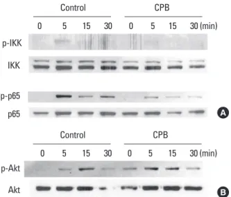
We next assessed the effect of CPB on the stimulation of NF-κB and Akt signaling pathways by RANKL. RANKL weak- ly stimulated the phosphorylation of IKK and CPB abolish- ed the IKK activation (Fig. 5A). RANKL also increased the phosphorylated form of p65 NF-κB. This activation of p65 by RANKL was greatly inhibited by the presence of CPB (Fig.
5A). The activation of Akt by RANKL was also observed in the Western blotting experiments with a phopho-Akt anti- body (Fig. 5B). Unlike IKK and p65, the activation of Akt by RANKL was not suppressed by CPB (Fig. 5B). Taken togeth- Fig. 3. Croton pycnanthus Benth. (CPB) decreased the c-Fos and nu-
clear factor-activated T cells c1 (NFATc1) protein levels. 2×10⁵ bone marrow-derived macrophages (BMM) cells were seeded in 6-well plates and treated with macrophage colony-stimulating factor (M- CSF; 30 ng/mL) and receptor activator of nuclear factor-kappa B (NF- κB) ligand (RANKL; 100 ng/mL) in the absence or presence of CPB (10 μg/mL). Thirty μg of protein samples were loaded to the sodium do- decyl sulfate polyacrylamide gel electrophoresis (SDS-PAGE) gels and transferred to the nitrocellulose membranes. Membranes were blotted with c-Fos, NFATc1 and actin primary antibodies for overnight at 4°C and then with secondary antibodies for 1 hr at room tempera- ture before detection with chemiluminescence reagents. CPB, Croton pycnanthus Benth.; NFATc1, nuclear factor-activated T cells c1.
d0 d1 d2
c-Fos
NFATc1
β-actin
- - CPB - CPB
Fig. 4. Croton pycnanthus Benth. (CPB) suppressed the receptor acti- vator of nuclear factor-kappa B (NF-κB) ligand (RANKL)-induced mito- gen-activated protein kinase (MAPK) activation. 1×10⁶ bone mar- row-derived macrophages (BMM) cells were plated in 60 mm dish.
After 3 hr of starvation, CPB (10 μg/mL) was added. After further in- cubation for another 3 hr, cells were treated with RANKL (500 ng/mL) for 5-30 min and subjected to Western blotting with indicated anti- bodies. CPB, Croton pycnanthus Benth.; ERK, extracellular signal-reg- ulated kinase; JNK, c-Jun-N-terminal kinase.
0 5 15 30 0 5 15 30 (min)
JNK p-JNK p38 p-p38 p-ERK
ERK
Control CPB
Fig. 5. Croton pycnanthus Benth. (CPB) suppressed the activation of nuclear factor-kappa B (NF-κB) pathway, but did not affect the Akt pathway. 1×10⁶ bone marrow-derived macrophages (BMM) cells were plated in 60 mm dish. After 3 hr of starvation, CPB (10 μg/mL) was added. After further incubation for another 3 hr, cells were treat- ed with receptor activator of NF-κB ligand (RANKL; 500 ng/mL) for 5-30 min and subjected to Western blotting analyses. Antibodies against p65, phospho-p65, inhibitor of kappaB kinase (IKK), and phos- pho-IKK were used to examine the NF-kB pathway (A), and antibodies against Akt and phospho-Akt were used to assess the Akt pathway (B). CPB, Croton pycnanthus Benth.; IKK, inhibitor of kappaB kinase.
p65 p-p65 p-IKK
IKK
0 5 15 30 0 5 15 30 (min)
Control CPB
A
Akt p-Akt
0 5 15 30 0 5 15 30 (min)
Control CPB
B
er, the inhibition of osteoclast differentiation by CPB is like- ly to be caused by suppression of the MAPK and NF-κB, but not Akt, signaling pathways of RANK.
DISCUSSION
In this paper, we screened several plant extracts obtain- ed from IBMRC to discover potential therapeutic agents with anti-osteoclastogenic activity. Among them, CPB show- ed inhibitory effects on osteoclast differentiation without cytotoxicity. CPB suppressed the induction of expression of c-Fos and NFATc1 by RANKL at both mRNA and protein levels. CPB also suppressed the RANKL-dependent activa- tion of MAPK and NF-κB, but not Akt, signaling pathways.
The significance of RANK signaling in osteoclast differen- tiation has been well studied. RANK deficient mice showed osteopetrotic phenotype, which resulted from the failure of osteoclast differentiation.[11] RANKL deficient mice are also well characterized by their osteopetrotic morphology.
[12] Therefore, it is important to focus on the regulation of RANKL-induced signaling pathways to control osteoclast differentiation.
RANK signaling promotes osteoclast differentiation throu- gh the MAPK and NF-κB pathways and osteoclast survival through the Akt pathway.[8] ERK that is stimulated by the Raf and MAPK kinase (MEK)1/2 axis eventually activates c- Fos while JNK activates c-Jun and activator protein-1 (AP- 1). c-Fos and AP-1 interact each other and induce the ex- pression of osteoclast marker genes. In case of p38, it acti- vates another transcription factor microphthalmia-associ- ated transcription factor (MITF), which also contributes to the expression of genes associated with osteoclastogene- sis.[13,14] CPB suppressed all three MAPK signaling path- ways (Fig. 4). The importance of NF-κB signaling in osteo- clast differentiation has been well studied. NF-κB p50/p52 double-deficient mice have osteopetrotic phenotype, which caused by defects in osteoclastogenesis.[15] RANKL stimu- lation activates IKK and eventually elevates the transcrip- tion activity of p50, p52, and p65.[16] In our study, CPB ef- fectively reduced the phosphorylation of IKK and p65 NF- κB (Fig. 5A). Therefore, it appears that the reduced osteo- clastogenesis by CPB is achieved by its interference with the activation of NF-κB as well as MAPK signaling pathways by RANKL.
In many studies including our current study, the expres-
sion of c-Fos and NFATc1 was elevated by RANKL both at mRNA and protein levels. While the explanation for the up- regulation of c-Fos mRNA by RANKL is elusive, the RANKL induction of NFATc1 mRNA has been suggested to be me- diated by c-Fos and NF-κB. NFATc1 protein in turn binds the promoter of its own gene and further stimulates NFATc1 mRNA expression.[7] CPB significantly reduced the levels of mRNA and protein of c-Fos and NFATc1, suggesting that CPB controlled the expression of these transcription fac- tors at the transcriptional level. As the activation of NFATc1 transcription factor requires calcium signaling, examining the effect of CPB on calcium oscillation during osteoclast differentiation may be an intriguing future investigation.
Akt has been known for its ability to regulate survival of many types of cells including differentiated osteoclasts.
[17] CPB did not affect to the activation of Akt signaling pathway by RANKL (Fig. 5B). It is consistent with the results of cytotoxicity assay in which CPB did not alter the viability of BMMs. Therefore, the negative regulation of CPB on os- teoclast differentiation is not likely to be caused by decreas- ed proliferation of BMMs or survival of differentiating os- teoclasts.
Multinucleation is a distinct feature of osteoclasts and make mature osteoclasts easily distinguishable from their precursors. The cell fusion process for multinucleation dur- ing osteoclastogenesis has been suggested to be mainly mediated by DC-STAMP.[18] The expression of DC-STAMP mRNA was reduced by CPB in our study (Fig. 2C). DC-STAMP deficient mice showed increased bone mass.[10] In Paget’s disease, which shows increased bone resorption, osteo- clasts have much more nuclei than those of normal ones.
[19] These reports therefore suggest that the extent of mul- tinucleation may be, to a certain level, proportional to the proficiency of osteoclasts and that DC-STAMP level may have a correlation with the bone-resorbing activity of os- teoclasts.
Various types of plant extracts have been developed as medicine. For example, Ayahuasca which grows naturally in Ecuador has been developed to a medicine, Da Vine, for cardiovascular disease.[20] Another example is Maca Lep- idium meyenii from Peru that has antiviral activity.[21] Tur- meric curcuma longa from India, applied as a salve, reduc- es inflammatory response.[22,23] As many government and enterprises have eyes on patents for plant medicine, it may be a useful strategy to screen extracts of plants from a
broad range of countries to find components that have anti- osteolytic or pro-osteoblastic activity.
Further investigations are clearly required to develop CPB as a therapeutic agent. Experiments to validate the in vivo efficacy of CPB as well as identification of the compo- nents in CPB that is responsible for the anti-osteoclasto- genic activity should be performed in future studies.
REFERENCES
1. Lacey DL, Timms E, Tan HL, et al. Osteoprotegerin ligand is a cytokine that regulates osteoclast differentiation and activation. Cell 1998;93:165-76.
2. Fuller K, Wong B, Fox S, et al. TRANCE is necessary and suf- ficient for osteoblast-mediated activation of bone resorp- tion in osteoclasts. J Exp Med 1998;188:997-1001.
3. Miyamoto T, Ohneda O, Arai F, et al. Bifurcation of osteo- clasts and dendritic cells from common progenitors. Blood 2001;98:2544-54.
4. Takayanagi H, Kim S, Koga T, et al. Induction and activation of the transcription factor NFATc1 (NFAT2) integrate RANKL signaling in terminal differentiation of osteoclasts. Dev Cell 2002;3:889-901.
5. Wagner EF, Eferl R. Fos/AP-1 proteins in bone and the im- mune system. Immunol Rev 2005;208:126-40.
6. Aliprantis AO, Ueki Y, Sulyanto R, et al. NFATc1 in mice re- presses osteoprotegerin during osteoclastogenesis and dissociates systemic osteopenia from inflammation in cheru- bism. J Clin Invest 2008;118:3775-89.
7. Asagiri M, Sato K, Usami T, et al. Autoamplification of NFATc1 expression determines its essential role in bone homeo- stasis. J Exp Med 2005;202:1261-9.
8. Lee ZH, Kim HH. Signal transduction by receptor activator of nuclear factor kappa B in osteoclasts. Biochem Biophys Res Commun 2003;305:211-4.
9. Takayanagi H. Osteoimmunology: shared mechanisms and crosstalk between the immune and bone systems.
Nat Rev Immunol 2007;7:292-304.
10. Yagi M, Miyamoto T, Sawatani Y, et al. DC-STAMP is essen- tial for cell-cell fusion in osteoclasts and foreign body gi- ant cells. J Exp Med 2005;202:345-51.
11. Dougall WC, Glaccum M, Charrier K, et al. RANK is essen-
tial for osteoclast and lymph node development. Genes Dev 1999;13:2412-24.
12. Kong YY, Yoshida H, Sarosi I, et al. OPGL is a key regulator of osteoclastogenesis, lymphocyte development and lymph- node organogenesis. Nature 1999;397:315-23.
13. Jimi E, Akiyama S, Tsurukai T, et al. Osteoclast differentia- tion factor acts as a multifunctional regulator in murine osteoclast differentiation and function. J Immunol 1999;
163:434-42.
14. Lee SE, Woo KM, Kim SY, et al. The phosphatidylinositol 3-kinase, p38, and extracellular signal-regulated kinase pathways are involved in osteoclast differentiation. Bone 2002;30:71-7.
15. Iotsova V, Caamaño J, Loy J, et al. Osteopetrosis in mice lacking NF-kappaB1 and NF-kappaB2. Nat Med 1997;3:
1285-9.
16. Wei S, Teitelbaum SL, Wang MW, et al. Receptor activator of nuclear factor-kappa b ligand activates nuclear factor- kappa b in osteoclast precursors. Endocrinology 2001;142:
1290-5.
17. Datta SR, Brunet A, Greenberg ME. Cellular survival: a play in three Akts. Genes Dev 1999;13:2905-27.
18. Zhang C, Dou CE, Xu J, et al. DC-STAMP, the key fusion-me- diating molecule in osteoclastogenesis. J Cell Physiol 2014;
229:1330-5.
19. Roodman GD, Windle JJ. Paget disease of bone. J Clin In- vest 2005;115:200-8.
20. Riba J, Valle M, Urbano G, et al. Human pharmacology of ayahuasca: subjective and cardiovascular effects, mono- amine metabolite excretion, and pharmacokinetics. J Phar- macol Exp Ther 2003;306:73-83.
21. Del Valle Mendoza J, Pumarola T, Gonzales LA, et al. Anti- viral activity of maca (Lepidium meyenii) against human influenza virus. Asian Pac J Trop Med 2014;7S1:S415-20.
22. Arora RB, Kapoor V, Basu N, et al. Anti-inflammatory stud- ies on Curcuma longa (turmeric). Indian J Med Res 1971;
59:1289-95.
23. Manhas A, Khanna V, Prakash P, et al. Curcuma oil reduces endothelial cell-mediated inflammation in postmyocardi- al ischemia/reperfusion in rats. J Cardiovasc Pharmacol 2014;64:228-36.