Copyright © 2019 The Korean Society for Bone and Mineral Research
This is an Open Access article distributed under the terms of the Creative Commons Attribution Non-Commercial Li- cense (http://creativecommons.org/licenses/by-nc/4.0/) which permits unrestricted non-commercial use, distribu- tion, and reproduction in any medium, provided the original work is properly cited.
Extracts of Flavoparmelia sp. Inhibit Receptor Activator of Nuclear Factor-κB Ligand-Mediated Osteoclast Differentiation
Kwang-Jin Kim1, Yongjin Lee1, Min-Hye Jeong2, Jae-Seoun Hur2,*, Young-Jin Son1,*
1Department of Pharmacy, Sunchon National University, Suncheon;
2Korean Lichen Research Institute, Sunchon National University, Suncheon, Korea
Background: Osteoporosis is a geriatric disease with diminished bone density. The in- crease in the number of patients and medical expenses due to a global aging society are recognized as problems. Bone loss is the most common symptom of bone disease, not only osteoporosis but Paget's disease, rheumatoid arthritis, multiple myeloma, and oth- er diseases. The main cause of this symptoms is excessive increase in the number and activity of osteoclasts. Osteoclasts are multinucleated giant cells that can resorb bone.
They are differentiated and activation from monocytes/macrophages in the presence of macrophage colony-stimulating factor and receptor activator of nuclear factor-κB ligand (RANKL). Methods: The effect of extract of Flavoparmelia sp. (EFV), a genus of lichenized fungi within the Parmeliaceae, on the differentiation of bone marrow-derived macro- phages (BMMs) into osteoclasts was examined by phenotype assay and the cell cytotox- icity was evaluated by cell counting kit-8. The osteoclast differentiation-related genes and proteins were investigated by real-time polymerase chain reaction and immunob- lotting. The functional activity of osteoclast in response to EFV treatment was evaluated by an Osteo Assay plate. Results: In this study, we found that EFV, a genus of lichenized fungi within the Parmeliaceae, inhibited osteoclast formation. And we investigated its inhibitory mechanism. EFV reduced RANKL-mediated osteoclast formation and activa- tion by inhibiting expression of nuclear factor of activated T cells 1, a key factor of osteo- clastogenesis. Conclusions: Taken together, our results show that EFV is a promising candidate for health functional foods or therapeutic agents that can help treat bone dis- eases such as osteoporosis.
Key Words: Flavoparmelia · Lichens · NFATC transcription factors · Osteoclasts · Osteopo- rosis
INTRODUCTION
Bones are an organ that changes over time during the process of production, growth, and absorption over a lifetime. Approximately 10% of an adult bone is re- placed with new bone each year.[1] The bone density is highest in people in their 20s to 30s and decreases gradually after that. In women, the bone mineral density decreases rapidly during the first 5 years of menopause.[2] Excessive bone loss is a major cause of osteoporosis, which is becoming a public health problem because of the increased frequency of osteoporotic fractures in elderly people.[3] Osteo- Corresponding author
Jae-Seoun Hur
Korean Lichen Research Institute, Sunchon National University, 255 Jungang-ro, Suncheon 57922, Korea
Tel: +82-61-750-3383 Fax: +82-61-750-3308 E-mail: [email protected] Young-Jin Son
Department of Pharmacy, Sunchon National University, 255 Jungang-ro, Suncheon 57922, Korea
Tel: +82-61-750-3755 Fax: +82-61-750-3028 E-mail: [email protected] Received: April 10, 2019 Revised: May 9, 2019 Accepted: May 21, 2019
* Jae-Seoun Hur and Young-Jin Son contributed equally to this work and should be considered co- corresponding authors.
porosis is an age-related disorder that is characterized by bone loss and deterioration of the bone structure, particu- larly affecting postmenopausal women.[4]
Bone homeostasis/remodeling is important for main- taining the bone mass and quality. This process requires a balanced action of bone-resorbing osteoclasts and bone- forming osteoblasts.[5] Typical bone diseases, such as os- teoporosis, Paget’s disease, and rheumatoid arthritis, have the symptoms of decreased bone mass because of the in- creased number and activity of osteoclasts.[6] Osteoclasts are large multinucleated cells (MNCs) that remove the old/
weakened bones by acid decalcification and proteolytic degradation.[7] They are differentiated from the mono- cyte-macrophage of a hematopoietic lineage. Osteoclast formation is controlled by 2 cytokines, macrophage colo- ny-stimulating factor (M-CSF) and receptor activator of nu- clear factor-κB ligand (RANKL), which are secreted by os- teoblasts/activated T cells.[8] M-CSF acts on osteoclast precursor cells to induce signaling related to cell survival and promotes the expression of RANKL receptor, RANK.[9]
RANKL is also known as the tumor necrosis factor-related activation-induced cytokine or osteoclast-differentiation factor. Binding of RANKL and RANK ultimately leads to the formation of osteoclasts by increasing the expression of nuclear factor of activated T cells 1 (NFATc1), a key transcrip- tion factor for osteoclastogenesis.[10]
Lichen is a symbiotic relationship between fungi (myco- biont) and photosynthetic organisms (photobionts) that use each other's metabolites to generate a range of sec- ondary metabolites.[11,12] Their metabolites have been discovered over the past decade and are being assessed to find new bioactive compounds.[13,14] Recently, various species of lichen were collected and bioactive extracts were obtained. In addition, the extracts of Flavoparmelia sp. (EFV) inhibited osteoclast formation. This study investigated the mechanisms by which EFV inhibits osteoclast formation.
METHODS
1. Preparation of Flavoparmelia sp. extracts
Flavoparmelia sp. was collected from Peru in May of 2017 during a field trip of San Tuario, Muruhuay, Acobamba, Junin organized by Prof. Rebeca Magdalena Pavlich Herrera at Peruvian University Cayetano Heredia, Peru. The field trip was conducted in the frame of an internal joint programbetween Korea and Peru, supported by Korea National Re- search Foundation. The field studies did not involve en- dangered or protected species. Duplicates were deposited at the Korean Lichen and Allied Bioresource Center in the Korean Lichen Research Institute, Sunchon National Uni- versity (SNCU), Korea. The air-dried Flavoparmelia sp. (10 g) were extracted twice with 2 L methanol at room tempera- ture for 48 hr using sonication. The extract was filtered then concentrated under vacuum at 40°C using a rotary evapo- rator. The extract was subjected to high performance liq- uid chromatography (HPLC) analyses (LC-20A; Shimadzu, Kyoto, Japan) on a YMC-PackTM ODS-A (150×3.9 mm I.D.;
YMC, Kyoto, Japan) reverse-phase column containing fully end-cap ped C18 material (particle size, 5 µm; pore size, 12 nm). Elution was performed at a flow rate of 1 mL/min un- der the following conditions before subsequent injection:
column temperature, 40°C; and solvent system, methanol:
water:phosphoric acid (80:20:1, v/v/v). The analyses were monitored using a photodiode array detector (SPD-M20A;
Shimadzu) over the range, 190 to 800 nm, throughout the HPLC run. The observed peaks were scanned between 190 and 400 nm.
2. Cell culture and osteoclast differentiation
This study was conducted in strict accordance with the recommendations contained in the Standard Protocol for Animal Study of SCNU. The protocol was approved by the SCNU Institutional Animal Care and Use Committee (IACUC) with Permit No. SCNU IACUC 2016-06. All efforts were made to minimize suffering.All cells were cultured in a 5% CO2 at 37°C. The culture medium was replaced with fresh medium every 3 days.
Bone marrow cells (BMCs) were isolated from the femurs and tibias of 5-week-old male ICR mice (n=2; RaonBio Inc., Yongin, Korea). The BMCs were incubated with 10 ng/mL M-CSF (PeproTech, Rocky Hill, NJ, USA) for 16 hr in α-mini- mum essential medium (MEM; Thermo Fisher Scientific Inc., Waltham, MA, USA) containing 10% fetal bovine se- rum (FBS; Thermo Fisher Scientific Inc.) and 100 U/mL pen- icillin/streptomycin (10% α-MEM) on a 10 cm culture dish.
The non-adherent cells were cultured with 30 ng/mL M- CSF in 10% α-MEM on a 10 cm Petri dish. After 3 days, the adhered cells were harvested and used as bone marrow- derived macrophages (BMMs). The BMMs were cultured with 10 ng/mL RANKL (R&D Systems, Minneapolis, MN, USA)
and 30 ng/mL M-CSF in 10% α-MEM for 4 days in the pres- ence of the vehicle (0.1% dimethyl sulfoxide [DMSO]) or EFV.
3. TRAP staining
The adherent cells were fixed with 10% formaldehyde for 5 min, permeabilized with 0.1% Triton X-100 for 10 min, and incubated with a tartrate-resistant acid phosphatase (TRAP)-staining solution (Sigma-Aldrich, St. Louis, MO, USA) at room temperature for 10 min. The TRAP-positive cells stained red and stained cells with 3 or more nuclei were counted as mature osteoclasts.
4. Cytotoxicity assay for extracts of
Flavoparmelia sp.BMMs were cultured with 30 ng/mL M-CSF in 10% α-MEM in the presence of the vehicle (0.1% DMSO) or EFV. After 3 days, the cell viability was assessed using a cell counting kit-8 (CCK-8; Dojindo Molecular Technologies, Kumamoto, Japan) according to the manufacturer’s protocols.
5. Real-time polymerase chain reaction (PCR)
Real-time PCR was performed, as described elsewhere.[15] BMMs were cultured with 10 ng/mL RANKL and 30 ng/mL M-CSF in 10% α-MEM for the indicated days in the presence of vehicle (0.1% DMSO) or EFV. The primer sets for real-time PCR were designed (Table 1) using the online primer3 program.[16] The total RNA was obtained using the TRIzol reagent (Thermo Fisher Scientific Inc.) according to the manufacturer's protocol. First-strand cDNA was modi- fied using a moloney murine leukemia virus cDNA Synthe- sis kit (Enzynomics, Daejeon, Korea) according to the man- ufacturer's instructions. Quantitative PCR (qPCR) was per- formed using the TOPreal qPCR 2×PreMIX (Bio-Rad, Her-
cules, CA, USA) in a Real-Time PCR Detection System (Bio- Rad). The relative levels of the tested genes were normal- ized to the level of glyceraldehyde-3-phosphate dehydro- genase and the data were analyzed using the 2−ΔΔCT meth- od.[17]
6. Western blot
Western blotting was performed, as described previous- ly.[18] BMMs were incubated in the same manner as real- time PCR assays. The cells were washed with phosphate- buffered saline and lysed in a lysis buffer (50 mM Tris-HCl, 150 mM NaCl, 5 mM ethylenediaminetetraacetic acid, 1%
Triton X-100, 1 mM sodium fluoride, 1 mM sodium vana- date, and 1% deoxycholate) supplemented with 1 mM phenylmethylsulfonyl fluoride (Bio Basic Inc., Amherst, NY, USA). The lysates were centrifuged at 20,000×g for 13 min at 4°C and the supernatant containing the proteins was collected. The proteins were subjected to 10% sodium do- decyl sulfate-polyacrylamide gel electrophoresis and transferred to a polyvinylidene difluoride membrane (Mil- lipore Corporation, Billerica, MA, USA). The membranes were blocked with 5% skim milk for 1 hr at room tempera- ture and incubated overnight at 4°C with the primary anti- body. They were then incubated with the secondary anti- body conjugated to horseradish peroxidase for 2 hr at room temperature. The membranes were developed with CLAROTM Mucho (Bio-D, Gwangmyeong, Korea) using a LAS-4000 lu- minescent image analyzer (Fuji Photo Film Co. Ltd., Tokyo, Japan).
7. Bone pit formation assay
A bone pit formation assay was performed, as described elsewhere.[19] BMMs were seeded on an Osteo Assay plate (24 well plate; Corning, Tewksbury, MA, USA) at a density of 3×10⁵ cells/well and cultured with 10 ng/mL RANKL and 30 ng/mL M-CSF in the presence of vehicle (0.1% DMSO) or EFV. After 4 days, the cells were removed completely with 5% sodium hypochlorite for 5 min, and the pit area was then observed by optical microscopy (magnification,
×50; Leica Microsystems, Wetzlar, Germany) and measured by ImageJ software (National Institutes of Health, Bethes- da, MD, USA).
8. Statistical analysis
All quantitative data are presented as the means±stan- Table 1. Primer sequences used in this study
Gene of interest
Primer sequence (5’→3’)
Sense Anti-sense
NFATc1 GGGTCAGTGTGACCGAAGAT GGAAGTCAGAAGTGGGTGGA Cathepsin K GGCCAACTCAAGAAGAAAAC GTGCTTGCTTCCCTTCTGG DC-STAMP CCAAGGAGTCGTCCATGATT GGCTGCTTTGATCGTTTCTC TRAP GATGACTTTGCCAGTCAGCA ACATAGCCCACACCGTTCTC GAPDH AACTTTGGCATTGTGGAAGG ACACATTGGGGGTAGGAACA NFATc1, nuclear factor of activated T cells 1; DC-STAMP, dendritic cell- specific transmembrane protein; TRAP, tartrate-resistant acid phospha- tase; GAPDH, glyceraldehyde-3-phosphate dehydrogenase.
dard deviation of 3 replicate experiments. Statistical differ- ences were analyzed using Student’s t-tests. Probability P<
0.05 were considered significant (P-values *<0.05, **<0.01, and ***<0.001).
RESULTS
1. The methanol extract of Flavoparmelia sp.
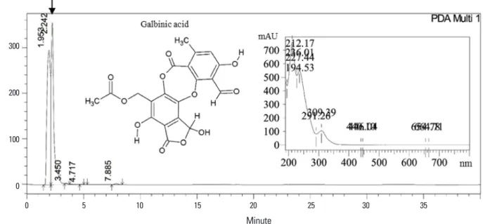
HPLC analysis of the methanol extract of Flavoparmelia sp. showed that galbinic acid was the main component of the extract (Fig. 1). The retention time (Rt=2.242) and the maximum absorption wavelengths (λmax) of the UV-spec- tra (the insert in Fig. 1) of the peak confirmed that the com- pound was well matched with galbinic acid.
2. EFV inhibits RANKL-mediated osteoclastogenesis
The potential role of EFV in osteoclastogenesis was eval- uated by examining the effects of EFV on the ability of RANKL to differentiate BMMs. BMMs were cultured with 0.1% DMSO (vehicle) or EFV (0, 1, 3, and 10 μg/mL) for 4 days in the presence of 10 ng/mL RANKL and 30 ng/mL M- CSF. Red stained TRAP-positive cells were induced by RANKL, but EFV reduced this induction (Fig. 2A). Moreover, EFV in-
hibited the number of TRAP-positive MNCs (nuclei ≥ 3) in a dose-dependent manner and the formation of MNCs was inhibited almost completely at 10 μg/mL (Fig. 2B).
3. EFV had no cytotoxic effect on BMMs
To determine if the anti-osteoclastogenesis efficacy of EFV is due to cytotoxicity, cell viability analysis was per- formed with a CCK-8 in BMMs. The BMMs were incubated with 0.1% DMSO (vehicle) or EFV (0, 1, 3, and 10 μg/mL) for 3 days in the presence of 30 ng/mL M-CSF. EFV had no cy- totoxic effects on the BMMs at the indicated concentration in this study (Fig. 2C).
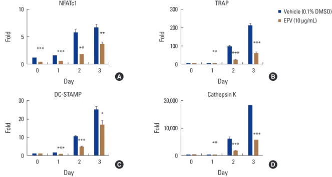
4. Effects of EFV on RANKL-induced gene expression
The effects of EFV on gene expression of NFATc1, a major transcriptional factor of osteoclast differentiation, were ex- amined. Real-time PCR showed that RANKL gradually in- creased the mRNA level of NFATc1, but EFV decreased the transcriptional level of NFATc1 significantly (Fig. 3A). More- over, EFV also reduced the transcriptional levels of TRAP, dendritic cell-specific transmembrane protein (DC-STAMP), and cathepsin K, the osteoclast differentiation marker genes regulated by NFATc1 (Fig. 3B-D).
Fig. 1. High performance liquid chromatography (HPLC) profiling of methanol extract from Flavoparmelia sp. methanol extract of Flavoparmelia sp.
was analyzed by HPLC with YMC-Pack ODS-A reversed-phase C18 column. The ultraviolet–visible-spectra of the maximum absorption wave- lengths (λmax) of galbinic acid are present in the insert. By comparing both the retention time and λmax with the standard, the main compound of the extract was identified to be galbinic acid. The molecular structure of galbinic acid is also presented in the inset.
300
200
100
0
mAU
0 5 10 15 20 25 30 35 Minute
Fig. 2. The extracts of Flavoparmelia sp. (EFV) inhibited osteoclast differentiation. (A) Bone marrow-derived macrophages (BMMs) were cultured with 10 ng/mL receptor activator of nuclear factor-κB ligand (RANKL) and 30 ng/mL macrophage colony-stimulating factor (M-CSF) for 4 days in the presence of the vehicle (0.1% dimethyl sulfoxide [DMSO]) or the indicated concentrations of EFV. The cells were fixed in 3.7% formalin, per- meabilized with 0.1% Triton X-100, and stained with tartrate-resistant acid phosphatase (TRAP) solution. (B) TRAP-positive multinucleated cells (3 or more nuclei) were counted as osteoclasts. (C) BMMs were cultured with 30 ng/mL M-CSF for 3 days in the presence of the vehicle (0.1% DMSO) or the indicated concentrations of EFV. The effects of EFV on the BMMs viability were assessed using a cell counting kit-8. *P<0.001 (n=3).
EFV (μg/mL)
RANKL (10 ng/mL)
0 0 1 3 10
- + + + + A
600
400
200
0 TRAP positive MNCs/well (n≥3)
0 1 3 10
EFV (μg/mL)
*
*
200 150 100 50 0
Survival (%)
0 1 3 10
EFV (μg/mL)
B C
Fig. 3. Effects of the extracts of Flavoparmelia sp. (EFV) on receptor activator of nuclear factor-κB ligand (RANKL)-mediated mRNA expressions of nuclear factor of activated T cells 1 (NFATc1). The bone marrow-derived macrophages were treated with the vehicle (0.1% DW) or EFV (10 μg/mL) and macrophage colony-stimulating factor (30 ng/mL) for 1 hr and then RANKL (10 ng/mL) were treated at the indicated times. The total RNA was then isolated using the TRIzol reagent, and the mRNA expression levels were evaluated by performing real-time polymerase chain reaction.
(A) NFATc1, (B) dendritic cell-specific transmembrane protein (DC-STAMP), (C) tartrate-resistant acid phosphatase (TRAP), and (D) cathepsin K.
Glyceraldehyde-3-phosphate dehydrogenase was used as the internal control. *P<0.05. **P<0.01. ***P<0.001.
10
5
0
Fold
0 1 2 3 Day
*** *** **
**
NFATc1
300
200
100
0
Fold
0 1 2 3 Day
** ***
***
TRAP
30
20
10
0
Fold
0 1 2 3 Day
***
***
* DC-STAMP
20,000
10,000
0
Fold
0 1 2 3 Day
** ***
***
Cathepsin K
Vehicle (0.1% DMSO) EFV (10 μg/mL)
A B
C D
5. EFV inhibited RANKL-induced protein expression of NFATc1
In the previous experiment, EFV reduced the mRNA ex- pression of NFATc1. Hence, this study examined whether EFV affected the protein level of NFATc1 by western blot- ting. The protein level of NFATc1 was increased significantly by RANKL but was reduced dramatically by EFV (Fig. 4). This suggests that EFV inhibits the translational expression of NFATc1 and suppresses osteoclast formation.
6. Effects of EFV on RANKL-mediated bone resorptive activity of osteoclasts
Experiments were conducted to determine if the osteo- clast formation inhibited by EFV also affects bone resorp- tion. The osteoclasts formed wide pit areas on the bone slice, but EFV reduced the pit areas markedly (Fig. 5).
DISCUSSION
Lichens are a complex organism, in which algae or cya- nobacteria (photobionts) live among the filaments of vari- ous fungi (mycobiont).[11] In addition, a lichen thallus of- ten contains diverse assemblages of microfungi and mi- croorganisms.[20] The combination of these various organ- isms allows the lichen to generate and use various second- ary metabolites,[12] such as alkaloids,[21] peptides,[22]
and terpenes.[23,24] Their metabolites have been studied to discover new bioactive compounds [25,26] and their bi-
ological activity is also being revealed.[27-29]
Osteoporosis is a representative geriatric disease that is becoming a major concern because of the aging popula- tion. The disease results in an increased risk of bone frac- tures by lowering the strength of the bone because the number and activity of osteoclasts are increased due to a range of causes.[30] In addition, investigations have shown that the number of fracture patients due to osteoporosis and the cost of treatment are increasing.[31-33] Therefore, it is necessary to study various means of preventing and treating bone diseases, such as osteoporosis. Osteoclasts are MNCs arising from hematopoietic stem cells/macro- phage lineage. Their differentiation and function is con- trolled by M-CSF and RANKL, which are produced in ma- Fig. 4. The extracts of Flavoparmelia sp. (EFV) abolishes receptor ac-
tivator of nuclear factor-κB ligand (RANKL)-mediated protein expres- sion of nuclear factor of activated T cells 1 (NFATc1). Bone marrow- derived macrophages were pretreated with the vehicle (0.1% dimeth- yl sulfoxide) or EFV (10 μg/mL) and macrophage colony-stimulating factor (30 ng/mL) for 1 hr prior to RANKL (10 ng/mL) stimulation for the indicated times. The cell lysates were resolved by sodium dodec- yl sulfate-polyacrylamide gel electrophoresis, and Western blotting was performed with anti-NFATc1, and anti-actin antibodies as indi- cated.
EFV (10 μg/mL)
RANKL 0 1 2 3 0 1 2 3 Day
NFATc1
Actin
Fig. 5. The extracts of Flavoparmelia sp. (EFV) inhibited bone resorp- tion by receptor activator of nuclear factor-κB ligand (RANKL)-induced osteoclasts. (A) Bone marrow-derived macrophages were plated on an Osteo Assay Plate and treated with 30 ng/mL macrophage colony- stimulating factor and 10 ng/mL RANKL in the presence of 10 μg/mL of EFV. After 4 days of culture, the cells attached to the Osteo Assay plate were removed and photographed under an optical microscope.
(B) Pit areas were quantified using the ImageJ program. *P<0.001 (n=3).
EFV (μg/mL)
RANKL (10 ng/mL)
0 10
+ +
A
100 80 60 40 20 0
Pit area (%)
0 10
EFV (μg/mL)
*
B
ture osteoblasts and stromal cells.[7,34] M-CSF is involved in the growth, survival, proliferation, and differentiation of both hematopoietic and non-hematopoietic cells.[35,36]
In particular, it promotes the expression of the RANKL re- ceptor, RANK, in osteoclastic progenitor cells.[37] RANKL is the most important cytokine during the osteoclast differ- entiation process.[10,37] The RANKL-RANK signaling path- way activates NFATc1. As a result, the osteoclast precursor is differentiated into TRAP-positive MNCs (mature osteo- clast).
In vitro screening experiments with the extract of various lichens extracts on RANKL-mediated osteoclast differentia- tion were performed prior to this study. The results showed that EFV inhibited the osteoclast differentiation. Subse- quently, this study examined the inhibitive mechanism of EFV against osteoclast formation. First, EFV was treated at concentrations of 0, 1, 3, and 10 μg/mL during osteoclast differentiation to select the optimal concentration of EFV in this study. EFV significantly reduced osteoclast forma- tion at concentrations ≥3 μg/mL and did not exhibit cyto- toxicity even at concentrations of 10 μg/mL. The cells were treated with EFV at a concentration of 10 μg/mL, which showed optimal efficacy in the experiment, and then ex- amined by real-time PCR and western blotting to identify the anti-osteoclastogenic mechanism.
NFATc1 is a master transcription factor on osteoclasto- genesis and is activated by the RANKL signaling pathway.
In this study, EFV inhibited RANKL-mediated mRNA and the protein expression level of NFATc1. In addition, EFV re- duced the levels of TRAP, DC-STAMP, and cathepsin K ex- pression, which are osteoclast formation and activation-re- lated molecules, by decreasing NFATc1.[37-41] And we confirmed that RANKL-induced bone resorption was de- creased by EFV in vitro. This means that EFV inhibits and reduces the formation and activity of osteoclasts. Overall, these results suggest that EFV had a substance that affect- ed osteoclast differentiation and function, and this sub- stance reduced the expression of NFATc1, a key molecule during osteoclastogenesis.
DECLARATIONS Acknowledgments
This research was supported by the R&D Program for Forest Science Technology (2017024A00-1919-BA01) pro-
vided by Korea Forest Service (Korea Forestry Promotion Institute), Sunchon National University Research (2018- 0190), and the National Research Foundation of Korea (NRF- 2016K1A3A1A12953760).
Ethics approval and consent to participate
Not applicable.Conflict of interest
No potential conflict of interest relevant to this article was reported. The founding sponsors had no role in the design of the study; in the collection, analyses, or interpre- tation of data; in the writing of the manuscript, and in the decision to publish the results.
ORCID
Jae-Seoun Hur http://orcid.org/0000-0001-8547-7075 Young-Jin Son http://orcid.org/0000-0002-6873-9181
REFERENCES
1. Manolagas SC. Birth and death of bone cells: basic regula- tory mechanisms and implications for the pathogenesis and treatment of osteoporosis. Endocr Rev 2000;21:115- 37.
2. Finkelstein JS, Brockwell SE, Mehta V, et al. Bone mineral density changes during the menopause transition in a multiethnic cohort of women. J Clin Endocrinol Metab 2008;93:861-8.
3. Kanis JA, Johnell O, Oden A, et al. Ten year probabilities of osteoporotic fractures according to BMD and diagnostic thresholds. Osteoporos Int 2001;12:989-95.
4. Tella SH, Gallagher JC. Prevention and treatment of post- menopausal osteoporosis. J Steroid Biochem Mol Biol 2014;
142:155-70.
5. Riggs BL, Parfitt AM. Drugs used to treat osteoporosis: the critical need for a uniform nomenclature based on their action on bone remodeling. J Bone Miner Res 2005;20:
177-84.
6. Rodan GA, Martin TJ. Therapeutic approaches to bone dis- eases. Science 2000;289:1508-14.
7. Boyle WJ, Simonet WS, Lacey DL. Osteoclast differentia- tion and activation. Nature 2003;423:337-42.
8. Feng X. RANKing intracellular signaling in osteoclasts.
IUBMB Life 2005;57:389-95.
9. Kansara M, Teng MW, Smyth MJ, et al. Translational biolo- gy of osteosarcoma. Nat Rev Cancer 2014;14:722-35.
10. Takayanagi H, Kim S, Koga T, et al. Induction and activa- tion of the transcription factor NFATc1 (NFAT2) integrate RANKL signaling in terminal differentiation of osteoclasts.
Dev Cell 2002;3:889-901.
11. Spribille T, Tuovinen V, Resl P, et al. Basidiomycete yeasts in the cortex of ascomycete macrolichens. Science 2016;353:
488-92.
12. Kellogg JJ, Raja HA. Endolichenic fungi: a new source of rich bioactive secondary metabolites on the horizon. Phy- tochem Rev 2017;16:271-93.
13. Chen GD, Chen Y, Gao H, et al. Xanthoquinodins from the endolichenic fungal strain Chaetomium elatum. J Nat Prod 2013;76:702-9.
14. Li XB, Zhou YH, Zhu RX, et al. Identification and biological evaluation of secondary metabolites from the endolichen- ic fungus Aspergillus versicolor. Chem Biodivers 2015;12:
575-92.
15. Kim KJ, Yeon JT, Choi SW, et al. Decursin inhibits osteoclas- togenesis by downregulating NFATc1 and blocking fusion of pre-osteoclasts. Bone 2015;81:208-16.
16. Rozen S, Skaletsky H. Primer3 on the WWW for general users and for biologist programmers. Methods Mol Biol 2000;132:365-86.
17. Livak KJ, Schmittgen TD. Analysis of relative gene expres- sion data using real-time quantitative PCR and the 2(-Del- ta Delta C(T)) Method. Methods 2001;25:402-8.
18. Kim H, Kim KJ, Yeon JT, et al. Placotylene A, an inhibitor of the receptor activator of nuclear factor-κB ligand-induced osteoclast differentiation, from a Korean sponge Placo- spongia sp. Mar Drugs 2014;12:2054-65.
19. Kim KJ, Lee Y, Hwang HG, et al. Betulin suppresses osteo- clast formation via down-regulating NFATc1. J Clin Med 2018;7.
20. Girlanda M, Isocrono D, Bianco C, et al. Two foliose lichens as microfungal ecological niches. Mycologia 1997;89:531-6.
21. Li XB, Li L, Zhu RX, et al. Tetramic acids and pyridone alka- loids from the endolichenic fungus tolypocladium cylin- drosporum. J Nat Prod 2015;78:2155-60.
22. Wu W, Dai H, Bao L, et al. Isolation and structural elucida- tion of proline-containing cyclopentapeptides from an endolichenic Xylaria sp. J Nat Prod 2011;74:1303-8.
23. Wijeratne EM, Bashyal BP, Liu MX, et al. Geopyxins A-E, ent-kaurane diterpenoids from endolichenic fungal strains
Geopyxis aff. majalis and Geopyxis sp. AZ0066: structure- activity relationships of geopyxins and their analogues. J Nat Prod 2012;75:361-9.
24. Wu YH, Chen GD, Wang CX, et al. Pericoterpenoid A, a new bioactive cadinane-type sesquiterpene from Periconia sp.
J Asian Nat Prod Res 2015;17:671-5.
25. Frisvad JC, Andersen B, Thrane U. The use of secondary metabolite profiling in chemotaxonomy of filamentous fungi. Mycol Res 2008;112:231-40.
26. Parrot D, Jan S, Baert N, et al. Comparative metabolite pro- filing and chemical study of Ramalina siliquosa complex using LC-ESI-MS/MS approach. Phytochemistry 2013;89:
114-24.
27. Kim GS, Ko W, Kim JW, et al. Bioactive α-pyrone derivatives from the endolichenic fungus dothideomycetes sp. EL00- 3334. J Nat Prod 2018;81:1084-8.
28. Kim JW, Ko W, Kim E, et al. Anti-inflammatory phomaliche- nones from an endolichenic fungus Phoma sp. J Antibiot (Tokyo) 2018;71:753-6.
29. Kim KJ, Jeong MH, Lee Y, et al. Effect of usnic acid on os- teoclastogenic activity. J Clin Med 2018;7.
30. Golob AL, Laya MB. Osteoporosis: screening, prevention, and management. Med Clin North Am 2015;99:587-606.
31. Blume SW, Curtis JR. Medical costs of osteoporosis in the elderly Medicare population. Osteoporos Int 2011;22:1835- 44.
32. Svedbom A, Hernlund E, Ivergård M, et al. Osteoporosis in the European Union: a compendium of country-specific reports. Arch Osteoporos 2013;8:137.
33. Wade SW, Strader C, Fitzpatrick LA, et al. Estimating preva- lence of osteoporosis: examples from industrialized coun- tries. Arch Osteoporos 2014;9:182.
34. Yasuda H, Shima N, Nakagawa N, et al. A novel molecular mechanism modulating osteoclast differentiation and function. Bone 1999;25:109-13.
35. Sherr CJ. Colony-stimulating factor-1 receptor. Blood 1990;
75:1-12.
36. Tanaka S, Takahashi N, Udagawa N, et al. Macrophage col- ony-stimulating factor is indispensable for both prolifera- tion and differentiation of osteoclast progenitors. J Clin Invest 1993;91:257-63.
37. Takayanagi H. Osteoimmunology: shared mechanisms and crosstalk between the immune and bone systems.
Nat Rev Immunol 2007;7:292-304.
38. Minkin C. Bone acid phosphatase: tartrate-resistant acid
phosphatase as a marker of osteoclast function. Calcif Tis- sue Int 1982;34:285-90.
39. Kim B, Kim HH, Lee ZH. α-Tocopheryl succinate inhibits osteolytic bone metastasis of breast cancer by suppress- ing migration of cancer cells and receptor activator of nu- clear factor-κB ligand expression of osteoblasts. J Bone Metab 2018;25:23-33.
40. Yagi M, Miyamoto T, Toyama Y, et al. Role of DC-STAMP in cellular fusion of osteoclasts and macrophage giant cells.
J Bone Miner Metab 2006;24:355-8.
41. Matsumoto M, Kogawa M, Wada S, et al. Essential role of p38 mitogen-activated protein kinase in cathepsin K gene expression during osteoclastogenesis through association of NFATc1 and PU.1. J Biol Chem 2004;279:45969-79.