Copyright © 2020 The Korean Society for Bone and Mineral Research
This is an Open Access article distributed under the terms of the Creative Commons Attribution Non-Commercial Li- cense (https://creativecommons.org/licenses/by-nc/4.0/) which permits unrestricted non-commercial use, distribu- tion, and reproduction in any medium, provided the original work is properly cited.
Inhibitory Effect of Rosae Multiflorae Fructus
Extracts on the Receptor Activator of NF-κB Ligand- Induced Osteoclastogenesis through Modulation of P38- and Ca 2+ -Mediated Nuclear Factor of Activated T-Cells Cytoplasmic 1 Expression
Keun Ha Park1, Dong Ryun Gu1, Min Seuk Kim2,3, Seoung Hoon Lee1,3
1Department of Oral Microbiology and Immunology, College of Dentistry, Wonkwang University, Iksan;
2Department of Oral Physiology, College of Dentistry, Wonkwang University, Iksan;
3Institute of Biomaterials and Implant, College of Dentistry, Wonkwang University, Iksan, Korea
Background: Rosae Multiflorae fructus (RMF), known to have anti-inflammatory and an- tioxidant properties, has been used as a traditional remedy for inflammatory diseases such as arthritis in Eastern Asia. However, its effect on osteoclasts, which play a crucial role in resorptive inflammatory bone diseases, is yet to be elucidated. Methods: The ef- fect of extract of RMF (RMF-E) on receptor activator of nuclear factor-κB ligand (RANKL)- mediated osteoclastogenesis was examined by tartrate-resistant acid phosphatase (TRAP) staining, real-time polymerase chain reaction and western blot analysis. In addition, RA- NKL-induced Ca2+-oscillation was also investigated. Results: RMF-E remarkably inhibited TRAP+-osteoclast and resorptive pit formation in a dose-dependent manner. In addition, the expression of c-Fos and nuclear factor of activated T-cells cytoplasmic, known as piv- otal transcription factors for osteoclast formation in vitro and in vivo, and that of the os- teoclast differentiation markers such as Acp5, Oscar, CtsK, Atp6v0d2, Tm7sf4, and Nfatc1 were significantly decreased by RMF-E treatment during osteoclastogenesis. The inhibi- tory effect of RMF-E on RANKL-induced osteoclastogenesis was caused by the suppres- sion of p38 mitogen-activated protein kinase activation, and RANKL-induced Ca2+-oscil- lation removal via inactivation of Bruton's tyrosine kinase (BTK), and subsequently phos- pholipase C-γ2. Conclusions: RMF-E negatively regulates osteoclast differentiation and formation. These findings suggest the possibility of RMF-E as a traditional therapeutic agent against osteoclast-related bone disorders such as osteoporosis, rheumatoid ar- thritis, and periodontitis.
Key Words: Osteoclasts · Bone diseases · NFATc1 · Calcium signaling · Osteogenesis
INTRODUCTION
Osteoclasts, which work to resorb the bone matrix, are responsible for main- taining bone homeostasis, and remodeling with osteoblasts, which make bone matrix. Osteoclasts originate from the monocyte/macrophage lineage, and are differentiated by the stimulation of the indispensable osteoclast differentiation Corresponding author
Seoung Hoon Lee
Department of Oral Microbiology and Immunology, College of Dentistry, Wonkwang University, 460 Iksandae-ro, Iksan 54538, Korea
Tel: +82-63-850-6981 Fax: +82-63-850-7313 E-mail: [email protected] Received: January 21, 2020 Revised: January 31, 2020 Accepted: February 5, 2020
Original Article
J Bone Metab 2020;27(1):53-63 https://doi.org/10.11005/jbm.2020.27.1.53 pISSN 2287-6375 eISSN 2287-7029
Keun Ha Park, et al.
cytokine receptor activator of nuclear factor-κB ligand (RANKL).[1] Complicated and delicate intracellular signal- ing pathways induced by the interaction of RANKL and its receptor molecule; receptor activator of nuclear factor-κB (RANK), have been identified.[2,3] RANKL to RANK interac- tion activates nuclear factor-κB (NF-κB) and mitogen-acti- vated protein kinases (MAPK; extracellular signal-regulated kinase [ERK], c-JUN N-terminal kinase [JNK], and p38), thr- ough tumor necrosis factor receptor-associated factor 6- dependent signaling, followed by c-Fos upregulation. In another pathway known as co-stimulatory signaling, RANKL/
RANK binding triggers the activation of immunoreceptor tyrosine-based activation motif (ITAM)-harboring adap- tors, Fc receptor common γ subunit (FcRγ), or DNAX-acti- vating protein of kDa 12 (DAP12), subsequently inducing the activation of Bruton’s tyrosine kinase (BTK) and phospho- lipase C-γ2 (PLCγ2).[2,4-6] PLCγ2 activation produces inosi- tol triphosphate (IP3) which binds to IP3 receptors, from phosphatidylinositol-4,5-bisphosphate, followed by gen- erating Ca2+-oscillation which is important for osteoclast differentiation, via Ca2+ release from the endoplasmic re- ticulum.[4,7,8] Both signaling pathways consequently in- duce the expression and activation of nuclear factor-acti- vated T cells c1 (NFATc1), known to be the most important transcription factor for osteoclast differentiation, activa- tion, and survival, both in vitro and in vivo.[9]
Rosae Multiflorae fructus (RMF) is the dried fruit of Rosa multiflora Thunberg (‘Youngsil’ in Korean, ‘Yingshi’ in Chi- nese, and ‘Eijitsu’ in Japanese), known as Multiflora rose;
originated from Korea, China, and Japan, and is known to have potential anti-inflammatory, pain-relief, and antioxi- dant properties.[10-14] RMF shows no critical toxicity, and has been used as tea, jam, and juice.[10,15] As a herbal remedy, RMF has been used traditionally for various dis- eases, including inflammatory disorders, cold, flu, edema, beriberi, and chronic pain.[12,13,16] Recently, in addition to its anti-inflammatory and analgesic effects in rodent models,[17] several reports indicate that RMF exerts its ef- fects on allergic inflammatory diseases like asthma, food allergy accompanying anaphylaxis, and allergic rhinitis (AR), via functional modulation of Th2 and mast cells, in- cluding Th2 cytokine production and histamine release, respectively.[12,18] Additionally, previous reports have shown the obvious inhibitory effects of herbal formula (RL) containing RMF and Lonicerae Japonica Flos, on Toll-like re-
ceptor (TLR) signaling and collagen-induced arthritis (CIA).
RL reportedly suppresses IκB-α and MAPK activation by in- hibiting the interleukin (IL)-1 receptor associated kinase/
transforming growth factor-β activated kinase 1 and TBK1/
interferon regulatory factor 3 (IRF3) pathways, resulting in inhibitory modulation of transcriptional factors such as ac- tivator protein-1 (AP-1), NF-κB, and IRF3, and reduction of various pro-inflammatory cytokine and chemokine pro- duction in lipopolysaccharide-stimulated RAW264.7 and THP-1 cells.[10,19] Moreover, RL also reportedly inhibited TLR-4 signaling, and significantly improved clinical symp- toms of CIA rats, including the suppression of bone erosion and osteophyte formation in joints.[11]
Although several reports on its anti-inflammatory effects exist, the specific effects of RMF on osteoclastogenesis re- main unknown. Here, we provide biological data regarding the inhibitory effects of RMF extract (RMF-E), via modula- tion of intracellular p38- and Ca2+-signaling, on RANKL-in- duced osteoclastogenesis.
METHODS
1. Experimental animals and reagents
The C57BL/6N mice used in this study were purchased from Orient Bio Inc. (Seongnam, Korea). All mouse studies were performed following the protocol (WKU16-87) ap- proved by the Animal Care and Use Committee of Wonk- wang University.
Cell culture agents, including media, fetal bovine serum (FBS), and supplements, were purchased from Hyclone (Pier- ce, Rockford, IL, USA). Soluble recombinant mouse RANKL and recombinant human macrophage colony-stimulating factor (M-CSF) were supplied by T Kim (KIOM, Daejeon, Ko- rea). Anti-NFATc1 and anti-c-Fos antibodies were purchased from Santa Cruz Biotechnology (Santa Cruz, CA, USA) and anti-actin antibody from Sigma-Aldrich (St. Louis, MO, USA).
Antibodies against other proteins used in this study (phos- pho-ERK [p-ERK; Thr202/Tyr204], ERK, phospho-JNK [Thr183/
Tyr185], JNK, p-p38 [Thr180/Tyr182], p-38, phospho-IκBα [Ser32], IκBα, phospho-BTK [Ser180], BTK, phospho-PLCγ2 [Tyr759], and PLCγ2) were purchased from Cell Signaling Technology (Danvers, MA, USA).
2. Preparation of ethanol RMF-E
RMF was purchased in May 2012 from the University
Inhibitory Effect of RMF on RANKL-Induced Osteoclatogenesis
Oriental Herbal Drugstore (Iksan, Korea). A voucher speci- men (No. NNMBS-2012-046) was deposited at the Herbari- um of the College of Pharmacy, Wonkwang University (Ik- san, Korea). Dried and pulverized RMF (50 g) was extracted with hot 70% aqueous ethanol (EtOH) for 2 hr, and filtered with filter paper. The filtrate was evaporated in vacuo, to produce a 70% EtOH extract (16.28 g). The extract was then suspended with distilled water (100 mL), followed by filtra- tion, to obtain the precipitate (1.13 g; NNMBS-2012-046).
Finally, RMF-E was dissolved in dimethyl sulfoxide and used in all experiments.
3. Cell viability assay
The EZ-Cytox enhanced cell viability assay kit (ItsBio, Seoul, Korea) was purchased for performing the cell viability as- say, following the manufacturer's instructions. Briefly, bone marrow (BM) cells from the tibiae and femora of 6 to 8 week- old C57BL/6N mice were cultured in α-minimal essential medium supplemented with 10% FBS and M-CSF (30 ng/
mL) for 3 days. The adherent cells, considered as BM-de- rived macrophage (BMMs), were plated in 96-well culture plates at a density of 1×10⁴ cells/well, with various RMF-E (0, 5, 10, 20, 30, and 40 μg/mL) concentrations, and cul- tured for 1 day with M-CSF (30 ng/mL), or 4 days with M- CSF under 30 μg/mL RMF-E treatment. The EZ-Cytox re- agent was added to cultured cells for 4 hr at 37°C, follow- ing the manufacturer's instructions. OD was measured at 450 nm, using a Sunrise™ enzyme-linked immunoassay (ELISA) plate reader (Tecan, Crailsheim, Germany).
4. Assay of in vitro osteoclast differentiation, actin ring, and pit formation
BMM preparation and osteoclast formation were done as previously described.[20] To assess osteoclastogenesis, BMMs were cultured in the presence of M-CSF (30 ng/mL) and RANKL (100 ng/mL), at various concentrations of RMF- E (0, 2, 5, 10, 20, and 30 μg/mL), for 4 days. Media contain- ing M-CSF, RANKL, and RME-F was replaced on day 3. Cells were fixed with 10% formalin, and permeabilized with Et- OH/Acetone (1:1). Cells were then stained with rhodamine- phalloidin from Molecular Probes (Eugene, OR, USA), to la- bel the F-actin ring, sequentially followed by tartrate-resis- tant acid phosphatase (TRAP) solution assay and TRAP stain- ing, as previously described.[21] The F-actin ring was mea- sured under a fluorescent inverted microscope from Leica
Microsystems Ltd. (Wetzlar, Germany). Total TRAP activity using p-nitrophenyl phosphate from Sigma-Aldrich, as sub- strate, was measured at an absorbance of 405 nm, and TRAP- positive multinuclear cells (TRAP+-MNCs) containing ≥3 nuclei were counted as osteoclasts. To measure bone re- sorption activity, BMMs were cultured in a Cosmo Bio 48- well bone resorption assay plate (Oriental Yeast Co., Ltd., Tokyo, Japan) under M-CSF and RANKL treatment with/
without RMF-E (20 μg/mL) for 7 days, and then cleared us- ing bleach. The resorption pit area was analyzed using Im- ageJ software (National Institutes of Health, Bethesda, MD, USA; NIH 1.52).
5. Real-time quantitative polymerase chain reaction
BMMs were cultured under M-CSF (30 ng/mL) and RANKL (100 ng/mL) for 4 days with/without RMF-E (20 μg/mL).
One microgram of total RNA, extracted using the Trizol re- agent from Invitrogen (Carlsbad, CA, USA) at the indicated time points, was transcribed to first strand cDNA with ran- dom primers using Maxima reverse transcriptase from Ther- mo Scientific (Seoul, Korea), following the manufacturer’s direction. Real-time polymerase chain reaction (PCR) was performed using the VeriQuest SYBR Green qPCR Master mix from Affymetrix (Santa Clara, CA, USA), and StepOneP- lus™ Real-Time PCR Systems from Applied Biosystems (For- ster City, CA, USA). All results were normalized to glyceral- dehyde 3-phosphate dehydrogenase (Gapdh). The primers used in this study were described in Table 1.
6. Western blot analyses
BMMs were cultured with M-CSF (30 ng/mL) and RANKL (100 ng/mL) in the presence or absence of RMF-E (20 μg/
mL), for specific time points. In some experiments, RMF-E (20 μg/mL) was pretreated in BMMs supplemented with M-CSF (30 ng/mL) for 2 hr, and the cells stimulated with RANKL (100 ng/mL). To prepare protein lysate, the cells washed with cold phosphate buffered saline were lysed in radioimmunoprecipitation assay buffer (25 mM Tris-HCl, pH 7.6, 150 mM NaCl, 1% NP-40, 1% sodium deoxycholate, and 0.1% sodium dodecyl sulfate [SDS]) containing 1 mM phenylmethylsulfonyl fluoride, protease-inhibitor cocktail from Roche (Mannheim, Germany), and phosphatase in- hibitor tablets from Thermo Scientific, and then the super- natants were collected by centrifugation at 14,000×g for
Keun Ha Park, et al.
10 min at 4°C. Thirty micrograms of total lysate were sub- jected to SDS-polyacrylamide gel electrophoresis (PAGE), and then transferred to Amersham Hybond-P PVDF mem- branes from GE-Healthcare Life Science (Amersham, UK).
Western blot analyses were sequentially performed with 1:1,000 dilutions of the primary antibody and 1:5,000 dilu- tions of HRP-conjugated IgG, as secondary antibody in TBST (TBS; 50 mM Tris-HCl, pH 7.6, 150 mM NaCl, and 0.1% Tween- 20). The enhanced chemiluminescence detection system from Thermo Scientific was used to detect immunoreac- tive proteins, following the manufacturer’s instructions.
The quantification of detected proteins was performed us- ing ImageJ software.
7. Measurement of Intracellular Ca
2+concentration ([Ca
2+]i)
[Ca2+]i was measured using the fluorescence Ca2+ indica- tor (Fura-2, AM), as previously described.[22] Briefly, BMMs seeded on coverslips were treated with RANKL (100 μg/mL) to induce osteoclast differentiation and maintained for ad- ditional 24 hr. After incubation, Fura-2 (5 μM) was loaded into cell culture for 50 min at 37°C. Prior to [Ca2+]i measure- ment, unloaded fura-2 was washed out by continuous per- fusion of N-2-hydroxyethylpiperazine-N’-2-ethanesulfonic acid (HEPES)-buffered medium containing 10 mM HEPES, pH 7.4, 140 mM NaCl, 5 mM KCl, 1 mM MgCl2, 1 mM CaCl2, and 10 mM glucose. RMF-E (20 μg/mL) diluted in HEPES- buffered medium was acutely treated on cells for specific times. Fura-2 inside cells was excited with dual wavelengths (340 and 380 nm), and the emitted wavelength (510 nm) was captured using a CCD camera. Captured images were digitized and presented as F340/380 ratio. MetaFluor soft- ware from the Molecular Devices Corporation (Downing- town, PA, USA), was used to analyze captured images.
8. Statistical Analyses
Data were analyzed using the Student’s t-test, and are presented as mean±standard deviation. A P value of less than 0.05 was considered statistically significant. All exper- iments were repeated at least twice, and representative data are shown.
RESULTS
1. Analyses of RMF-E effect on cell viability
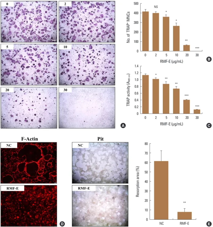
Prior to RMF-E experimental use, its cytotoxicity on os- teoclast precursors had been verified. When BMMs were cultured with various RMF-E concentrations, no cell cyto- toxicity was detected all tested concentrations, up to 40 μg/mL RMF-E (Fig. 1A). In addition, BMMs were treated with 30 μg/mL RMF-E for 4 days, with daily cell viability measurements. No significant difference was observed be- tween the daily cell viabilities of the control (0 µg/mL) and RMF-E-treated (30 µg/mL) cells (Fig. 1B). Therefore, follow- ing these cytotoxicity results, 20 µg/mL RMF-E was select- ed for most experiments in this study.2. Inhibitory effect of RMF-E on osteoclast differentiation and resorption activity
Initially, we investigated the effect of RMF-E on osteo- clast differentiation and formation. BMMs were treated with various concentrations of RMF-E in the presence of M- CSF and RANKL for 4 days. The TRAP staining and TRAP so- lution assay was performed to measure osteoclast differ- entiation and formation. As shown in Figure 2A and B, RMF- E dramatically inhibited TRAP+-MNCs containing ≥3 nuclei and ≥100 μm in diameter (counted as mature osteoclasts), in a dose-dependent manner. The decrease in TRAP+-MNCs formation commenced in 5 μg/mL RMF-E, and rarely formed Table 1. The nucleotide sequences of primers used in this study
Gene Primer
Sense Antisense
Acp5 (Trap) 5´-ctggagtgcacgatgccagcgaca-3´ 5´-tccgtgctcggcgatggaccaga-3´
Oscar 5´-ggggtaacggatcagctccccaga-3´ 5´-ccaaggagccagaacgtcgaaact-3´
CtsK (Cathepsin K) 5´-acggaggcattgactctgaagatg-3´ 5´-gttgttcttattccgagccaagag-3´
Tm7sf4 (Dc-stamp) 5´-tggaagttcacttgaaactacgtg-3´ 5´-ctcggtttcccgtcagcctctctc-3´
Atp6v0d2 5´-tcagatctcttcaaggctgtgctg-3´ 5´-gtgccaaatgagttcagagtgatg-3´
Nfatc1 5´-ctcgaaagacagcactggagcat-3´ 5´-cggctgccttccgtctcatag-3´
Gapdh 5´-tgccagcctcgtcccgtagac-3´ 5´-cctcaccccatttgatgttag-3´
Inhibitory Effect of RMF on RANKL-Induced Osteoclatogenesis
in 30 μg/mL RMF-E. Moreover, total TRAP activity from TRAP+- mono-, di-, and multi-nuclear osteoclasts, was also signifi- cantly dose-dependently diminished by RMF-E treatment (Fig. 2C). To assess if RMF-E affects osteoclast bone resorb- ing activity, we measured in vitro F-actin ring and pit for- mation in osteoclast cultured with/without RMF-E treat- ment. The F-actin ring structure, which is required for bone matrix resorption in vitro and in vivo, was rarely formed in RMF-E-treated osteoclasts, as opposed to the untreated control (Fig. 2D). Eventually, RMF-E-treated osteoclasts show- ed a dramatically decreased pit formation, compared with the untreated control (Fig. 2E). The suppression of resorp- tion activity via F-actin ring and pit formation is attribut- able to decreased TRAP+-MNC formation in RMF-E-treated osteoclasts. These data demonstrate that RMF-E conve- niently represses osteoclast differentiation and activity.
3. Inhibitory effect of RMF-E on the expression of osteoclast differentiation factors
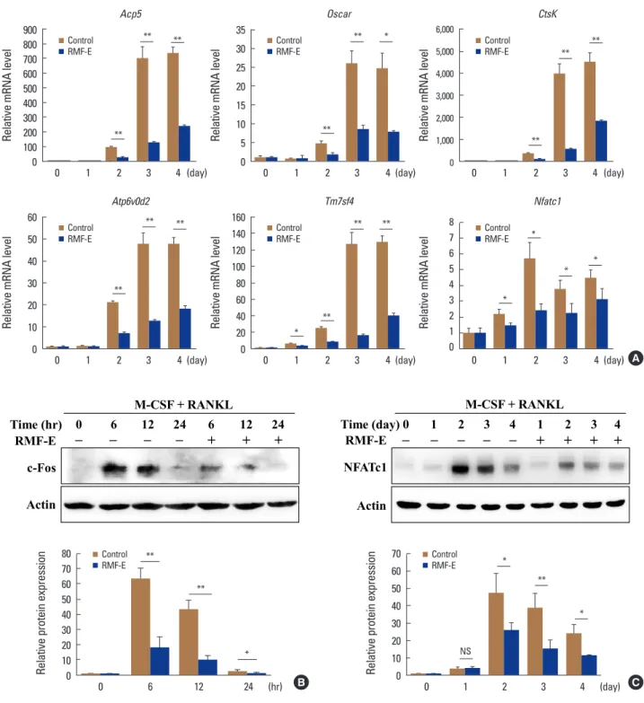
To verify RMF-E inhibitory effect on osteoclast differenti- ation at the molecular level, the expression of osteoclast differentiation marker genes (Acp5, Oscar, CtsK, Atp6v0d2, Tm7sf4, and Nfatc1) and osteoclast differentiation critical transcription factors (c-Fos and NFATc1) was measured us- ing real-time PCR and/or immunoblot analyses, during RA- NKL-mediated osteoclastogenesis. The expression of all tested marker genes, including Nfatc1, was significantly in- hibited in RMF-E-treated osteoclasts (Fig. 3A). Additionally, c-Fos and NFATc1 expression were considerably suppressed by RMF-E treatment (approximately 35% and 55%, respec-
tively), compared with those of the control cells (Fig. 3B, C).
These results support the previous results (Fig. 2) regard- ing RMF-E inhibitory effect on RANKL-induced osteoclast differentiation and formation.
4. Inhibitory mechanism of RMF-E on RANKL- induced osteoclastogenesis
As observed, RMF-E suppressed the expression of c-Fos and NFATc1, pivotal osteoclastogenesis transcription fac- tors (Fig. 3). Therefore, we attempted to examine how RMF- E down-regulates c-Fos and NFATc1 expression, underlying osteoclast differentiation and formation. First, the activa- tion of NF-κB and MAPKs, the main signaling pathway in- duced by the RANKL/RANK interaction, were examined.
BMMs were pre-cultured with/without RMF-E (20 μg/mL) under M-CSF treatment, and RANKL-induced MAPKs and IκBα activation in measured using Western blot analyses.
The activation of p38 looked like a little repressed, but over- all the activation of IκBα and MAPKs seem not affected by RMF-F treatment (Fig. 4A).
The RANKL/RANK interaction is also capable of stimulat- ing Ca2+ signaling via adaptor protein containing ITAM such as DAP12 and FcRγ, followed by modulation of NFATc1 ex- pression and activation. Next, we examined if RMF-E affects RANKL-induced Ca2+-oscillation. BMMs cultured under RA- NKL stimulation were acutely treated with RMF-F. Before RMF-E treatment, RANKL-stimulated cells exhibited typical Ca2+-oscillation. However, as soon as RMF-F treatment com- menced, intracellular Ca2+ concentration frequency imme- diately disappeared (Fig. 4B). We then examined if RMF-E Fig. 1. Effects of Rosae Multiflorae fructus extract (RMF-E) on cell viability. (A) Bone marrow-derived macrophages (BMMs) were cultured with the indicated concentrations of RMF-E, in the presence of macrophage colony-stimulating factor (M-CSF) (30 ng/mL) for 1 day. (B) BMMs were cultured with/without 30 μg/mL RMF-E, in the presence of M-CSF, for 4 days. Cell viability was measured as described in the materials and meth- ods. Data are presented as the mean±standard deviation of 3 independent experiments. NS, not significant.
140 120 100 80 60 40 20 0
Cell viability (%)
0 5 10 20 30 40 RMF-E (μg/mL)
NS Control
RMF-E 120
100 80 60 40 20 0
Cell viability (%)
1 2 3 4
Culture (Day)
NS NS NS NS
A B
Keun Ha Park, et al.
Fig. 2. Effect of Rosae Multiflorae fructus extract (RMF-E) on osteoclast differentiation and resorption activity. Bone marrow-derived macro- phages were cultured with the indicated concentrations of RMF-E, under receptor activator of nuclear factor-κB ligand (100 ng/mL) and macro- phage-colony stimulating factor (30 ng/mL) treatment, for 4 days. (A) Osteoclasts were fixed and stained for tartrate-resistant acid phosphatase (TRAP). (B) TRAP+-multinuclear cells with ≥3 nuclei were counted as mature osteoclasts. (C) Total TRAP activity from TRAP+-mono-, di-, and multi-nuclear cells were measured at an absorbance of 405 nm (A405 nm). (D) F-actin rings were stained with rhodamine-phalloidin in osteoclasts treated with/without RMF-E (20 μg/mL). (E) The resorption activity of osteoclasts treated with/without RMF-E (20 μg/mL) was measured by pit formation on hydroxyapatite-coated plates, after 7 days. Data are expressed as the mean±standard deviation and are representative of 3 inde- pendent experiments. *P<0.05, **P<0.01, and ***P<0.001 vs. the non-treated control (NC, 0 μg/mL RMF-E) (Scale bar=200 μm). NS, not sig- nificant.
Fig. 2 K.H. Park, et al.
D
NC
RMF-E
Resorption area (%)
40
NC RMF-E
**
NC
A
No. of TRAP+ MNCs
0 0.2 0.4 0.6 0.8 1.0 1.2 1.4
TRAP activity (A405nm)
0 2 5 10 20
0 100 200 300 400
RMF-E
2
5 10
20 30
0
*
500
F-Actin Pit
(μg/mL) 30
0 2 5 10 20
RMF-E
(μg/mL) 30
**
*
***
** **
***
***
*
NS
B
C
70 60 50 80
20 10 0 RMF-E 30
E
500 400 300 200 100 0 No. of TRAP+ MNCs
0 2 5 10 20 30 RMF-E (μg/mL)
NS
*
*
**
***
1.4 1.2 1.0 0.8 0.6 0.4 0.2 0 TRAP activity (A405 nm)
0 2 5 10 20 30 RMF-E (μg/mL)
* **
**
** ***
***
80 70 60 50 40 30 20 10 0
Resorption area (%)
NC RMF-E
**
A
B
C
D E
Inhibitory Effect of RMF on RANKL-Induced Osteoclatogenesis
Fig. 3. Effects of Rosae Multiflorae fructus extract (RMF-E) on the expression of osteoclast differentiation marker genes and transcription factors.
Bone marrow-derived macrophages were cultured with receptor activator of nuclear factor-κB ligand (RANKL) (100 ng/mL) and macrophage colo- ny-stimulating (M-CSF) treatment in the presence or absence of RMF-E (20 μg/mL) for 4 days or the indicated times (for c-Fos). (A) Osteoclast dif- ferentiation marker gene expression was examined by real-time polymerase chain reaction. Messenger RNA (mRNA) levels were normalized with Gapdh and expressed as fold change of mRNA level. (B, C) Whole lysate (30 μg) was subjected to sodium dodecyl sulfate–polyacrylamide gel electrophoresis and analyzed by immunoblotting. c-Fos (B) and nuclear factor-activated T cells c1 (NFATc1) (C) expression were detected using the anti-c-Fos and NFATc1 antibody, respectively. Fold change normalized by actin is presented in the lower panel. Data are expressed as the mean±standard deviation and are representative of 3 independent experiments. *P<0.05 and **P<0.01 vs. the control group (0 μg/mL RMF-E).
Control RMF-E 900
800 700 600 500 400 300 200 100 0
Relative mRNA level
0 1 2 3 4 (day) Acp5
**
** **
Relative mRNA level
Control RMF-E 6,000
5,000 4,000 3,000 2,000 1,000
0 0 1 2 3 4 (day) CtsK
**
**
**
Control RMF-E 35
30 25 20 15 10 5 0
Relative mRNA level
0 1 2 3 4 (day) Oscar
**
** *
Control RMF-E 60
50 40 30 20 10 0
Relative mRNA level
0 1 2 3 4 (day) Atp6v0d2
**
** ** Control
RMF-E 8
7 6 5 4 3 2 1 0
Relative mRNA level
0 1 2 3 4 (day) Nfatc1
*
*
* *
Control RMF-E 160
140 120 100 80 60 40 20 0
Relative mRNA level
0 1 2 3 4 (day) Tm7sf4
**
*
** **
A
Fig. 3
K.H. Park, et al.
Atp6v0d2
Acp5 Oscar CtsK
Nfatc1 Tm7sf4
Relative mRNA level Relative mRNA level Relative mRNA levelRelative mRNA level Relative mRNA levelRelative mRNA level
0 1 2 3 4 d
**
** **
* *
*
** *
** **
**
** **
*
** **
** **
**
0 800 600 400 200
0 35 30 25 20
10 5
0 6000 5000 4000 3000 2000 1000
0 50 60
30
10 40 20
0 20 60 120 80 40 100 140
0 8 7 6 4 2 5 3 1 Control
RMF-E 900
700
300 100 500
0 1 2 3 4 d
15
0 1 2 3 4 d
160
0 1 2 3 4 d 0 1 2 3 4 d
*
0 1 2 3 4 d
B C
c-Fos
M-CSF + RANKL
6 12 12
0 24 24
Time (hr) 6
Actin
RMF-E – – – – + + + Time (day)0 1 2 3 4 1 2 3 4
NFATc1 Actin
– – –
RMF-E – – + + + +
M-CSF + RANKL
A
0 10 20 30 40 70 60 50 80
Relative protein expression
6 12
0 24 h
Control RMF-E
**
**
*
0 10 20 30 40 70 60 50
Relative protein expression
0 1 2 3 4 d
*
**
*
NS
Fig. 3
K.H. Park, et al.
Atp6v0d2
Acp5 Oscar CtsK
Nfatc1 Tm7sf4
Relative mRNA level Relative mRNA level Relative mRNA levelRelative mRNA level Relative mRNA levelRelative mRNA level
0 1 2 3 4 d
**
** **
* *
*
** *
** **
**
** **
*
** **
** **
**
0 800 600 400 200
0 35 30 25 20
10 5
0 6000 5000 4000 3000 2000 1000
0 50 60
30
10 40 20
0 20 60 120 80 40 100 140
0 8 7 6 4 2 5 3 1 Control
RMF-E 900
700
300 100 500
0 1 2 3 4 d
15
0 1 2 3 4 d
160
0 1 2 3 4 d 0 1 2 3 4 d
*
0 1 2 3 4 d
B C
c-Fos
M-CSF + RANKL
6 12 12
0 24 24
Time (hr) 6
Actin
RMF-E – – – – + + + Time (day)0 1 2 3 4 1 2 3 4
NFATc1 Actin
– – –
RMF-E – – + + + +
M-CSF + RANKL
A
0 10 20 30 40 70 60 50 80
Relative protein expression
6 12
0 24 h
Control RMF-E
**
**
*
0 10 20 30 40 70 60 50
Relative protein expression
0 1 2 3 4 d
*
**
*
NS Control RMF-E 70
60 50 40 30 20 10 Relative protein expression 0
0 1 2 3 4 (day)
NS
*
**
* Control
RMF-E 80
70 60 50 40 30 20 10 Relative protein expression 0
0 6 12 24 (hr)
**
*
**
B C
Keun Ha Park, et al.
affects RANKL-stimulated BTK and PLCγ activation respon- sible for intracellular Ca2+-oscillation in upstream RANKL- signaling. BTK and PLCγ were normally activated by RANKL
stimulation. Although RMF-E pretreated cells showed a slight increase at 5 min, BTK and PLCγ2 phosphorylation were gradually suppressed in RMF-E pretreated cells, com- Fig. 4. Effects of Rosae Multiflorae fructus extract (RMF-E) on receptor activator of nuclear factor-κB ligand (RANKL)-induced intracellular signal- ing. Bone marrow-derived macrophages (BMMs) were pretreated with/without RMF-E (20 μg/mL) for 2 hr in the presence of macrophage colony- stimulating factor (M-CSF) (30 ng/mL), and then RANKL (100 ng/mL)-treated, to stimulate intracellular signaling, at indicated times. Lysate (30 μg) was subjected to sodium dodecyl sulfate–polyacrylamide gel electrophoresis and analyzed by immunoblotting. (A) The activation of mitogen-acti- vated protein kinases (extracellular signal-regulated kinase [ERK], c-JUN N-terminal kinase [JNK], and p38) and IκBα was examined using their respective antibodies. (B) Twenty four-hr RANKL-stimulated BMMs were acutely treated with RMF-E (20 μg/mL). RANKL-stimulated Ca2+-oscilla- tion by was measured using the fluorescence Ca2+ indicator (Fura-2, AM). (C) Bruton’s tyrosine kinase (BTK) and phospholipase C-γ2 (PLCγ2) acti- vation were detected using the anti p-BTK/BTK and p-PLCγ2/PLCγ2 antibody, respectively. Fold change normalized by their non-phosphorylated proteins is presented in the right panel. Data are expressed as mean±standard deviation and are representative of 3 independent experiments.
*P<0.05 and **P<0.01 vs. the control group (0 μg/mL RMF-E).
Fig. 4
K.H. Park, et al.
A
B C
p-p38 p38 p-ERK Time (min) RMF-E
p-IκB IκB p-JNK JNK ERK
–
RANKL
5 15 5
0 30 15 30
– – 60 60
+
– – + + +
Actin
–
M-CSF + RANKL
5 15 5
0 30 15 30
– –
Time (min) 60 60
RMF-E – – + + + +
p-Btk Btk p-PLCγ2 PLCγ2 Actin
Relative protein expression
2 3
0 4 5 6
5 15
0 30 60 min
1
p-PLCγ2/PLCγ2
Relative protein expression
1 1.5
0 2 2.5 3
5 15
0 30 60 min
0.5
p-Btk/Btk
** **
* NS 5 15
0 30 60 min 0 5 15 30 60 min
Relative protein expression
5 10
0 15 20 25
4 8
0 12 16
Relative protein expression
5 10 0 15 20 25 30
Relative protein expression Relative protein expression
40 80
0 120 140
5 15
0 30 60 min 0 5 15 30 60 min
p-ERK/ERK
Control RMF-E
p-JNK/JNK
p-p38/p38 p-IκB/IκB
* *
*
NS
NS NS NS
NS
NS
NS NS NS
NS
NS NS NS
Control RMF-E 7 **
**
* * 2 min
Ratio (0.1)
RMF-E RANKL
Ratio (0.1)
2 min
A
B C
p-PLCγ2/PLCγ2 (min)
(min) (min)
(min)
(min) (min)
Inhibitory Effect of RMF on RANKL-Induced Osteoclatogenesis
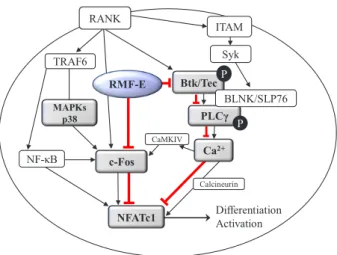
Fig. 5. Schematic diagram of the effect of Rosae Multiflorae fructus extract (RMF-E) on receptor activator of nuclear factor-κB ligand (RANKL)-induced osteoclastogenesis. The RANKL/RANK interaction may lead to mitogen-activated protein kinases (MAPKs) activation, followed by induction of c-Fos expression, and it also leads to Bru- ton’s tyrosine kinase (BTK) and phospholipase C-γ2 (PLCγ2) activation inducing calcium signaling, followed by induction of nuclear factor- activated T cells c1 (NFATc1) expression and activation. RMF-E may inhibit NFATc1 induction via suppression of c-Fos and modulation of the BTK/PLCγ signaling pathways regulating Ca2+ signaling, resulting in the inhibition of RANKL-induced osteoclastogenesis. The red line indicates the inhibition pathway of RMF-E. TRAF6, tumor necrosis fac- tor receptor associated factor 6; ITAM, immunoreceptor tyrosine- based activation motif.
K.H. Park, et al.
Fig. 5
Differentiation Activation NFATc1
TRAF6
Btk/Tec PLCγ
Ca2+
c-Fos
BLNK/SLP76 Syk P
P RMF-E
MAPKs p38 NF-κB
CaMKIV
Calcineurin
RANK ITAM
pared with those of untreated control cells (Fig. 4C).
Collectively, these data demonstrate that RMF-E regu- lates MAPKs, specifically p38, as well as BTK/PLCγ2 activa- tion and RANKL-induced Ca2+-oscillation, which are all im- portant factors for c-Fos and NFATc1 induction in RANKL- stimulated osteoclast differentiation and formation (Fig. 5).
DISCUSSION
For several decades, a good number of studies have dem- onstrated the medicinal effectiveness of extracts or con- stituents from various plants used as traditional medicines against resorptive bone diseases, including osteoporosis.
[23] Although their potential anti-bone loss effects have been suggested, only some of these plants have been ap- propriately examined for their physiological and pharma- cological properties, and their action mechanism. The pre- sented medicinal plants have been reported to modulate the differentiation of osteoclasts, osteoblasts, or both, by
diverse effects and action mechanisms.[23] Although mo- lecular action mechanisms of medicinal plant extracts have been known and found responsible for the regulation of osteoclast differentiation, intracellular signaling for the ac- tivation of MAPKs as well as NF-κB is dominantly modulat- ed by medicinal plants, followed by inhibition of c-Fos and NFATc1, known as critical osteoclastogenic transcription factors.[24-27]
In Eastern Asian countries like Korea and China, RMF has been traditionally used to calm inflammatory disorders such as arthritis.[13] In western countries including both Europe and the United States, the rosehips of Rosa canina, another wild rose like multiflora rose, have also been clini- cally used as osteoarthritis remedies.[28,29] Although RMF has been linked to inflammatory bone disease treatment, the precise underlying mechanism of its effect on bone cells, especially osteoclasts, remains unelucidated. In this study, we investigated the effect of RMF-E on RANKL-me- diated osteoclast differentiation and activation. RMF-E in- hibited RANKL-induced TRAP+-mature osteoclast forma- tion and bone resorption activity, accompanied by repres- sion of osteoclast differentiation markers, in a dose-depen- dent manner (Fig. 2, 3A). In addition, c-Fos and NFATc1 ex- pression were decreased by RMF-E treatment during RANKL- mediated osteoclastogenesis (Fig. 3B). These RMF-E inhibi- tory effects appeared to be partially caused by downregu- lation of p38 MAPK activation.
In terms of RANKL-RANK interaction-induced osteoclas- togenesis, the Ca2+signaling pathway via FcRγ-DAP12/BTK/
PLCγ signaling, also affects the expression of transcription factors such as c-Fos and NFATc1.[4,6,8] Our previous re- ports already showed that the inhibitory effects of the EtOH extracts of some herbal plants (Chrysanthemum zawadskii Herbich var. and Glechoma hederacea) on RANKL-induced osteoclastogenesis was due to Ca2+ signaling pathway mod- ulation, followed by downregulation of NFATc1.[22,30] In the present study, RMF-E also exerted its inhibitory effect by modifying Ca2+ signaling via BTK/PLCγ2 pathway inacti- vation (Fig. 4B, C). Overall, it appears RMF-E can regulate the canonical signal pathway by MAPK activation, and the co-stimulatory signal pathway by Ca2+-oscillation, during RANKL-mediated osteoclastogenesis. Although many oth- er signaling pathways are capable of regulating osteoclas- togenesis, those related to reactive oxygen species pro- duction and inflammation, and the role of RMF-E in anti-