저작자표시-비영리-동일조건변경허락 2.0 대한민국 이용자는 아래의 조건을 따르는 경우에 한하여 자유롭게
l 이 저작물을 복제, 배포, 전송, 전시, 공연 및 방송할 수 있습니다. l 이차적 저작물을 작성할 수 있습니다.
다음과 같은 조건을 따라야 합니다:
l 귀하는, 이 저작물의 재이용이나 배포의 경우, 이 저작물에 적용된 이용허락조건 을 명확하게 나타내어야 합니다.
l 저작권자로부터 별도의 허가를 받으면 이러한 조건들은 적용되지 않습니다.
저작권법에 따른 이용자의 권리는 위의 내용에 의하여 영향을 받지 않습니다. 이것은 이용허락규약(Legal Code)을 이해하기 쉽게 요약한 것입니다.
Disclaimer
저작자표시. 귀하는 원저작자를 표시하여야 합니다.
비영리. 귀하는 이 저작물을 영리 목적으로 이용할 수 없습니다.
동일조건변경허락. 귀하가 이 저작물을 개작, 변형 또는 가공했을 경우 에는, 이 저작물과 동일한 이용허락조건하에서만 배포할 수 있습니다.
의학 석사 학위논문
Establishment and
characterization of breast cancer patient-derived cell lines, xenograft, and organoids
유방암 환자 유래 이종이식 종양 세포주 및 오가노이드 수립과 특성 분석
2020 년 8 월
서울대학교 대학원 의과학과 의과학전공
김 가 혜
i
Abstract
Ga-Hye Kim Major in Biomedical Sciences The Graduate School Seoul National University
Breast cancer is the most common cancer in woman, and although many targeted agents have been developed for last 2 decades, breast cancer still remains one of the leading causes of death in women. Breast cancer is heterogeneous disease which could be divided into three major subtypes and endocrine treatment and many targeted agents are currently developing to control this disease. Twenty pleural effusion-derived breast cancer cell lines and one ascites-derived breast cancer cell line were newly established. In addition, three pairs of cell line-organoid were established from the tumor tissue of breast cancer patient derived xenograft (PDX). Cellular and molecular properties of a total of new 24 cell lines and 3 organoids were analyzed. Genetic characteristics were revealed through screening for mutant
ii
genes that are found to have a large variation in breast cancer such as TP53, PIK3CA, PTEN, ERBB2(HER2), BRCA1/2, RB1, MAP3K. The hormone receptor expression status including estrogen receptor (ER), progesterone receptor (PR) and human epithelial growth factor receptor 2 (HER2) were confirmed. and the new breast cancer cell lines and organoids were grouped and divided according to ER, PR, and HER2 status. Next, we measured sensitivity to various drugs that are widely used in breast cancer treatment, research and clinical trials. The results showed corresponding outcomes with the presence or absence of target mutations known to affect the reactivity or expression of drug targets previously identified. Even if they were from the same patient origin, each cell line showed some different results, and two pairs of cell line-organoid showed a noticeable difference in reactivity despite having similar mutational profiles.
The results of gene screening based on the database proved that pleural effusion could be used for constructing an experimental model in breast cancer. In addition, the differences between patients and the similarity and diversity between cell lines from same patient can be the basis for clinical treatment planning.
These results suggest that the accumulation of various
iii
characters and types of experimental models will greatly contribute to improving the accuracy and suitability of the database.
Keywords : Pleural effusion, Ascites, patient-derived xenograft (PDX), breast cancer cell line, breast cancer organoid, characterization analysis, Drug sensitivity, Fusion gene
학 번 : 2017-23206
iv
Contents
Abstract ... i
Contents ... ⅳ List of tables ... ⅵ List of figures ... ⅷ Introduction... 1
Material and Methods Establishment and maintenance of human breast cell lines ... 4
Establishment and maintenance of breast cancer organoids ... 5
Growth properties and morphology in vitro ... 6
Genomic DNA extraction and DNA fingerprinting analysis ... 7
Genomic DNA Mycoplasma detection test ... 8
Drug sensitivity test ... 9
Protein extraction and Western blotting ... 10
v
Confocal analysis of immunofluorescence staining... 12
FFPE block production and H&E staining ... 13
Whole Exome Sequencing ... 14
RNA Sequencing - fusion gene analysis ... 17
Results Sample origin and identity verification ... 19
Culture characteristics ... 24
Mutational traits ... 32
Anticancer drug response ... 48
Fusion gene analysis ... 60
Discussion ... 65
Acknowledgements ... 79
References ... 80
Abstract in Korean ... 89
vi
List of tables
Table 1. Chemotherapeutic agents and targeted agents for breast cancer used in this study ... 18 Table 2. Origin and
in vivo
characteristics of21 human breast cancer cell lines and
3 paired cell line-organoid sets derived from
same patient-derived xenograft (PDX) tumor.. 20
Table 3. DNA fingerprinting analysis using 15 STR loci
and amelogenin for newly established
21 breast cancer cell lines and
3 matched patient-derived xenograft (PDX)
cell line-organoid pairs ... 22
Table 4.
in vitro
characteristics of24 breast cancer cell lines ... 31
Table 5. Major mutational profile table ... 37
vii
Table 6. Classification of cell lines and organoids
based on ER and HER2 expression ... 47
Table 7. AUC of 23 Breast cancer cell lines
and 3 organoids ... 55
Table 8. Reported fusion genes of
11 established breast cancer cell lines ... 62
viii
List of figures
Figure 1. Mycoplasma detection test ... 26
Figure 2. Microscopic images of 24 breast cancer cell lines
and 3 breast PDX organoids ... 27
Figure 3. Images of confocal microscopy and
FFPE block H&E staining of
3 organoids derived from PDXT ... 29
Figure 4. Mutational landscape of the established
breast cancer cell lines and PDX organoids ... 36
Figure 5. Western blot analysis ... 41
Figure 6. Drug sensitivity AUC result heatmap of
breast cancer cell lines and organoids ... 51
Figure 7. AUC heat map divided into groups according to
patient information and ER/HER2 expression .... 53
1
1. Introduction
For both sexes combined, Breast cancer is a type of cancer with a high incidence rate, which is almost the same as that of lung cancer with the most frequently diagnosed cancer (11.6%).
Among females, breast cancer is the most common cancer and leading cause of cancer death [1]. Human breast cancers are heterogeneous, and breast cancer patient outcomes and responses to therapy are extremely varied.
Subtype of breast cancer was based on the following basic classification criteria : histological type, tumor grade, lymph node status, the presence or/and absence of hormonal (estrogen, progesterone) receptors (ER, PR) and human epidermal growth factor receptor2 (HER2), expression of a marker of proliferation.
Advances in immunohistochemical techniques and molecular biological methods have enabled in-depth studies of various forms identification of breast cancer. The genetic features of breast cancer are also highly diverse. Genes that are most frequently mutated in breast cancer include TP53, PIK3CA, MYC, CCND1, PTEN, ERBB2, GATA3, RB1 and MAP3K1 [2].
HER2 is encoded by the ERBB2 gene. In particular, HER2 is
2
amplified in 20% of breast cancers. It is known that an intrinsic activating mutation leads to a valine-to-leucine substitution at codon 777 within the HER2 kinase domain (HER2 V777L), which induces trastuzumab resistance [3]. The tumor suppressor genes BRCA1 and BRCA2 are mutated in hereditary breast- ovarian cancer syndrome. Patients with this variant are likely to have a higher probability of lifetime risk, but no conclusive conclusion has yet been reached [4]. High-throughput molecular profiling studies have confirmed that breast cancer has spatial and temporal intra-tumor heterogeneity beyond our expectations [5]. Clinical approaches and management of the disease comprises are tailored to these various characteristics, including morphological assessments (size, grade), three subtypes based on immunohistochemical staining of breast cancer tissues and genomic features [6, 7]. Naturally, the coexistence of multiple subclone with different genetic variations and relative difference of drug sensitivities might not be effective against anticancer drugs that target predominant aberrations [5].
Therefore, the accumulation of data on as many and various characteristics as possible is necessary, and the more diverse the breast cancer research platform is, the more effective the
3
study is expected to be applied to clinical applications.
Various types of cancer research models have been developed and used. Immortalized cell lines are used commonly to study breast cancer, are easy to manipulate, and are ideal for examining the molecular mechanism of tumor cell biology [8]. Patient- derived xenograft (PDX) models have an advantage in that it preserves heterogeneity and microenvironment included.
Organoid culture not only retains heterogeneity, but also enables high-throughput screening [9]. In this way, each platform has its own advantages and limitations, and experimenters use a suitable model for their research purpose. Patient-derived cell lines, PDX-derived cell lines and organoids were established and characterization was performed for various type of breast cancer research and proceeding to present the direction of personalized treatment. Several types of experimental models with various characteristics are expected to be greatly helpful in future breast cancer research.
4
2. Materials and Methods
2.1. Establishment and maintenance of breast cancer cell lines Twenty Cell lines were established from malignant pleural effusion and one cell line was established from malignant ascites sample. Three cell lines were gained from patient derived xenograft (PDX) tumor tissue sample. All samples were from patients in Seoul National University Hospital. Suspended cells were gathered by spinning down. Gathered cell pellet were seeded into T-25㎠ or T-75㎠ flasks. Cancer cells were initially cultured in Opti-MEMⅠ (Thermo Fisher Scientific, MA, USA) with 5% fetal bovine serum (FBS). Confined-area trypsinization or scraping method was used to attain achieve a pure tumor cells when stromal cells like mesothelial cells or fibroblasts grew in the initial culture. After primary culture, established cell lines were sustained in RPMI 1640 (Thermo Fisher Scientific, MA, USA) with 10% fetal bovine serum and 1%
(v/v) penicillin and streptomycin (10,000U/ml). Incubated flasks in humidified incubators at 37℃ in an atmosphere of 5% CO2 and 95% air.
5
2.2. Establishment and maintenance of breast cancer organoids 2.2.1 Tumor isolation and culture
PDX tumor tissue was cut finely with scissors for about 5 min.
The enzyme solution consisting of CollagenaseⅡ(1.5mg/ml), Hyaluronidase (20ug/ml) and Ly27632 (10μM) was added to the chopped tissue and incubated for at least 4hr at 37℃ while spinning. FCS was added for neutralization and the mixture was filtered through 100μM cell strainer (SPL, #93100) to remove large chunks and impurities that were not cut well. Spun down pellet at 1,000 rpm for 3 min. In the case of a pleural effusion sample, centrifugation was performed immediately without any enzyme digestion. The supernatant was suctioned and then plated in an appropriate amount of BME gel (Gibco, A14132-02).
When the BME gel hardened, the HBEC medium (Basal culture medium with 50% Wnt conditioned medium, 20% R-Spondin conditioned medium, 10% Noggin conditioned medium, 1x B27 (Gibco, 17504-044), 1.25mM n-Acetyl cysteine (Sigma, A7250), 5mM Nicotinamide, 5nM Neuregulin(Peprotech, 100- 03), 500nM A83-01, 500nM SB202190(Sigma, S7067), 5mM Ly27632, 5ng/ml human EGF (Peprotech,AF-100-15), 20ng/ml human FGF-10 (Peprotech, 100-26), 5ng/ml human FGF-
6
7(Peprotech, 100-19) and 50μg/ml Primocin (Thermo, ant- pm-1))was added and incubated at 37℃.
2.2.2 Organoid Culture
When passaging, first remove the media and pipet off the BME gel. Collect the loose gel in a tube. Mixture of the organoids and gel was centrifuged at 1,000 rpm for 3min and the medium suctioned. About 5 ml Triple Express (Invitrogen) was added and the mixture was incubated at 37℃ for approximately 10 min.
After 5 minutes, the size of organoids was checked and the gel was removed every minute. Care should be taken not to treat organoids in the Triple Express for too long. To neutralize, FCS and medium were added and loose cells were spun down at 1,500 rpm for 3 min. After mixing the pellet with the appropriate amount of gel, mixture was plated in droplets of 50-100μL each. Leave for 10 min to allow the BME gel to solidify and then fill the HBEC medium. The media is usually changed every week.
2.3. Growth properties and morphology in vitro
To acquire each tumor population’s doubling time, 5x10 to 2x10 viable cells from each cell line were seeded into 12–24
7
identical well of 96 well plate and cell viability was calculated daily for 5-12 days. Since the first cell seeding, in every 24 hours, 10ul EZ-Cytox solution (Daeil Lab, Seoul, Korea) was added to well of each seeded lung cancer cells in triplicate. After 2 hour-incubation at 37℃, Optical density of EZ-Cytox- treated cells was calculated by Multiskan™ GO Microplate Spectrophotometer (Thermo Fisher Scientific, MA, USA).
Growth rate values were measured by GraphPad Prism 5 (GraphPad Software, CA, USA). Growth rates are values to multiply 10 by days that cell population were duplicated. To observe cell line’s morphology, each cell line was cultured in 75cm2 culture flasks and then pictured daily by phase-contrast microscopy.
2.4. Genomic DNA extraction and DNA fingerprinting analysis Genomic DNA (gDNA) extraction was performed by using QIAamp DNA Mini kit (Qiagen). gDNA extracted from each breast cancer cell line and organoid was amplified using an AmpFlSTR identifier Polymerase Chain Reaction (PCR) Amplification Kit (Applied Biosystems, CA, USA). A single cycle of PCR amplified 15 short tandem repeat markers (CSF1PO,
8
D2S1338, D3S1358, D5S818, D7S820, D8S1179, D13S317, D16S539, D18S51, D19S433, D21S11, FGA, TH01, TPOX and VWA) and an amelogenin gender-determining marker containing highly polymorphic microsatellite markers. Amplified PCR products were analyzed by an ABI 3500XL Genetic analyzer (Applied Biosystems).
2.5. Genomic DNA Mycoplasma detection test
gDNA extracted from each breast cancer cell line and organoid was amplified using an TaKaRa Polymerase Chain Reaction (PCR) Mycoplasma Detection Set (TAKARA BIO INC., Shiga, JAPAN).
a primer set was designed to detect the presence of Mycoplasma which might contaminate biological materials such as cultured cells. This primer set allows sensitive and specific detection of several different species of Mycoplasma (M. fermentans, M.
hyorhinis, M. arginini, M. orale, M. salivarium, M. hominis, M.
pulmonis, M. arthritidis, M. neurolyticum, M. hyopneumoniae, M.
capricolum) and one species of Ureaplasma (U. urealyticum).
Amplify the spacer regions in the rRNA operon (for example, the region between the 16S and 23S genes) using two primers (F1
9
and R1), which were designed based on the DNA encoding the 16S and 23S rRNAs. Perform Nested PCR using two primers, F2, based on the conserved region, and R2, based on the 23S gene.
2.6. Drug sensitivity test
2.6.1 2D cell lines seeding/treatment procedure
2x10 to 4x10 viable cells from each cell line were seeded into well of 96 well white plate (SPL, #30196) in triplicate to measure sensitivity of several drugs. A day after, all cell lines and organoids were respectively treated for proper concentration of Drugs. If the cell line was adherent cell, adherent state was confirmed. After 72 hours-incubation at 37 ° C, 10ul Cell-titer glo solution was added to well of each seeded breast cancer cells and organoids. After 20 minute-incubation at 37°C, optical density of Cell-titer glo treated cells was calculated by Multiskan™ Ascent Microplate Luminometer (Thermo Fisher Scientific). These steps were repeated in duplicate.
10
2.6.1 3D organoids seeding/treatment procedure
Cleaved organoids were placed around the rim of the well of 96 well white plates (SPL, #30196) in a 1:1 mixture of HBEC medium and RGF basement membrane matrix (Gibco, A14132- 02). Plates are incubated at 37℃ with 5% CO2 for 15 minutes to solidify the gel. After solidation, 20 ul of pre-warmed HBEC medium to each well. 96 hours later, 20 ul of serial diluted drug solution is added to each well. The mixture of HBEC medium and drug-solvent solution is added for the control well. After 20 minute-incubation at 37°C, optical density of Cell-titer glo treated cells was calculated by Multiskan™ Ascent Microplate Luminometer (Thermo Fisher Scientific). These steps were repeated in duplicate.
2.7. Protein extraction and Western blotting
Cultivated cells that had full confluency were harvested with cell scrapper. Cell pellet was treated by EzRIPA buffer (ATTO Co., Tokyo, Japan) after washed by cool PBS. Whole protein was extracted by this step. Protein concentration of each cell line was determined by Pierce™ BCA Protein Assay Kit (Thermo
11
Scientific). Proteins that fixed into equal concentration were loaded on a 4-15% Polyacrylamide gel (Bio-Rad) at 50 volts for 3 hours and then proteins on loaded gel were transferred to a Trans-Blot®Turbo™ Transfer Pack PVDF membrane (Bio-rad) by Trans-Blot®Turbo™ Transfer system (Bio-Rad). Proteins of transferred membrane was blocked by incubating in 1.5% to 2.0% skim milk and 0.05% Tween 20-TBS buffer including 1mM MgCl2 for an hour at room temperature. Primary antibodies were used against E-cadherin, N-cadherin, Snail, ER-α, PR A/B, EGFR, phospho-EGFR, HER2, pan Akt, phospho-Akt, mTOR, phospho-mTOR, IGF-1Rβ, MEK 1/2, phospho-MEK1/2, PTEN and B-actin. Those antibodies were Abcam products (Abcam, Cambridge, UK) and CST products (Cell Signaling Technology, MA, USA) except for exon 19 E746-A750 deleted EGFR that was Cell signaling product (Cell signaling, MA, USA). Mouse or rabbit IgG 2nd antibody (Jackson Immunoresearch, PA, USA) (1:5000) conjugated with peroxidase that matched with used 1st antibody was added to membrane. After chemiluminescent working solution, SuperSignalTM West Pico PLUS Chemiluminescent Substrate (Thermo Scientific) was treated to the membrane, the membrane was exposed to Fuji RX film
12
(Fujifilm, Tokyo, Japan) for 0.5 - 10 minutes.
2.8. Confocal analysis of immunofluorescence staining
Cells were seeded on chambered coverglass (Thermo Fisher Scientific, MA, USA) with a desirable cell confluency. The chambered coverglass was designed to be hydrophilic and no ECM component was treated before seeding. 72 hours after cell seeding, cells were washed with cold DPBS for 15 minutes three times. Then, cells were fixed and permeabilized with BD Cytofix/Cytoperm™ (BD science, CA, USA). After cells were washed with washing solution (BD science), DPBS containing 2%
FBS (GE Healthcare Life Sciences, Buckinghamshire, UK) was applied for an hour for blocking. After cells were washed with cold DPBS, HER2 (Santa Cruz Biotechnology, CA, USA) and E- cadherin antibody (Abcam, Cambridge, United Kingdom) diluted in 0.05% of PBS.T was applied for an 1.5 hours in room temperature. Thereafter, cells were washed with 0.05% of PBS.T, and Alexa 488 and Alexa 594 secondary antibodies (Thermo Fisher Scientific, MA, USA) diluted in 0.05% of PBS.T were applied for an hour in room temperature. 1x DAPI (Sigma- Aldrich, MO, USA) were diluted in distilled water and applied
13
for 30 minutes in room temperature. The cells were washed with DPBS three times, and pictured under confocal microscope.
LSM800 Confocal Laser Scanning Microscope and ZEN software (Carl Zeiss, Oberkochen, Germany) was used to examine cells. Diverse magnifications were used for different growth patterns and sizes of cells. The intensity of each channel was fixed for the comparison of target protein expression between samples. Digital resolution, scan speed and the number of pictures averaged were set to 1024 x 1024, 40 seconds per one channel, and 8 pictures respectively. The pictures were focused on the very bottom of the fixed cells for investigating protruding region of cell colonies and the location of HER2 and E-cadherin.
.
2.9. FFPE block production and H&E staining
The gel dome containing the organoid was scraped out the flask floor with a pipette tip that has been cut off. PBS was added and transferred to the 1.5mL tube. After a short centrifugation, the process of suctioning the supernatant is repeated so that the gel disappears and only the organoids remain as much as possible.
Collected pellets were embedded into low melting (2% diluted in
14
PBS) agarose gel (iNtRON Biotechnology, Seongnam, Korea).
Solidified agarose gel was fix 10% paraformaldehyde (PFA) for 30 minutes at room temperature. The agarose blocks were processed before being embedded into paraffin. Sections (4μm) of organoids were subjected to routine haematoxylin and eosin (H&E) staining.
2.10. Whole Exome Sequencing 2.10.1. Analysis of 3 organoids
SureSelect sequencing libraries were prepared according to manufacturer’s instructions (Agilent sureselect all Exon kit 50 Mb) using the Bravo automated liquid handler. Three micrograms of genomic DNA were fragmented to a median size of 150 bp using the Covaris-S2 instrument (Covaris, Woburn, MA). The adapter ligated DNA was amplified by PCR, and the PCR product quality was assessed by capillary electrophoresis (Bioanalyzer, Agilent). The hybridization buffer and DNA blocker mix were incubated for 5 minute at 95°C and then for10 minutes at 65°C in a thermal cycler. The hybridization mixture was added to the bead suspension and incubated for 30 minutes at RT while mixing. The beads were washed, and DNA was
15
eluted with 50 ml SureSelect elution buffer (Agilent). The flow cell loaded on HISEQ 2500 sequencing system (Illumina).
2.10.2. Analysis of 24 cell lines
DNA should be as intact as possible, with an OD260/280 ratio of 1.8–2. we checked quality of DNA by 1% agarose gel electrophoresis and PicoGreen® dsDNA Assay(Invitrogen).
SureSelect sequencing libraries were prepared according to the manufacturer’s instructions (Agilent SureSelectXT Human All Exon V4) using The Bravo automated liquid handler. 200ng of genomic DNA in 50 ul EB buffer was fragmented to a median size of 150 bp using the Covaris-S2 instrument (Covaris) with the following settings: duty cycle 10%, intensity 5, cycles per burst 200, and mode frequency sweeping for 360 s at 4℃. The fragmentation efficiency was evaluated by capillary electrophoresis on DNA1000 chips (Bioanalyzer, Agilent).
Sequencing adapters were ligated on the DNA fragments following the manufacturer’s protocol (Agilent). The adapter ligated DNA was amplified by PCR. The quality of the PCR products was assessed by capillary electrophoresis (Bioanalyzer, Agilent). SureSelect hyb #1, #2, #3, and #4 reagents (Agilent) were mixed to prepare the hybridization buffer. The amplified
16
DNA fragments were concentrated 750 ng in 3.4 ul. SureSelect block #1, #2, and #3 reagents (Agilent) were added to the 750 ng of DNA. The hybridization buffer and the DNA blocker mix were incubated for 5 min at 95℃ and then for 10 min at 65℃ in a thermal cycler. RNase block (Agilent) was added to the SureSelect oligo capture library (Agilent). The capture library was incubated for 2 min at 65℃. First the hybridization buffer, and then the DNA blocker mix were added to the capture library and the mixture was incubated for 24 hours at 65℃ in a thermal cycler. Fifty ul of streptavidin coated the Dynal MyOne Streptavidin T1 (Invitrogen) were washed three times with 200 ml SureSelect binding buffer (Agilent) and resuspended in 200 ml of the binding buffer. The hybridization mixture was added to the bead suspension and incubated for 30 min at RT with mixing.
The beads were washed with 500 ml SureSelect wash buffer #1 (Agilent) for 15 min at RT, and three times with 500 ml SureSelect wash buffer #2 (Agilent) for 10 min at 65℃. DNA was eluted with 30 ul nuclease-free water. The captured library was amplified to add index tags using Herculase II Fusion DNA Polymerase (Finnzymes). The quality of the amplified libraries was verified by capillary electrophoresis (Bioanalyzer, Agilent).
17
After QPCR using SYBR Green PCR Master Mix (Applied Biosystems), we combined libraries that index tagged in equimolar amounts in the pool. Sequencing is performed using an Illumina Novaseq 6000 system following provided protocols for 2x100 sequencing.
2.10. RNA Sequencing - Fusion gene analysis
Both Fusionmap (Version:8.0.2.32) and Chimerascan (version0.4.5) were used to analyze fusion genes. The Fusionmap data were needed for the data filtering step based on the recommended filtering options in the sample: Seed Count >=
3, splice Pattern Class = Canonical Pattern [Major] or Canonical Pattern [Minor], Filter = Empty. The Chimerascan data were already filtered according to the recommended filtering option.
All the RNA sequencing data were visualized using the integrative genomics viewer (IGV) version 2.4 and the fusion gene candidates were confirmed, respectively. The candidates of fusion gene were sorted reliably based on the intersection of gene sets obtained using Defuse, Fusionmap and Chimerascan methods.
18
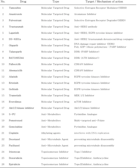
Table 1. Chemotherapeutic agents and targeted agents for breast cancer used in this study
* DDR : DNA Damage Response
No. Drug Type Target / Mechanism of action
1 Tamoxifen Molecular Targeted Drug Selective Estrogen Receptor Modulator(SERM)
2 Anastrozole Molecular Targeted Drug Aromatase Inhibitor
3 Fulvestrant Molecular Targeted Drug Selective Estrogen Receptor Degrader(SERD)
4 Trastuzumab Molecular Targeted Drug Anti-HER2 antibody
5 Lapatinib Molecular Targeted Drug Anti-HER2, EGFR tyrosine kinase inhibitor
6 DS-8201a Molecular Targeted Drug Anti-HER2 (trastuzumab deruxtecan)drug conjugate
7 Olaparib Molecular Targeted Drug DNA damage repair inhibitor (DDRi) Poly ADP-ribose polymerase : PARP Inhibitor 8 Talazoparib Molecular Targeted Drug DDRi (PARP Inhibitor)
9 BAY1895344 Molecular Targeted Drug DDRi (ATR Inhibitor))
10 Palbociclib Molecular Targeted Drug CDK4/6 Inhibitor
11 Abemaciclib Molecular Targeted Drug CDK4/6 Inhibitor
12 Afatinib Molecular Targeted Drug EGFR tyrosine kinases Inhibitor
13 Erlotinib Molecular Targeted Drug EGFR tyrosine kinases Inhibitor
14 Gefitinib Molecular Targeted Drug EGFR tyrosine kinases Inhibitor
15 Trametinib Molecular Targeted Drug MEK 1/2 Inhibitor
16 Everolimus Molecular Targeted Drug mTOR Inhibitor
17 Akt1/2 kinase inhibitor Molecular Targeted Drug Akt1/2 kinase Inhibitor
18 5-FU Anti-Metabolites Pyrimidine Analogue
19 Pemetrexed Anti-Metabolites Multi-targeted anti-Folate
20 Gemcitabine Anti-Metabolites Pyrimidine Analogue
21 Cisplatin Alkylating agents interferes with DNA replication
22 Docetaxel Anti-Microtubule Agent preventing microtubule disassembly
23 Paclitaxel Anti-Microtubule Agent preventing microtubule disassembly
24 Irinotecan Topoisomerase Inhibitor TopoⅠInhibitor
25 Doxorubicin Topoisomerase Inhibitor TopoⅡInhibitor, Anthracycline
26 Epirubicin Topoisomerase Inhibitor TopoⅡInhibitor, Anthracycline
19
3. Results
3.1. Sample origin and identity verification
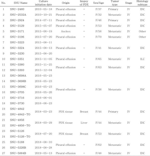
Within the 24 newly established cell lines, 20 were derived from pleural effusion, one was established from ascites, and 3 were from PDX tissues. Three organoids were established from three identical PDX tissues each derived from a cell line. For convenience, patient 1 (a set of SNU-3223, SNU-3224, SNU- 3230), Patient 2 (a set of SNU-3380, SNU-3393), Patient 3 (a set of SNU-3698A, SNU-3698B, SNU-3698C, SNU-3705, SNU-3716, SNU-3730) and patient 4 (a set of SNU-5188, SNU-5226B) were named. In addition, the cell line-organoid pairs were referred to as set 1 (a pair of SNU-4842, SNU- 4842-TO), set 2 (a pair of SNU-4856, SNU-4856-TO) and set 3 (a pair of SNU-5126, SNU-5126-TO). DNA fingerprinting revealed a heterogeneous distribution of 15 tetranucleotide repeat loci and an amelogenin gender determining marker in each cell line and organoids. This confirmed 15 unique and unrelated cell lines, and that it was a different cell line between same patients. It also proved that the 3 cell lines and organoids are from the same PDX tissue.
20
Table 2. Origin and in vivo characteristics of 21 human breast cancer cell lines and 3 paired cell line-organoid sets derived from same patient-derived xenograft (PDX) tumor
No. SNU Name Culture
initiation date Origin biopsy
of PDX Sex/Age Tumor
type Stage Histology subtype 1 SNU-2480 2010-05-18 Pleural effusion - F/37 Primary IV IDC 2 SNU-2532A 2010-10-21 Pleural effusion - F/32 Metastatic IV IDC 3 SNU-2924 2014-07-11 Pleural effusion - F/46 Primary IV IDC 4 SNU-3129 2015-05-07 Pleural effusion - F/53 Metastatic IV IDC 5 SNU-3171 2015-06-19 Ascites - F/56 Metastatic IV Other 6 SNU-3196 2015-07-20 Pleural effusion - F/70 Metastatic IV Other 7 SNU-3223 2015-08-11
Pleural effusion - F/41 Metastatic IV IDC 8 SNU-3224 2015-08-13
9 SNU-3230 2015-08-20
10 SNU-3351 2015-11-05 Pleural effusion - F/65 Metastatic IV ILC 11 SNU-3380 2015-11-25
Pleural effusion - F/63 Metastatic IV IDC 12 SNU-3393 2015-12-04
13 SNU-3698A 2016-05-23
Pleural effusion - F/36 Metastatic IV IDC 14 SNU-3698B 2016-05-23
15 SNU-3698C 2016-05-23 16 SNU-3705 2016-05-26 17 SNU-3716 2016-06-01 18 SNU-3730 2016-06-23 19 SNU-4842
2018-03-23 PDX tissue Breast F/44 Primary IV IDC 20 SNU-4842-TO
21 SNU-4856
2018-03-29 PDX tissue Liver F/44 Metastatic IV IDC 22 SNU-4856-TO
23 SNU-5126
2018-07-20 PDX tissue Breast F/53 Metastatic IV IDC 24 SNU-5126-TO
25 SNU-5188 2018-08-10
Pleural effusion - F/62 Metastatic IV IDC 26 SNU-5226B 2018-08-28
27 SNU-5884B 2019-05-13 Pleural effusion - F/48 Metastatic IV IDC
* - : not recorded ** IDC : Invasive Ductal Carcinoma *** ILC : Invasive Lobular Carcinoma
21
Continued
No. SNU Name Subtype ER PR HER2 Ki-67
1 SNU-2480 HR-HER2- Negative Negative Negative 60
2 SNU-2532A HR-HER2- Negative Negative 1+ -
3 SNU-2924 HR-HER2- Negative Negative Negative 50 4 SNU-3129 HR-HER2- Negative Negative Negative 5
5 SNU-3171 HR+HER2- 60 Negative 2+(FISH-) -
6 SNU-3196 HR+HER2- Focal weak - Negative -
7 SNU-3223
HR-HER2+ Negative Negative 3+ 60 8 SNU-3224
9 SNU-3230
10 SNU-3351 HR+HER2- 90 50 2+ -
11 SNU-3380
HR-HER2+ Negative Negative 3+ -
12 SNU-3393 13 SNU-3698A
HR-HER2+ Negative Negative 3+ -
14 SNU-3698B 15 SNU-3698C 16 SNU-3705 17 SNU-3716 18 SNU-3730 19 SNU-4842
HR-HER2+ Negative Negative Negative 30 20 SNU-4842-TO
21 SNU-4856
HR+HER2- Negative 70 Negative 10 22 SNU-4856-TO
23 SNU-5126
HR+HER2- 95 Negative Negative -
24 SNU-5126-TO 25 SNU-5188
HR+HER2- 80 5 Negative 7
26 SNU-5226B
27 SNU-5884B HR-HER2- <1% Negative Negative -
22
Table 3. DNA fingerprinting analysis using 15 STR loci and amelogenin for newly established 21 breast cancer cell lines and 3 matched patient-derived xenograft (PDX) cell line-organoid pairs
No. Cell-Name D8S1179 D21S11 D7S820 CSF1PO D3S1358 TH01 D13S317 D16S539
1 SNU-2480 10, 13 30, 31 9, 11 11 15 7 8 11
2 SNU-2532A 13 29 10, 12 9, 11 15, 16 6, 9 8 11
3 SNU-2924 12 30, 33.2 10 12 15 7 8 9, 11
4 SNU-3129 8, 12 31.2 8, 10 10 15 7 11 9, 10
5 SNU-3171 14, 15 30 11 11 15 9 8 11, 12
6 SNU-3196 11, 12 30, 31 11, 12 12 15, 18 10 9 9
7 SNU-3223 13,16 30 10, 12 12 15, 16 9 8, 10 10, 11
8 SNU-3224 13, 16 30 10, 12 12 15, 16 9 8, 10 10, 11
9 SNU-3230 13, 16 30 10, 12 12 15, 16 9 8, 10 10, 11
10 SNU-3351 13 31.2, 32.2 11, 12 11, 12 14 6, 8 8, 11 13
11 SNU-3380 10, 13 30, 31 11 12 15 9.3 11 11
12 SNU-3393 10, 13 30, 31 11 12 15 9.3 11 11
13 SNU-3698A 11, 16 30 11 12 15 9 8 9
14 SNU-3698B 11, 16 30 11 12 15 9 8 9
15 SNU-3698C 11, 16 30 11 12 15 9 8 9
16 SNU-3705 11, 16 30 11 12 15 9 8 9
17 SNU-3716 11, 16 30 11 12 15 9 8 9
18 SNU-3730 11, 16 30 11 12 15 9 8 9
19 SNU-4842 14, 15 31, 32.2 10, 12 12 17 9 8 10, 11
20 SNU-4842-TO 14, 15 31, 32.2 10, 12 12 17 9 8 10, 11
21 SNU-4856 10, 12 29, 30 10, 12 11, 12 15, 16 8, 9 8, 10 12 22 SNU-4856-TO 10, 12 29, 30 10, 12 11, 12 15, 16 8, 9 8, 10 12
23 SNU-5126 10, 12 29, 32.2 11, 12 10 15, 16 8, 9 8, 10 12
24 SNU-5126-TO 10, 12 29, 32.2 11, 12 10 15, 16 8, 9 8, 10 12
25 SNU-5188 12, 16 30 11, 12 9, 10 16, 17 9, 9.3 12 9
26 SNU-5226B 12, 16 30 11, 12 9, 10 16, 17 9, 9.3 12 9
27 SNU-5884B 13, 14, 16 31.2 11 10, 11 15, 17 9 10 9, 10
23
Continued
No. Cell-Name D2S1338 D19S433 VWA TPOX D18S51 Amelogenin D5S818 FGA
1 SNU-2480 17 13, 14 16, 17 11 12, 16 X, X 12 26
2 SNU-2532A 20 14.2 20 10, 11 13 X, X 12 26
3 SNU-2924 17 13 16 11, 12 18 X, X 9 21
4 SNU-3129 19, 25 12, 14.2 18 8, 11 11, 13 X, X 13 22
5 SNU-3171 19 13, 13.2 14 8, 9 10, 21.2 X, X 10 23
6 SNU-3196 23, 24 14 14, 18 11 19 X, X 10 24
7 SNU-3223 17, 20 14, 14.2 14 9 13, 16 X, X 11 20, 21
8 SNU-3224 17, 20 14, 14.2 14 9 13, 16 X, X 11 20, 21
9 SNU-3230 17, 20 14, 14.2 14 9 13, 16 X, X 11 20, 21
10 SNU-3351 19 14, 15.2 14, 19 8, 11 14 X, X 11, 12 22, 22
11 SNU-3380 23, 25 13, 15.2 14, 19 8, 11 16 X, X 11, 12 22, 23 12 SNU-3393 23, 25 13, 15.2 14, 19 8, 11 16 X, X 11, 12 22, 23
13 SNU-3698A 23 13, 14.2 16, 17 8, 11 15 X, X 11, 13 22
14 SNU-3698B 23 13, 14.2 16, 17 8, 11 15 X, X 11, 13 22
15 SNU-3698C 23 13, 14.2 16, 17 8, 11 15 X, X 11, 13 22
16 SNU-3705 23 13, 14.2 16, 17 8, 11 15 X, X 11, 13 22
17 SNU-3716 23 13, 14.2 16, 17 8, 11 15 X, X 11, 13 22
18 SNU-3730 23 13, 14.2 16, 17 8, 11 15 X, X 11, 13 22
19 SNU-4842 23 14 16, 17 9, 11 14, 16 X, X 10 22, 23
20 SNU-4842-TO 23 14 16, 17 9, 11 14, 16 X, X 10 22, 23
21 SNU-4856 19 12, 13 14, 18 8, 11 13, 17 X, X 10, 13 19, 25 22 SNU-4856-TO 19 12, 13 14, 18 8, 11 13, 17 X, X 10, 13 19, 25
23 SNU-5126 17 12, 15.2 16 9, 11 13 X, X 12 22, 24
24 SNU-5126-TO 17 12, 15.2 16 9, 11 13 X, X 12 22, 24
25 SNU-5188 17 13, 15.2 14, 19 8, 10 13, 20 X, X 10, 12 24
26 SNU-5226B 17 13, 15.2 14, 19 8, 10 13, 20 X, X 10, 12 24 27 SNU-5884B 17, 24 15.2 14, 18 11 12, 17 X, X 11, 12 19, 25
24 3.1. Culture characteristics
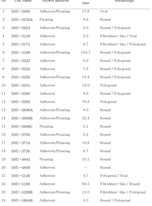
Majority of the established cell lines were in adherent form or coexisted with floating cells. Although derived from pleural effusion and ascites, only SNU-2532A, SNU-3698C cell lines maintained floating morphology. Of the three cell lines derived from PDX tissues, SNU-4842 was only floating pattern, the cells were tightly aggregated into a single lump like a matched organoid. The morphology of the whole cell line was classified into four types: polygonal, oval, fibroblast-like, round. Even in the same patient-derived cell lines, the more floating and clumped form, the slower it grew. As described above, growth patterns and characteristics of cell lines of the same origin were slightly different. SNU-3393, SNU-3698B and SNU-5188 were particularly slow in growth rate than other cell lines of the same patient. Overall, it was SNU-3196 with the longest doubling time and SNU-3129 with the shortest. It has been observed that all three organoids grow in various forms: dense and round, hollow, polygonal. Organoid sizes were mostly around 100-200㎛. The results of FFPE slide H&E staining also showed well-organized round-shaped organoids. In the initial passage, the size was larger, but over the passage, it did not
25
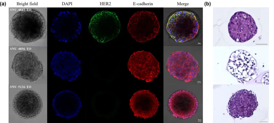
appear to be greatly larger than a certain level. When taking three organoid confocal images, it was noticeable that SNU- 4842-TO only HER2 green was overexpressed under the same conditions and brightness correction. All cell lines and organoids were confirmed to be free of contamination from either bacteria or mycoplasma and without cross-contamination.
26 Figure 1. Mycoplasma detection test
Mycoplasma was not detected in all cell lines and organoids. (a) 1st PCR result - Sample control : 810bp, Mycoplasma detection : 700-300bp (b) 2nd PCR result - Sample control : 590bp, Mycoplasma detection : 250-150bp
27
28
Figure 2. Microscopic images of 24 breast cancer cell lines and 3 breast PDX organoids
(a) Phase-contrast microscopy of newly established 24 breast cancer cell lines. The magnification is 100X and scale bar is 50
㎛. (b) Morphology of 3 breast cancer organoids taken through EVOS™ FL Auto 2 Imaging System (Thermo Fisher Scientific, MA, USA). The magnification is 200X and scale bar is 200㎛.
29
Figure 3. Images of confocal microscope and FFPE H&E staining of 3 organoids derived from PDXT
30
(a) Confocal analysis of immunofluorescence staining of 3 organoids. The scale bar size is 20㎛. Blue : DAPI, Green : HER2, Red : E-cadherin (b) H&E images from FFPE block sections of 3 organoids. The magnification is 100X and taken by EVOS™ FL Auto 2 Imaging System (Thermo Fisher Scientific, MA, USA).
The scale bar size is 100㎛.
31
Table 4. in vitro characteristics of 24 breast cancer cell lines
No Cell Name Growth patterns Doubling
time Morphology 1 SNU-2480 Adherent/Floating 17.6 Oval
2 SNU-2532A Floating 9.9 Round
3 SNU-2924 Adherent/Floating 6.0 Round / Polygonal 4 SNU-3129 Adherent 2.3 Fibroblast-like / Oval 5 SNU-3171 Adherent 8.7 Fibroblast-like / Polygonal 6 SNU-3196 Adherent/Floating 163.7 Round / Polygonal
7 SNU-3223 Adherent 6.0 Round / Polygonal
8 SNU-3224 Adherent 7.6 Round / Polygonal
9 SNU-3230 Adherent/Floating 18.9 Round / Polygonal
10 SNU-3351 Adherent 19.0 Polygonal
11 SNU-3380 Adherent 8.0 Round / Polygonal
12 SNU-3393 Adherent 76.5 Polygonal
13 SNU-3698A Adherent/Floating 8.0 Round 14 SNU-3698B Adherent/Floating 23.4 Round
15 SNU-3698C Floating 5.1 Round
16 SNU-3705 Adherent/Floating 5.8 Round 17 SNU-3716 Adherent/Floating 10.9 Round 18 SNU-3730 Adherent/Floating 6.7 Round
19 SNU-4842 Floating 10.1 Round
20 SNU-4856 Adherent - Round
21 SNU-5126 Adherent 4.7 Polygonal / Oval 22 SNU-5188 Adherent 60.5 Fibroblast-like / Round 23 SNU-5226B Adherent/Floating 12.0 Fibroblast-like / Polygonal 24 SNU-5884B Adherent 4.3 Round / Polygonal
32 3.2. Mutational traits
Whole-exome sequencing (WES) was performed for genetic characterization of newly established breast cancer cell lines and organoids. Among the overall results, 52 genes known for many genetic aberrations in breast cancer were analyzed. In the mutations reported in the Clinvar database (https://www.ncbi.nlm.nih.gov/clinvar), the results for the effect predictable mutations and BRCA, ERBB2 genes considered important in breast cancer are summarized in Table 5. Out of the 52 screened genes, organoids tend to have more mutations than cell lines. The least mutation burden was SNU-2532A.
Throughout all cell lines and organoids, the gene with the highest number of mutations was WNK2, and in almost all cases, the gene with aberrations was TP53. On the other hand, CHEK2, PBXW7, MEN1, NIBEAL2, NOTCH1, PBRM1, PTEN, and USP9X were meaningful mutant genes that appeared only in one cell line or organoid. More than half of the many genetic variations in breast cancer mentioned above were also identified in out samples:
TP53, PIK3CA, PTEN, HER2, RB1 and MAP3K. Different cell lines from one patient were found to have the same mutational profiles overall. In patient 1 (SNU-3223, SNU-3224, SNU-
33
3230) cell lines, some genes, such as MAP3K1 or FOXO3, had different mutational status, but most of them were known to have no significant effect, and the aberrations of the most important BRCA gene in breast cancer was completely the same. Patient 2 cell lines (SNU-3380, SNU-3393), Patient 3 cell liens (SNU- 3698A, SNU-3698B, SNU-3698C, SNU-3705, SNU-3716, SNU-3730) and patient 4 cell lines (SNU-5188, SNU-5226B) were analyzed to have the same major mutational status within each set. On the other hand, the distinction between cell line and organoid sets seemed to be clearly different. However, as with differences between cell lines of the same origin, most were not considered to have a major impact. There were some notable genes in the mutant state with some differences. In particular, set 2 (SNU-4856, SNU-4856-TO) had a lot of mutations in organoids, some of which are still controversial but thought to be pathogenic or to have a specific effect. Set 3 organoid (SNU- 5126-TO) had TP53 aberration which is thought to be likely pathogenic. But, this mutation was not found in set 3 cell line (SNU-5126). This result was confirmed by the IGV (Integrative Genomics Viewer, Broad Institute and the Regents of the University of California) program, as the mutation detection may
34
be due to the difference in the number of nucleotide transition copies. Although the HER2 mutation that was immediately identified in the set 1 organoid (SNU-4842-TO) was not immediately identified in the cell line (SNU-4842), it was confirmed that the same mutation exists even though the number of copies was small in the cell line using the above program. In western blot, there was no significant difference between samples from the same patient as a whole. Patient 3 cell lines (SNU-3698A, SNU-3698B, SNU-3698C, SNU-3705, SNU- 3716, SNU-3730), however, are clearly differentiated within the same patient origin set. Among ER, PR and HER2, which are widely used as criteria for dividing breast cancer, the whole cell lines and organoids were grouped based on ER and HER2, which clearly showed results. The results are summarized in Table6.
Set 1 (SNU-4842, SNU-4842-TO) was classified as ER+HER2+ (ER positive, HER2 positive). Patient 1 cell lines (SNU-3223, SNU-3224, SNU-3230), Patient 2 cell lines (SNU-3380, SNU-3393) and Patient 3 cell lines (SNU-3698A, SNU-3698B, SNU-3698C, SNU-3705, SNU-3716, SNU- 3730) were categorized in ER-HER2+ (ER negative, HER2 positive). ER+HER2- (ER positive, HER2 negative) includes
35
set2 and patient 4 cell lines (SNU-5188, SNU-5226B). In addition, when the overall results of western blot were analyzed, SNU-3129 was not able to confirm the expression of most proteins or the expression level was quite low, whereas SNU- 3716 was fairly high in protein expression in the set or as a whole.
36
Figure 4. Mutational landscape of the established breast cancer cell lines and PDX organoids
37 Table 5. Major mutational profile table
Gene nt change a.a. change Effect Gene nt change a.a. change Effect
SNU-2480 CDKN1B 443G>T Cys148Phe Uncertain significance
SNU-2532A BRCA1
3548A>G Lys1183Arg Uncertain significance
PIK3CA 3140A>G His1047Arg Pathogenic 3113A>G Glu1038Gly CIP
2612C>T Pro871Leu CIP
SNU-2924 BRCA1
3548A>G Lys1183Arg Uncertain significance TP53 215C>G Pro72Arg Uncertain significance 3113A>G Glu1038Gly CIP TNXB 9946G>A Ala3316Thr Uncertain significance
2612C>T Pro871Leu CIP ERBB2 3508C>G Pro1170Ala -
SNU-3129 BRCA2 1114A>C Asn372His - TP53 215C>G Pro72Arg Uncertain significance
SNU-3171 BRCA1
3548A>G Lys1183Arg Uncertain significance
TP53
742C>T Arg248Trp CIP
3113A>G Glu1038Gly CIP 215C>G Pro72Arg Uncertain significance
2612C>T Pro871Leu CIP
SNU-3196 TP53 215C>G Pro72Arg Uncertain significance
BRCA2 1114A>C Asn372His -
BRCA1 3627dupA Glu1210fs Pathogenic
* nt : nucleotide ** a.a. : amino acid *** CIP : Conflicting interpretations of pathogenicity **** - : not reported
38
Continued
Gene nt change a.a. change Effect Gene nt change a.a. change Effect
SNU-3223 BRCA1 3548A>G Lys1183Arg Uncertain significance
BRCA1 2612C>T Pro871Leu CIP 3113A>G Glu1038Gly CIP
SNU-3224 BRCA1
3548A>G Lys1183Arg Uncertain significance
BRCA1 2612C>T Pro871Leu CIP 3113A>G Glu1038Gly CIP
SNU-3230 BRCA1 3548A>G Lys1183Arg Uncertain significance
BRCA1 2612C>T Pro871Leu CIP 3113A>G Glu1038Gly CIP
SNU-3351 PIK3CA 3140A>G His1047Arg Pathogenic ERBB2 3508C>G Pro1170Ala -
CTNNA1 770A>G Asn257Ser Uncertain significance CDKN1B 326T>G Val109Gly CIP SNU-3380 TP53 743G>A Arg248Gln Likely pathogenic ERBB2 3508C>G Pro1170Ala - SNU-3393 TP53 743G>A Arg248Gln Likely pathogenic ERBB2 3508C>G Pro1170Ala - SNU-3698A PIK3CA 3145G>C Gly1049Arg Likely pathogenic ERBB2 3508C>G Pro1170Ala - SNU-3698B PIK3CA 3145G>C Gly1049Arg Likely pathogenic ERBB2 3508C>G Pro1170Ala - SNU-3698C PIK3CA 3145G>C Gly1049Arg Likely pathogenic ERBB2 3508C>G Pro1170Ala - SNU-3705 PIK3CA 3145G>C Gly1049Arg Likely pathogenic ERBB2 3508C>G Pro1170Ala - SNU-3716 PIK3CA 3145G>C Gly1049Arg Likely pathogenic ERBB2 3508C>G Pro1170Ala - SNU-3730 PIK3CA 3145G>C Gly1049Arg Likely pathogenic ERBB2 3508C>G Pro1170Ala -