• 검색 결과가 없습니다.

Regulation of SLITRK1 gene by neuron restrictive silencer factor in NMB cells

N/A
N/A
Protected

Academic year: 2021

Share "Regulation of SLITRK1 gene by neuron restrictive silencer factor in NMB cells"

Copied!
10
0
0

로드 중.... (전체 텍스트 보기)

전체 글

(1)

Introduction

Neuron restrictive silencer factor (NRSF), also called the repressor element 1 (RE-1) silencing transcription factor (REST), is a major transcriptional repressor for neuron-specific genes in non-neuronal cells and neuronal progenitor cells [1-

3]. NRSF/REST mediates transcription repression through the association of its amino terminal repression domain with the mSin3/histone deacethylase 1/2 (HDAC 1/2) complex [4-6].

and through the association of its carboxy terminal repression domain with the CoREST/HDAC complex [7,8]. NRSF/

REST bind to a 21 bp consensus sequence of neural restrictive silencer element (NRSE), also known as the RE-1, in the regulatory regions of many neuron-specific genes: ion channels, neurotranssmitter receptor, neurotrophic receptor, neuronal cell adhesion molecules, neuronal growth factors, neuronal cytoskeleton, and others [9,10].

The SLIT and NTRK-like family is a family of structurally related transmembrane proteins with an extracellular leucine-

Regulation of SLITRK1 gene by neuron restrictive silencer factor in NMB cells

Myung Mi Kim 1,† , Jin-Soo Kim 1,† , Sung-Min Moon 1 , Mi Suk Choi 1 , Bo Ram Park 1 , Dong-Seol Lee 2 , Shin-Yeob Mo 2 , Seon-Ho Cho 2 , Chun Sung Kim 1, *

1

Department of Oral Biochemistry,

2

Oral Biology Research Institute, School of Dentistry, Chosun University, Gwangju, Korea

ABSTRACT

Purpose: The Slitrk family are neuronal transmembrane proteins that participate in the regulation of neurite outgrowth and are selectively expressed in the brains of adult mice. Neuron restrictive silencer factor (NRSF), also called RE1-silencing transcription factor, was originally discovered as a transcriptional repressor in non-neuronal cells. However, in this study, we show that the neuron restrictive silencer element (NRSE) of SLITRK1 functions as a critical regulator to repress SLITRK1 gene expression in NMB cells.

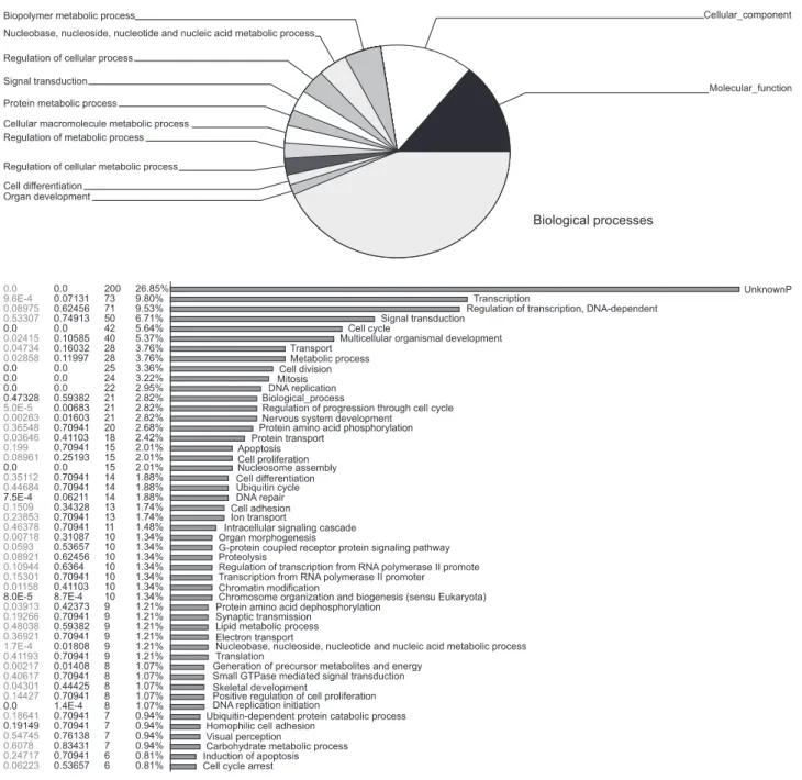
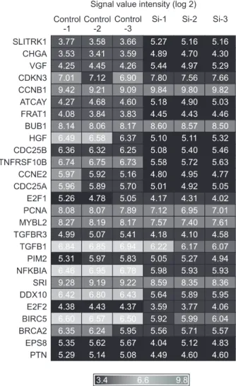
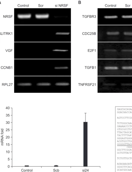
Materials and Methods: To screen NRSF/NRSE-regulated specific genes, we performed microarray analysis of NRSF siRNA- transfected NMB cells. We selected eight genes and confirmed their expression by RT-PCR analysis.

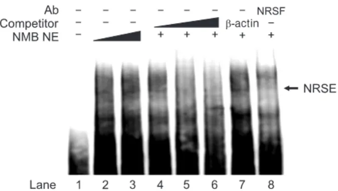
Results: Expression levels of SLITRK1, VGF, and CCNB1 were up-regulated in NRSF siRNA-transfected NMB cells compared with control NMB cells, whereas TGFBR3, CDC25B, E2F1, TGFB1, and TNFRSF21 were slightly down-regulated or showed no change. Especially, we identified three putative NRSF-binding sequences in the SLITRK1 gene, and its transcription was significantly up-regulated in NRSF siRNA-transfected NMB cells as measured by real-time PCR. Using co-transfection studies, we determined that the second NRSE of the SLITRK1 gene was functional in NMB cells. In addition, NRSF bound to the second NRSE of the SLITRK1 gene in a sequence-specific manner as confirmed by supershift assay. Our data suggest that NRSF interacts with and represses expression of the SLITRK1 gene.

Conclusion: NRSF plays an important role in regulating the growth and development of neuronal genes via SLITRK1 gene transcription.

Key Words: RE1-silencing transcription factor, Neuron restrictive silencer element, Transcriptional repressor, SLITRK1

Received Jun 19, 2013; Revised version received Jul 31, 2013 Accepted Jul 31, 2013

Corresponding author: Chun Sung Kim

Department of Oral Biochemistry, School of Dentistry, Chosun University, 309 Pilmun-daero, Dong-gu, Gwangju 501-759, Korea Tel: 82-62-230-7088, Fax: 82-62-224-3706

E-mail: [email protected]

These authors contributed equally to this work.

(2)

rich repeat domain homologous to SLIT and a short intracellular domain lacking the tyrosine phosphorylation motifs that are found among the other members of SLITRK family [11].

SLITRK family is consisted of six members (SLITRK1-6), which are expressed predominantly in neural tissue and have neurite-modulating activity in cultured neuronal cells [11].

SLITRK transcripts were selectively expressed in the brain of normal adult human. SLITRK1, SLITRK2, SLITRK3, SLITRK4, and SLITRK5 were expressed highly in the cerebral cortex and the fetal brain, whereas SLITRK6 was expressed in the putamen, the lung and the liver [12].

It was reported that SLIT and NTRK-like family, member 1 (SLITRK1) is related to the Tourette syndrome (TS) which is a potentially debilitating developmental neuropsychiatric disorder characterized by the presence of both motor and vocal tics and it has comorbid occurrence with obsessive-compulsive disorder and attention deficit hyperactivity disorder [13-16]. Recent studies, SLITRK1 enhanced the dentritic growth of the neuron by the interaction of its phosphorylated form and 14-3-3 proteins which are highly expressed in nervous system [17,18]. However, the mechanism of the expression regulation of the SLITRK1 is not known. Therefore, the aim of this study was to investigate the transcriptional regulation of SLITRK1 by the NRSF which is the well known as a transcriptional repressor.

Materials and Methods

Cell culture

The human neuroblastoma cell line NMB cells were grown in RPMI 1640 medium (Lonza, Walkersvile, MD, USA) supplement with 10% heat-inactivated fetal bovine serum, 100 U/mL penicillin, 100 μg/mL streptomycin (Gibco, Rockville, MD, USA) at 37

o

C in a humidified atmosphere of 5% CO

2

. siRNA transfection

NMB cells were plated in 6-well plates at a density of 7×10

5

cells in each well. After overnight incubation, cell were transfected with NRSF/NRSE siRNA using Lipofectamine 2000 transfection reagent (Invitrogen, Carlsbad, CA, USA) according to the supplier’s protocol. NRSF siRNA and Lipofectamine 2000 were each diluted in 250 μL Opti-MEM I and incubated for 5 minutes at room temperature. The diluted NRSF/REST siRNA and Lipofectamine 2000 were combined and incubated for 20 minutes at room

temperature. After incubation, NRSF/NRSE siRNA/Lipofectamin 2000 mixture were added to each well. The cells were incubated at 37

o

C in a humidified atmosphere of 5% CO

2

.

Microarray

For control and test RNAs, the synthesis of target cRNA probes and hybridization were performed using Agilent’s Low RNA Input Linear Amplification Kit PLUS (Agilent Technology, Santa Clara, CA, USA) according to the manufacturer’s instructions. Briefly, each 0.5 μg total RNA was mixed with the diluted Spike mix and T7 promoter primer mix and incubated at 65°C for 10 minutes. cDNA master mix (5× First strand buffer, 0.1 M DTT, 10 mM dNTP mix, RNase-Out, and MMLV-RT) was prepared and added to the reaction mixer. The samples were incubated at 40°C for 2 hours and then the RT and dsDNA synthesis was terminated by incubating at 65°C for 15 minutes. The transcription master mix was prepared as the manufacturer’s protocol (4× Transcription buffer, 0.1 M DTT, NTP mix, 50% PEG, RNase-Out, inorganic pyrophosphatase, T7-RNA polymerase, and Cyanine 3/5-CTP). Transcription of dsDNA was performed by adding the transcription master mix to the dsDNA reaction samples and incubating at 40°C for 2 hours. Amplified and labeled cRNA was purified on RNase Mini Spin Columns (Qiagen, Hilden, Germany) according to the manufacturer’s protocol. Labeled cRNA target was quantified using ND-1000 spectrophotometer (NanoDrop Technologies, Wilmington, DE, USA). After checking labeling efficiency, each 750 ng of cyanine 3-labeled and cyanine 5-labeled cRNA target were mixed and the fragmentation of cRNA was performed by adding 10× blocking agent and 25×

fragmentation buffer and incubating at 60°C for 30 minutes.

The fragmented cRNA was resuspended with 2× hybridization buffer and directly pipetted onto assembled Agilent Whole Human Genome Oligo Microarray. The arrays hybridized at 65°C for 17 hours using Agilent Hybridization Oven. The hybridized microarrays were washed as the manufacturer’s washing protocol (Agilent Technology).

Microarray data analysis

The hybridization images were analyzed by Agilent DNA

microarray Scanner and the data quantification was performed

using Agilent Feature Extraction software. The average

fluorescence intensity for each spot was calculated and local

(3)

background was subtracted. All data normalization and selection of fold-changed genes were performed using GeneSpring GX 7.3 (Agilent Technology). Genes were filtered with removing flag-out genes in each experiment. Intensity-dependent normalization (Lowess) was performed, where the ratio was reduced to the residual of the Lowess fit of the intensity vs. ratio curve. The averages of normalized ratios were calculated by dividing the average of normalized signal channel intensity by the average of normalized control channel intensity.

Semi-quantitative reverse transcription-polymerase chain reaction

Total RNA was isolated according to the supplier’s protocol (TRI Reagent; Molecular Research Center, Inc.). For reverse transcription-polymerase chain reaction (RT-PCR), 1 μg of total RNA was reverse transcribed and polymerase chain reactions (PCRs) were carried out with specific primers (Table 1) at the same tube using one-step RT-PCR reagent (Qiagen) in a My Genie 96 Thermal Block PCR machine (Bioneer, Daejeon, Korea). The PCR products were seperated in a 1% agarose gel with TAE beffer.

Real-time polymerase chain reaction

Total RNA was extracted using the TRIzol regeant (Invitro- gen) from NMB cells. Real-time PCR was performed as described by One Step SYBR

®

PrimeScript

TM

RT-PCR Kit (Perfect Real Time) protocol (Tacara, Seoul, Korea). Briefly, one step RT-PCR reagents were carried out in a 20 μL final volume of reaction buffer containing 2× One Step SYBR

®

RT- PCR Buffer III, TaKaRa Ex Taq

TM

HS (5 U/µL), PrimeScript

TM

RT enzyme Mix II, total RNA, RNase Free dH

2

O. The SLITRK1 forward primer (5'-ACTTCATGTACCTCCCGGT-3') and SLITRK1 reverse primer (5'-TACAGCTGAGGGCAGATCT-3') were added to the reaction buffer. The real-time PCR cycle

conditions for SLITRK1 consisted of 95

o

C for 30 seconds, 60

o

C for 30 seconds, 72

o

C for 30 seconds, followed by a 5 minutes extension at 72

o

C (40 cycle).

Cloning

The SLITRK1 was cloned by polymerase chain reaction amplification. PCR was performed using genomic DNA from NMB cells as a template and an upstream sense oligonucleotide (5'-GGGGTACCGGACAACCACAAAACGC AG-3'), containing a Hind III site, and downstream antisense oligonucleotide 1 (5'-GGGAAGCTTAGCCCAGACGCCA GTCAAT-3' containing a Kpn1 site, downstream antisense oligonucleotide 2 (5'-GGGAAGCTTAGTGTTGTTCTCTAC AGTAGC-3') containing a Kpn1 site, downstream antisense oligonucleotide 3 (5'-GGGAAGCTTCTCACACTTGAGGT CGCTC-3') containing a Kpn1 site. The amplified DNAs were cloned into PGL3-basic vector.

Luciferase assay

2×10

5

cells/well were cultured overnight before transfection.

NRSF binding sites were inserted upstream of the SLITRK1 promoter (−647 to +1,477), which had been inserted into the Kpn1-Hind III sites of pGL3 basic vector. Cells were transfected with PGL3/SLITRK1 promoter using FuGENE HD transfection reagent according to the manufacturer’s instruction. In brief, PGL3/SLITRK1 promoter and FuGENE HD transfection reagent were each dilution in 100 μL culture medium and incubation 15 minutes at room temperature. After incubation, PGL3/

SLITRK1 promoter/FuGENE HD transfection reagent mixture were added to each well. The cell were incubated at 37

o

C in a humidified atmosphere of 5% CO

2

for 24 hours. Cells grown to confluence were washed once with 1× phosphate buffered saline and lysed with prepared using lysis buffer (Promega, Madison, WI, USA). pCH110 (β-galactosidase; GE Healthcare

Table 1. Specific Primers for Reverse Transcription-Polymerase Chain Reaction SLITRK1

VGF CCNB1 TGFBR3 CDC25B E2F1 TGFBI TNFRSF21

5'ACTTCATGTACCTCCCGGT 5'ACCAGCTGTCTCCGGCAGC 5'GAAGGAAGCAAAACCTTCAG 5'GCCTCCGAGGCAGTTTGAA 5'GGAGCAGACGTTTGAACAGG 5'GGAAACTGACCATCAGTACCT 5'AACCACATCTTGAAGTCAGCT 5'CCACCCAGCTGGAAACTGA

5'TACAGCTGAGGGCAGATCT

5'CACGCCCTGGAAAAGCTCT

5'ATCAGAGAAAGCCTGACACA

5'AGTTGTGCCTCTGCTGGCA

5'TGCCAGAGCATGGGTGGAGC

5'TCCAGCTGCTGCTCGCTCT

5'TCCAGAGAGATGATTGCCGA

5'TGTCATCAAAGATAGGCTGC

(4)

Life Science, Piscataway, NJ, USA) was also co-transfected and measured to normalize transfection efficiency. The luciferase and β-galactosidase activities were determined according to the manufacture's instruction (Promega).

Electrophoretic mobility shift assay

The preparation of unclear extracts from NMB cells. Cyto- plasmic extract were prepared by lysing the cells for 15 minutes on ice with batter A (pH 7.9, 10 mM HEPES, 10 mM KCl, 0.1

mM EDTA, 0.1 mM EGTA, 1 mM DTT, 0.5 mM PMSF, 10 mM NaF, 2 mM Na

3

VO

4

, protease inhibitor). Add Nonidet P-40 at the end of incubation to the mixture at a concentration of 0.6%.

Centrifuge at 13,000 rpm for 1min and take the supernatant.

Resuspend in 40 µL of buffer B (pH 7.9 20 mM HEPES, 0.4 M NaCl, 1 mM EDTA, 1 mM EGTA, 1 mM DTT, 1 mM PMSF, 10 mM NaF, 1 mM Na

3

VO

4

, protease inhibitor) and vortex in the cold room for 20 minutes. Cell lysates were centrifuge at 13,000 rpm for 1 minute and take the supernatant was kept −80

o

C

Fig. 1. Biological function analysis of target gene by neuron restrictive silencer factor siRNA transfected and control NMB cells using microarray analysis.

(5)

prior to use. Biotin-labeled double-stranded oligonucleotides of the SLITRK1 promoter containing the NRSE were generated by oligonucleotide synthesis (upstream : 5'-GCTAACAGTTT ACCCTGCCCTGGGGGCTGCAGCTGAGTTTACCCTGC CCTGGGGGCTGCAGCTGCGACCA-3' downstream : 5'-TG GTCGCAGCTGCAGCCCCCAGGGCAGGGTAAACTCAG CTGCAGCCCCCAGGGCAGGGTAAACTGTTAGC-3') and electrophoretic mobility shift assay was performed as described by LightShift

®

Chemiluminescent EMSA Kit (Thermo Fisher Scientific Inc., Waltham, MA, USA).

Results

Gene expression profile in neuron restrictive silencer factor siRNA transfected NMB cells by microarray analysis To screen the genes which are regulated by NRSF expression in NMB cells, microarray analysis was performed by NRSF siRNA transfected. As shown in Fig. 1, 26% and 9% of genes were involved in transcription and its regulation, respectively.

Base on the microarray data, we selected 27 genes which are differential expression genes by NRSF siRNA transfection (Fig.

2). Among these genes, the expression level of SLITRK1, VGF, CCNB1, CDC25B, E2F1, TNFRSF21, TGFBR3, and TGFB1 were confirmed by RT-PCR analysis.

Gene expression by neuron restrictive silencer factor siRNA transfected NMB cells

To confirm the mircroarray data, RT-PCR was carried out.

The expression level of NRSF was significantly reduced in NRSF siRNA-transfected cells relative to negative siRNA-transfected cells. This data indicated that NRSF siRNA was successful in the knock-down of NRSF transcripts level. When NRSF expression was knock-downed, SLITRK1, VGF and CCNB1 were significantly increased but, CDC25B was decreased in NMB cells. In contrast, E2F1 and TNFRSF21, microarray based down-regulated genes, were significantly increased in NRSF knock-down NMB cells. However, TGFBR3 and TGFB1 were not altered in NRSF siRNA transfected NMB cells (Figs.

2 and 3). Base on this result, we analyzed the gene sequences of these genes to identify the NRSE region, NRSF binding site, and found 3 putative NRSE regions in SLITRK1 gene (Fig.

4). We also performed expriment to investgate whether the transcriptional repression of endogenous SLITRK1 gene can be

relieved after NRSF siRNA transfection in NMB cell. As shown Fig. 5, the expression level of SLITRK1 gene was significantly increased by NRSF siRNA transfection compared with scrambled siRNA control. This result indicated that the NRSF protein regulates the endogenous SLITRK1 gene expression at the transcription level.

Neuron restrictive silencer factor represses SLITRK1 transcription through a neuron restrictive silencer element-dependent mechanism

To determine the NRSF-binding site in the SLITRK1 promoter region, we analyzed promoter region of SLITRK1. As a result, there was three NRSF-binding sites; −624-−600, +1,015-+1,036, and +1,527-+1,548 (Fig. 5). Interestingly, the last two NRSF-

Fig. 2. The expression value of individual target gene by neuron

restrictive silencer factor siRNA transfected and control NMB cells

using microarray analysis.

(6)

binding sites are in the first exon (Fig. 5).

To investigate the main silencer site(s) of the NRSF, promoter assay was performed with luciferase expression plasmid constructs which contained various NRSF-binding site(s) (Fig.

6). The shortest construct, SLITRK1 −647/−529, had relatively high promoter activity in NMB cells. Deletion of SLITRK1

−647/+1,477 from 3' end to +1,461 and SLITRK1 −647/+1,477 showed significant loss of promoter activity. Deletion of 5'upstream region of SLITRK1 −647/+1,477 from −647 toward

−162 also significantly decreased the promoter activity in NMB cells. These data revealed that the region of pNRSE2 contributes

Fig. 3. Quantification of target genes of neuron restrictive silencer factor (NRSF) in NRSF knock-down NMB cells. NMB cells were transfected with NRSF siRNA and negative control siRNA (scr), and total RNA was isolated 24 hours post- transfection. Up-regulation (A) and down-regulation (B) of targets by NRSF knock-down in NMB cells. RPL27 was used as an internal control.

Fig. 4. SLITRK1 transcripts were increased by Knock-down of neu- ron restrictive silencer factor (NRSF). NMB cells were transfected with either NRSF siRNA or scrambled (scb) siRNA, and total RNAs were isolated 24 hours after transfection. SLITRK1 transcripts were analyzed by real-time polymerase chain reaction.

Fig. 5. The nucleotide sequence of the 5'flanking region and the first exon of human SLITRK1 gene. Neuron restrictive silencer factor ( NRSF) binding sites and start codon are marked with bold letters.

Several factor binding sites such as NF-Y, CREB, E2F1, NF-kB, PPAR-

g, and p300 are marked.

(7)

to the basal promoter activity of SLITRK1.

Binding of neuron restrictive silencer factor to the neuron restrictive silencer element cis-acting element of SLITRK1 gene

The NRSF gene was cloned on the basis of its ability to bind to NRSEs in the MOR and SCG10 genes. We investigated whether this NRSF protein was able to interact with the NRSE of the SLITRK1 gene.

We performed a super gel shift assay with nuclear ex- tracts from NMB cells expressing hight levels of the NRSF endogenously using the NRSE probe of the SLITRK1 promoter.

As shown in Fig. 6, one major DNA-binding complex from NMB cell nuclear extracts were observed (Fig. 7). The upper complex was abolished in the presence of monoclonal NRSF antibody (Fig. 7). Competitive binding experiments were also conducted in this electophoretic mobility shift assay using a 20-fold molar excess of a self-NRSE cold competitor and nonspecific β-actin

competitor (Fig. 7). The self-NRSE competitor for protein-DNA interac tion, whereas the nonspecific β-actin competitor did not affect the binding, indicating the specific interaction. These data demonstrated that the NRSF protein binds to the NRSE DNA element of the SLITRK1 promoter.

Discussion

It was well known that REST/NRSF plays an important role in repression of the neuronal specific gene expression of non- neuronal cells [1,2]. However, it functions the participation in the several cellular pathway as well as transcription repressor. NRSF affects as an oncogene in medulloblastoma [19], neuroblastoma [20] and functions as a tumor suppressor of brest cancer [21], colorectal cancer [22]. In addition, NRSF are related to cell survival, apoptosis, and translational regulation.

To investigate the other target genes of NRSF in neuronal cells,

the siRNA assay and microarray assay were performed in this

Fig. 6. NRSF represses SLITRK1 promoter activity (A). Promoter

activity of wild type and mutated NRSE in SLITRK1 gene (B). NMB

cells were transfected with those constructs, and whole protein were

prepared 24 hours post-transfection. Luciferase activity was normal-

ized to β-galactosidase activity measured with the control plasmid

(pCH110) which was co-transfected as internal standard. Data are

expressed as means±SEM from three independent experiments.

(8)

study. The microarray result showed that the expression level of SLITRK1, VGF, and CCNB1 were increased and TGFBR3, CDC25B, E2F1, TGFBI and TNFRSF21 were decreased in NRSF knock-downed NMB cells.

SLITRK1 has the ability of the enhances neuronal dendrite outgrowth [17]. CCNB1 is the key regulator fo cell division in all eukaryotes, where it binds to the cyclin-dependent kinase 1 to form the cyclin B/cdk1 complex, which initiates the mitotic program via phosphorylation of select proteins. CCNB1 is overexpressed in several human carcinomas, and its correlate in some cases with tumor aggressiveness. Neuropeptide VGF, a secreted neuropeptide, has antidepressant-like actions in rodents [23,24]. However, the significance and role of VGF in human psychiatric disorders remains to be elucidated. The expression of these genes is consistent with the microarray results were verified by RT-PCR in NRSF siRNA transfected NMB cells (Fig. 2). These results indicate that NRSF will serve as a repressor of these genes.

TGFBR3 is the major mediator of TGF-β signaling pathways [25] and also functions as a BMP cell-surface receptor.

Particularly, TGFBR3 can modulate the biological function of BMP2 [26]. E2F1 stimulates the transcription of several genes in the apoptosis pathway [27]. E2F1 is stabilized and activated by DNA damage in an ATM-dependent manner, resulting in the activation of a subset of E2F1 targets [28,29]. TGFBI is transforming growth factor B induced gene and expressed

predominantly on the external surface of corneal epithelial cells, mutated in corneal dystrophies, major component of abnormal deposits. CDC25 phosphatase function as key regulators in cell cycle progression by activating the CDK complexes. They also have an essential role in the DNA damage [30]. TNSFRSF21 is a death domain-containing receptor of the tumour necrosis factor-receptor family [31,32]. TNFRSF21 expression is increased in cancerous tissue biopsies from patients with late stage prostate or breast cancer compared with levels in normal tissue [33]. When performed RT-PCR, TGFBR3 and CDC25B were consistent with the results of microarray and TGFBI was not altered. However, E2F1 and TNFRSF21 were increased (Fig. 2).

SLITRK1 plays an important role in growth and development of neuronal tissue. It was reported that SLITRIK1 is closely related to the TS and Trichotillomania (TTM) [17,34]. TS is a potentially debilitating developmental neuropsychiatric disorder characterized by chronic phonic and motor tics [18]. TTM is a chronic behavioral disorder characterized by the irresistible urge to pull out one's hair [34]. The expression of SLITRIK1 was increased in case of knock-down of NRSF in NMB cells (Figs. 3, 4). These results indicate that NRSF act a transcriptional repressor for SLITRIK1 in NMB cells.

The promoter assay result showed that the pNRSE2, +1,015- +1,036 located in exon1 were the main regulatory regions acted an silencer (Figs. 5, 6). This was confirmed by gel shift assay (Fig. 7).

In summary, NRSF was interacted with SLITRK1 gene result in repressed the its gene transcription. So NRSF play an important role to regulate the growth and development of neuronal gene via SLITRK1 gene trnascription.

Acknowledgments

This study was supported (in part) by research funds from Education and Cultural Foundation of College of Dentistry, Chosun University, 2010.

References

1. Schoenherr CJ, Anderson DJ: The neuron-restrictive silencer factor (NRSF): a coordinate repressor of multiple neuron- specific genes. Science 267:1360-1363, 1995.

Fig. 7. Binding of NRSF protein to the SLITRK1 NRSE. A supershift assay was performed with nuclear extracts from NMB cells. The double-stranded biotin-labeled NRSE was used as a probe. lane 1, probe alone; lanes 2-8, probe plus 10 μg of NMB cell nuclear extracts;

lane 4, 20-fold molar excess of unlabeled self-competitor; lane 5, 40-

fold molar excess of unlabeled self-competitor; lane 6, 80-fold molar

excess of unlabeled self-competitor; lane 6, nonspecific β-actin; lane 7,

rabbit monoclonal NRSF antibody. NRSF: neuron restrictive silencer

factor, NRSE: neuron restrictive silencer element.

(9)

2. Chong JA, Tapia-Ramírez J, Kim S, Toledo-Aral JJ, Zheng Y, Boutros MC, Altshuller YM, Frohman MA, Kraner SD, Mandel G: REST: a mammalian silencer protein that restricts sodium channel gene expression to neurons. Cell 80:949-957, 1995.

3. Jones FS, Meech R: Knockout of REST/NRSF shows that the protein is a potent repressor of neuronally expressed genes in non-neural tissues. Bioessays 21:372-376, 1999.

4. Grimes JA, Nielsen SJ, Battaglioli E, Miska EA, Speh JC, Berry DL, Atouf F, Holdener BC, Mandel G, Kouzarides T: The co- repressor mSin3A is a functional component of the REST- CoREST repressor complex. Biol Chem 275:9461-9467, 2000.

5. Roopra A, Sharling L, Wood IC, Briggs T, Bachfischer U, Paquette AJ, Buckley NJ: Transcriptional repression by neuron- restrictive silencer factor is mediated via the Sin3-histone deacetylase complex. Mol Cell Biol 20:2147-2157, 2000.

6. Naruse Y, Aoki T, Kojima T, Mori N: Neural restrictive silencer factor recruits mSin3 and histone deacetylase complex to repress neuron-specific target genes. Proc Natl Acad Sci U S A 96:13691-13696, 1999.

7. You A, Tong JK, Grozinger CM, Schreiber SL: CoREST is an integral component of the CoREST- human histone deacetylase complex. Proc Natl Acad Sci U S A 98:1454-1458, 2001.

8. Ballas N, Battaglioli E, Atouf F, Andres ME, Chenoweth J, Anderson ME, Burger C, Moniwa M, Davie JR, Bowers WJ, Federoff HJ, Rose DW, Rosenfeld MG, Brehm P, Mandel G: Regulation of neuronal traits by a novel transcriptional complex. Neuron 31:353-365, 2001.

9. Lunyak VV, Burgess R, Prefontaine GG, Nelson C, Sze SH, Chenoweth J, Schwartz P, Pevzner PA, Glass C, Mandel G, Rosenfeld MG: Corepressor-dependent silencing of chromosomal regions encoding neuronal genes. Science 298:1747-1752, 2002.

10. Bruce AW, Donaldson IJ, Wood IC, Yerbury SA, Sadowski MI, Chapman M, Göttgens B, Buckley NJ: Genome-wide analysis of repressor element 1 silencing transcription factor/neuron- restrictive silencing factor (REST/NRSF) target genes. Proc Natl Acad Sci U S A 101:10458-10463, 2004.

11. Aruga J, Mikoshiba K: Identification and characterization of Slitrk, a novel neuronal transmembrane protein family controlling neurite outgrowth. Mol Cell Neurosci 24:117-129, 2003.

12. Aruga J, Yokota N, Mikoshiba K: Human SLITRK family genes:

genomic organization and expression profiling in normal brain and brain tumor tissue. Gene 315:87-94, 2003.

13. Stillman AA, Krsnik Z, Sun J, Rasin MR, State MW, Sestan N, Louvi A: Developmentally regulated and evolutionarily conserved expression of SLITRK1 in brain circuits implicated in Tourette syndrome. J Comp Neurol 513:21-37, 2009.

14. Singer H: Tourette's syndrome: from behaviour to biology.

Lancet Neurol 4:149-159, 2005.

15. Carter AS, O'Donnell DA, Schultz RT, Scahill L, Leckman JF, Pauls DL: Social and emotional adjustment in children affected with Gilles de la Tourette's syndrome: associations with ADHD

and family functioning. Attention Deficit Hyperactivity Disorder. J Child Psychol Psychiatry 41:215-223, 2000.

16. Lombroso PJ, Scahill L: Tourette syndrome and obsessive- compulsive disorder. Brain Dev 30:231-237, 2008.

17. Abelson JF, Kwan KY, O'Roak BJ, Baek DY, Stillman AA, Morgan TM, Mathews CA, Pauls DL, Rasin MR, Gunel M, Davis NR, Ercan-Sencicek AG, Guez DH, Spertus JA, Leckman JF, Dure LS 4th, Kurlan R, Singer HS, Gilbert DL, Farhi A, Louvi A, Lifton RP, Sestan N, State MW: Sequence variants in SLITRK1 are associated with Tourette's syndrome. Science 310:317-320, 2005.

18. Kajiwara Y, Buxbaum JD, Grice DE: SLITRK1 binds 14-3-3 and regulates neurite outgrowth in a phosphorylation-dependent manner. Biol Psychiatry 66:918-925, 2009.

19. Lawinger P, Venugopal R, Guo ZS, Immaneni A, Sengupta D, Lu W, Rastelli L, Marin Dias Carneiro A, Levin V, Fuller GN, Echelard Y, Majumder S: The neuronal repressor REST/NRSF is an essential regulator in medulloblastoma cells. Nat Med 6:826-831, 2000.

20. Palm K, Metsis M, Timmusk T: Neuron-specific splicing of zinc finger transcription factor REST/NRSF/XBR is frequent in neuroblastomas and conserved in human, mouse and rat.

Brain Res Mol Brain Res 72:30-39, 1999.

21. Lv H, Pan G, Zheng G, Wu X, Ren H, Liu Y, Wen J: Expression and functions of the repressor element 1 (RE-1)-silencing transcription factor (REST) in breast cancer. J Cell Biochem 110:968-974, 2010.

22. Westbrook TF, Martin ES, Schlabach MR, Leng Y, Liang AC, Feng B, Zhao JJ, Roberts TM, Mandel G, Hannon GJ, Depinho RA, Chin L, Elledge SJ: A genetic screen for candidate tumor suppressors identifies REST. Cell 121:837-848, 2005.

23. Hunsberger JG, Newton SS, Bennett AH, Duman CH, Russell DS, Salton SR, Duman RS: Antidepressant actions of the exercise-regulated gene VGF. Nat Med 13:1476-1482, 2007.

24. Thakker-Varia S, Krol JJ, Nettleton J, Bilimoria PM, Bangasser DA, Shors TJ, Black IB, Alder J: The neuropeptide VGF produces antidepressant-like behavioral effects and enhances proliferation in the hippocampus. J Neurosci 27:12156-12167, 2007.

25. Horner A, Kemp P, Summers C, Bord S, Bishop NJ, Kelsall AW, Coleman N, Compston JE: Expression and distribution of transforming growth factor-beta isoforms and their signaling receptors in growing human bone. Bone 23:95-102, 1998.

26. Kirkbride KC, Townsend TA, Bruinsma MW, Barnett JV, Blobe GC: Bone morphogenetic proteins signal through the transforming growth factor-beta type III receptor. J Biol Chem 283:7628-7637, 2008.

27. Nahle Z, Polakoff J, Davuluri RV, McCurrach ME, Jacobson MD, Narita M, Zhang MQ, Lazebnik Y, Bar-Sagi D, Lowe SW:

Direct coupling of the cell cycle and cell death machinery by E2F. Nat Cell Biol 4:859-864, 2002.

28. Lin WC, Lin FT, Nevins JR: Selective induction of E2F1 in

response to DNA damage, mediated by ATM-dependent

(10)

phosphorylation. Genes Dev 15:1833-1844, 2001.

29. Stevens C, Smith L, La Thangue NB: Chk2 activates E2F-1 in response to DNA damage. Nat Cell Biol 5:401-409, 2003.

30. Dalvai M, Mondesert O, Bourdon JC, Ducommun B, Dozier C:

Cdc25B is negatively regulated by p53 through Sp1 and NF-Y transcription factors. Oncogene 30:2282-2288, 2011.

31. Locksley RM, Killeen N, Lenardo MJ: The TNF and TNF receptor superfamilies: integrating mammalian biology. Cell 104:487-501, 2001.

32. Pan G, Bauer JH, Haridas V, Wang S, Liu D, Yu G, Vincenz C,

Aggarwal BB, Ni J, Dixit VM: Identification and functional characterization of DR6, a novel death domain-containing TNF receptor. FEBS Lett 431:351-356, 1998.

33. Kasof GM, Lu JJ, Liu D, Speer B, Mongan KN, Gomes BC, Lorenzi MV: Tumor necrosis factor-alpha induces the expression of DR6, a member of the TNF receptor family, through activation of NF-kappaB. Oncogene 20:7965-7975, 2001.

34. Zuchner S, Cuccaro ML, Tran-Viet KN, Cope H, Krishnan RR,

Pericak-Vance MA, Wright HH, Ashley-Koch A: SLITRK1

mutations in trichotillomania. Mol Psychiatry 11:887-889, 2006.

수치

Table 1. Specific Primers for Reverse Transcription-Polymerase Chain Reaction SLITRK1 VGF CCNB1 TGFBR3 CDC25B E2F1 TGFBI TNFRSF21 5'ACTTCATGTACCTCCCGGT  5'ACCAGCTGTCTCCGGCAGC  5'GAAGGAAGCAAAACCTTCAG 5'GCCTCCGAGGCAGTTTGAA5'GGAGCAGACGTTTGAACAGG 5'GGAAACTGACC
Fig. 1. Biological function analysis of target gene by neuron restrictive silencer factor siRNA transfected and control NMB cells using microarray analysis.
Fig. 2. The expression value of individual target gene by neuron  restrictive silencer factor siRNA transfected and control NMB cells  using microarray analysis.
Fig. 3. Quantification of target genes of  neuron restrictive silencer factor (NRSF)  in NRSF  knock-down NMB cells
+2

참조

관련 문서

In the present study GSEA and hierarchical clustering of gene expression profile demonstrated that the PBMCs did not carry the signs of TNF-α activation (i.e. “TNF-α signature”)

For the data collection, we generated our gene expression profiles of Illumina platform and our gene expression profile demonstrated higher expression and connectivity than

According to the dose-related gene expression analysis, heat shock 27 kDa protein (HSP27), neurodegeneration-associated protein 1 (Neurodap 1) genes were significantly up-regulated

Therefore, we characterized a genetic network of genes induced by ER stress using cDNA microarray and gene set expression coherence analysis (GSECA), and identified gene function

In our previous study, a neuron-specific gene expression system (NSE) was used to enhance the gene expression efficiency in the neural lineage, such as mouse adult

Experimental Design: Microarray technologies were used to generate and analyze gene expression profiling data from 65 gastric cancer patients to identify biomarker genes associated

In order to validate the results from the microarray analy- sis, we selected four genes, which are C1R, CCL2, CXCL6 and HLA-A, and compared their gene expression in porcine

A subset of genes that showed down-regulation of 2.5-fold or higher by wild-type melanocortin-1 receptor (WT-Mc1r) compared to mutant (MT)-Mc1r in microarray assay and a