Combined Muscle Motor and Somatosensory Evoked Potentials for Intramedullary Spinal Cord Tumour Surgery
Il Choi,1 Seung-Jae Hyun,2 Joong-Koo Kang,3 and Seung-Chul Rhim1
Departments of 1Neurosurgery and 3Neurology, Asan Medical Center, University of Ulsan College of Medicine, Seoul;
2Department of Neurosurgery, Spine Center, Seoul National University Bundang Hospital, Seoul National University College of Medicine, Seongnam, Korea.
Received: November 6, 2013 Revised: November 6, 2013 Accepted: November 26, 2013
Corresponding author: Dr. Seung-Chul Rhim, Department of Neurosurgery,
Asan Medical Center,
University of Ulsan College of Medicine, 88 Olympic-ro 43-gil, Songpa-gu, Seoul 138-736, Korea.
Tel: 82-2-3010-3554, Fax: 82-2-476-6739 E-mail: [email protected]
∙ The authors have no financial conflicts of interest.
© Copyright:
Yonsei University College of Medicine 2014 This is an Open Access article distributed under the terms of the Creative Commons Attribution Non- Commercial License (http://creativecommons.org/
licenses/by-nc/3.0) which permits unrestricted non- commercial use, distribution, and reproduction in any medium, provided the original work is properly cited.
Purpose: To evaluate whether intraoperative neurophysiologic monitoring (IONM) with combined muscle motor evoked potentials (mMEPs) and somatosensory evoked potentials is useful for more aggressive and safe resection in intramedul- lary spinal cord tumour (IMSCT) surgery. Materials and Methods: We reviewed data from consecutive patients who underwent surgery for IMSCT between 1998 and April 2012. The patients were divided into two groups based on whether or not IONM was applied. In the monitored group, the procedures were performed under IONM using 75% muscle amplitude decline weaning criteria. The control group was comprised of patients who underwent IMSCT surgery without IONM.
The primary outcome was the rate of gross total excision of the tumour on mag- netic resonance imaging at one week after surgery. The secondary outcome was the neurologic outcome based on the McCormick Grade scale. Results: The two groups had similar demographics. The total gross removal tended to increase when intraoperative neurophysiologic monitoring was used, but this tendency did not reach statistical significance (76% versus 58%; univariate analysis, p=0.049; mul- tivariate regression model, p=0.119). The serial McCormick scale score was simi- lar between the two groups (based on repeated measure ANOVA). Conclusion:
Our study evaluated combined IONM of trans-cranial electrical (Tce)-mMEPs and SEPs for IMSCT. During IMSCT surgery, combined Tce-mMEPs and SEPs using 75% muscle amplitude weaning criteria did not result in significant improvement in the rate of gross total excision of the tumour or neurologic outcome.
Key Words: Intraoperative monitoring, spinal cord neoplasm, sensitivity and specificity
INTRODUCTION
Intramedullary spinal cord tumours (IMSCT) are rare neoplasms that account for only 2--4% of central nervous system tumours and 15% of all primary intradural tumours in adults.1,2 Surgery is the most effective treatment for the majority of in- tramedullary tumours including most ependymomas and haemangioblastomas, al- though whether aggressive surgical resection of astrocytomas is beneficial remains
including sex, age, body mass index (BMI), location of the tumour, the tumour size, combination with syrinx, and his- tological diagnosis. For sub-category analyses, we divided the histological diagnosis into 5 categories, such as astrocy- toma, ependymoma, haemangioblastoma, cavernous hae- mangioma, and others. We also divided the patients based on the lesion site: cervical, thoracic, and lumbar. The size of the tumour was calculated based on the maximum diameter of the lesion.
Methodology of Tce-mMEP/SEP intraoperative neurophysiologic monitoring
Anaesthesia
Anaesthesia was maintained with continuous infusion of propofol (10 mg/kg/h) and remifentanil (0.25 mg/kg/min).
A single bolus of non-depolarising short-acting muscle re- laxant (rocuronium) was administered at induction to facili- tate tracheal intubation and ventilation.
No paralytic agents were used after induction and intuba- tion. The level of neuromuscular block was monitored by recording the compound muscle action potentials to a train of four stimuli. Invasive blood pressure, electrocardiogram, end-tidal carbon dioxide concentration, pulse oximetry, and temperature were all monitored. The bispectral index was used for monitoring the depth of sedation.17
Intraoperative SEPs
Stimulation of SEPs was accomplished using square wave electrical pulses of 0.3 ms in duration and a maximum inten- sity of 25 mA at a frequency of 4.7 Hz. Surface-stimulating electrodes were placed over each median nerve at the wrist and over each posterior tibial nerve at the ankle. Evoked po- tentials were recorded in a referential fashion from the C3’
(2 cm posterior to C3, right median nerve stimulation), C4’
(2 cm posterior to C4, left median nerve stimulation), and Cz’ (2 cm posterior to Cz, right and left tibial nerve stimula- tion) positions and from a reference electrode at FPZ (inter- national 10--20 system). The low-filter setting was typically 30 Hz, but in some cases, 50 Hz was used. The high-filter setting was 3000 Hz.
Intraoperative MEPs
Multi-pulse transcranial electrical stimulation was performed using a commercially available intraoperative monitoring (IOM) electrical stimulator (NeuropackMEB-9200K; Nihon Kohden Co., Tokyo, Japan). Nine-millimetre disc elec- controversial.1-3 Most intramedullary spinal cord neoplasms
are low-grade lesions and are well circumscribed. Thus, gross total tumour resection can lead to long-term survival by controlling long-term tumour recurrence and by preserv- ing neurological function.4-6 Intraoperative neurophysiolog- ic monitoring (IONM) is one of the modalities that assist in the goal of maximising tumour resection and minimising neurological morbidity.7-10 Since the application of somato- sensory evoked potential (SSEP) in scoliosis surgery was reported in 1977, trans-cranial electronic motor-evoked po- tentials (MEPs), electromyography (EMG), and direct wave monitoring (D-wave) have been introduced and used in IMSCT surgery.8,11-13 The types of IONM modalities or weaning criteria that provide the most reliable results for IMSCT surgery remain unclear. Recently, the combined D- wave and muscle MEPs (mMEPs) monitoring generated notable results. However, combined mMEPs and SSEP monitoring without D-wave has rarely been reported upon for IMSCT surgery.8,9,14-16
The aim of our study was to determine whether IONM with combined mMEP using an amplitude of 75% weaning criteria and SSEP is useful for more aggressive and safer resections for IMSCT surgery.
MATERIALS AND METHODS
Study design and setting
We reviewed consecutive data such as clinical charts, opera- tive notes, neurophysiologic data, and neuro-imaging stud- ies from patients who underwent surgery for IMSCT be- tween 1998 and April 2012. The patients were divided into two groups based on whether IONM was applied or not.
The monitored group (Group A) consisted of patients who underwent IMSCT surgery using trans-cranial electrical (Tce)-mMEP/SEP IONM between July 2006 and April 2012. The control group (Group B) consisted of patients who underwent IMSCT surgery without IONM between 1998 and July 2006. All operations were performed by one senior spine neurosurgeon (S.C.R.) within a single institute.
Our study was conducted with the approval of the Institu- tional Review Board (local institute IRB number S2012- 1952-0001).
Evaluating baseline demographic characteristics following clinical and radiologic findings
We compared demographic factors between the two groups
nance images for total excision at 1 week after the surgery.
The neuro-radiologist confirmed tumour excision and resid- ual tumour. The secondary outcome was neurologic out- come. We used the McCormick Grade scale to assess the neurologic outcome.2 Patients were graded a total of three times: upon admission, at postoperative week 1, and during the outpatient clinic stage. Neurologic examinations were conducted by a neurosurgery or rehabilitation medicine res- ident during the immediate postoperative week (week 1).
The follow-up assessments were conducted by attending neurosurgeons during clinic visits.
Statistical analyses
Data were imported into SPSS (version 20.1; SPSS Inc., Chicago, IL, USA) for analysis. Based on the characteristics of the variables, we used unpaired Student’s t-tests or chi- squared tests to determine significant differences between the two groups in sex, age, body mass index, location of tu- mour, tumour size (max, diameter), syrinx combined or not, and histological diagnoses. We performed repeated measure ANOVA to compare the effect of monitoring in real time for IMSCT surgery and to compare outcomes based on the McCormick scale during the preoperative period, the im- mediate post-op (one week) period, and during the outpa- tient clinic stage after surgery. Uni-variate regression analy- ses were performed to assess the individual effects of sex, age, tumour size, tumour location, and histological diagno- sis on the rate of total excision. A stepwise multiple linear regression was then performed using backward elimination to select an appropriate model. The p value >0.10 was used for removal.
RESULTS
We enrolled a total of 76 patients who underwent surgery for IMSCT between 1998 and April 2012. The monitored group consisted of 50 patients who underwent IMSCT sur- gery with IONM between July 2006 and April 2012. The control group included 26 patients operated on between 1998 and July 2006. The mean follow-up duration was 18.2 months in the monitored group and 31.3 months in the un- monitored group.
Patient baseline characteristics
Baseline characteristics are compared in Table 1. Overall, both cohorts were similar. The mean age of the study popu- trodes were attached to the scalp using colloid ion 6 cm an-
terior to Cz and at C3 and C4 (international 10--20 system).
trains of 6 pulses (individual stimulus duration: 50 ms) with inter-stimulus intervals of 3 ms were used with a period of at least 30 seconds between two successive trains. Stimulus in- tensity was increased gradually (50 V increments from 100 V to a maximum of 600 V) until MEP amplitudes were maxi- mised above a minimum of 20 mV. If response amplitudes of at least 20 mV were not obtained from either leg, MEP monitoring was abandoned. MEPs were recorded simulta- neously from the tibialis anterior and abductor hallucis muscles of both legs and from the abductor pollicis muscles of both arms using a pair of non-insulated subcutaneous needle electrodes inserted 3 cm apart in each muscle. The time base was 100 ms. The typical low-filter setting used was 30 Hz, but in some cases, 50 Hz was used. The high- filter setting used was 3000 Hz.
Alarming criteria and surgical corresponding
A drop in amplitude of 50% or a prolongation in SSEP la- tency of 10% was considered a significant finding. If mean- ingful SEP changes occurred prior to myelotomy, the pro- cedure was stopped temporarily and the corresponding the change was addressed. However, if the SSEP change oc- curred during the myelotomy, the myelotomy continued re- gardless of the SSEP finding.
A drop in mMEP of 75% or complete loss from baseline amplitude was considered a significant finding. If any signifi- cant changes in mMEP occurred, the surgical procedure was stopped temporarily. We then checked the mean blood pres- sure (BP) and increased the BP to at least above 60 mm Hg.
Additionally, we irrigated the surgical field using warm sa- line solution. If possible, we temporally skipped the site causing the alarm. We performed procedures at other tu- mour marginal sites. After all corrective methods, the sur- gery withdraw in selected cases regarding tumour histologi- cal findings, the patient’s preoperative neurological status.
We monitored MEP and SEP changes regularly, particu- larly during the following steps: 1) positioning the patient, 2) laminectomy, 3) dura opening, 4) tumour resection, 5) dura closure, 6) after laminar insertion. In addition, we per- formed MEP whenever the surgeon checked for spinal cord injury.
Outcome measurement
The primary outcome was the rate of gross total excision of the tumour. We evaluated postoperative magnetic reso-
ly associated with gross total resection. The use of monitor- ing did not result in a statistically significant effect [odds ratio (OR): 3.14, 95% confidence interval (CI), 0.75--13.33, p-value=0.119].
The neurologic outcome based on the McCormick scale score was not significantly different between the two groups.
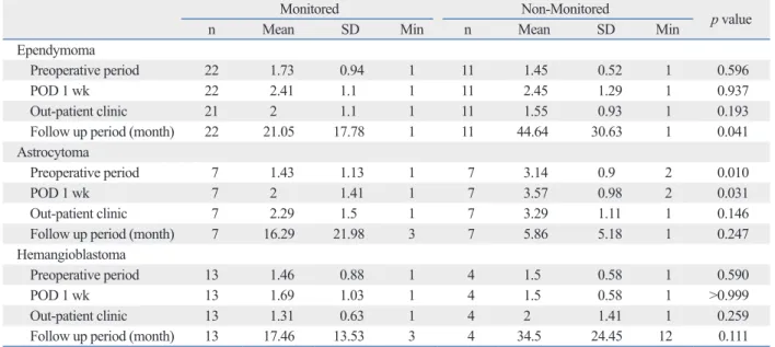
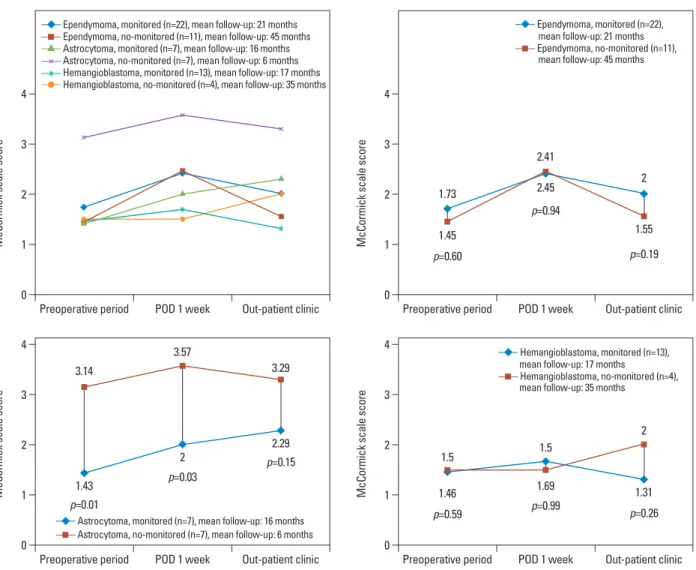
At the preoperative baseline, both groups showed a similar McCormick scale score (1.64 versus 2.07, p=0.083). After surgery, the outcome score worsened during the immediate postoperative stage (2.10 versus 2.65, p=0.076). During the follow-up period, the scores tended to improve (1.82 versus 2.31, p=0.115). Over time, the McCormick Grade score of each group showed a tendency to worsen during the imme- diate post-operative stage compared with the preoperative stage and a tendency to improve during the outpatient clinic stage compared with the immediate post-operative period (Table 4, Fig. 1). Moreover, when histological diagnosis (astrocytoma, ependymoma, haemangioblastoma) was in- cluded, this trend was maintained (Table 5, Fig. 2).
Verification of mMEP 75% criteria
The sensitivity and specificity of predicting postoperative neurologic deficit was 94% (17/18) and 94% (30/32). The positive predictive value and negative predictive value were 89% (17/19) and 97% (30/31). Within the 50 cases, false positives (2 cases) and a false negative (1 case) were observed. The false negative case was a 67-year-old man with ependymoma from C7 to T1. IONM revealed mMEP lation was 42.7 years in the monitored group and 40.1 years
in the non-monitored group. In the monitored group, the tu- mour size was slightly smaller than in the unmonitored group. Combined syrinx was observed more frequently in the monitored group. With respect to histological diagno- ses, the rate of ependymoma was 44% and 42% in the monitored and non-monitored group, respectively. In the unmonitored group, patients with astrocytoma were more likely to enroll (14% versus 27%) and the number of pa- tients with haemangioblastoma was lower (26% versus 15%), but there were no statistically significant differences between the groups. There was no statistically significant difference in sex, age, BMI, location of tumour, tumour size, combination with syrinx, or histological diagnoses.
Outcome data
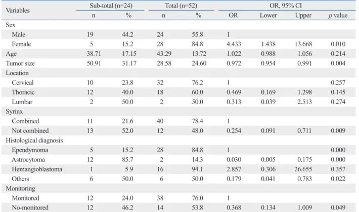
In the IONM group, the gross total tumour excision rate was greater than in the non-IONM group, but there was no significant difference (76% versus 58%; univariate analy- sis, p=0.049; multivariate regression model, p=0.119). We first performed univariate regression analyses to assess the factors affecting gross total excision. The female sex, tu- mour size, combination with syrinx, histological diagnosis, and being monitored showed an association with gross total excision (Table 2). In the final multiple linear regression model (Table 3), we built a multivariate logistic model to adjust for confounding factors. In the final model, the histo- logical diagnosis was the only variable that was significant-
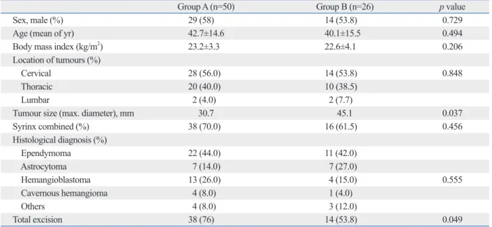
Table 1. Baseline Demographic Characteristics, Following Clinical and Radiologic Findings (Group A: Monitored, Group B:
Non-Monitored)
Group A (n=50) Group B (n=26) p value
Sex, male (%) 29 (58) 14 (53.8) 0.729
Age (mean of yr) 42.7±14.6 40.1±15.5 0.494
Body mass index (kg/m2) 23.2±3.3 22.6±4.1 0.206
Location of tumours (%)
Cervical 28 (56.0) 14 (53.8) 0.848
Thoracic 20 (40.0) 10 (38.5)
Lumbar 2 (4.0) 2 (7.7)
Tumour size (max. diameter), mm 30.7 45.1 0.037
Syrinx combined (%) 38 (70.0) 16 (61.5) 0.456
Histological diagnosis (%)
Ependymoma 22 (44.0) 11 (42.0)
Astrocytoma 7 (14.0) 7 (27.0)
Hemangioblastoma 13 (26.0) 4 (15.0) 0.555
Cavernous hemangioma 4 (8.0) 1 (4.0)
Others 4 (8.0) 3 (12.0)
Total excision 38 (76) 14 (53.8) 0.049
Categorical variable: chi-square test & Fisher’s exact test continuous variable: Mann-Whitney U test.
pedicle screws.11,12 However, intramedullary spinal cord tu- mours are rare, and for years, reports on the use of IOM techniques during their surgical removal have remained an- ecdotal. Before the advent of MEP during IMSCT surgery, SEPs were used alone with the assumption that changes in SEPs specifically represented spinal cord dysfunction.18 False negative results occurred during surgeries monitored only with SEPs. The patient woke up with a new parapare- sis that would not have been recognised if the patient were using SEPs alone.19 The proposal to have neurophysiologic parameters as a major outcome predictor of spinal cord sur- amplitude changes between 50% and 75%, but this change
did not satisfy our criteria. After surgery, the patient showed paraparesis (Gr. 4), but at 6 months, his condition had im- proved (Gr. 4→5).
DISCUSSION
IONM for safe spine surgery of scoliosis was first reported in 1977. SSEP, MEP and EMG have since been introduced and applied for correction of deformities or insertion of
Table 2. Univariate Analysis Showing the Individual Effects of Sex, Age, Tumour Size, Tumour Location, Histological Diagno- sis, Monitoring on Total Excision Rate
Variables Sub-total (n=24) Total (n=52) OR, 95% CI
n % n % OR Lower Upper p value
Sex
Male 19 44.2 24 55.8 1
Female 5 15.2 28 84.8 4.433 1.438 13.668 0.010
Age 38.71 17.15 43.29 13.72 1.022 0.988 1.056 0.214
Tumor size 50.91 31.17 28.58 24.60 0.972 0.954 0.991 0.004
Location
Cervical 10 23.8 32 76.2 1 0.257
Thoracic 12 40.0 18 60.0 0.469 0.169 1.298 0.145
Lumbar 2 50.0 2 50.0 0.313 0.039 2.513 0.274
Syrinx
Combined 11 21.6 40 78.4 1
Not combined 13 52.0 12 48.0 0.254 0.091 0.711 0.009
Histological diagnosis
Ependymoma 5 15.2 28 84.8 1 0.000
Astrocytoma 12 85.7 2 14.3 0.030 0.005 0.175 0.000
Hemangioblastoma 1 5.9 16 94.1 2.857 0.306 26.655 0.357
Others 6 50.0 6 50.0 0.179 0.041 0.783 0.022
Monitoring
Monitored 12 24.0 38 76.0 1
No-monitored 12 46.2 14 53.8 0.368 0.134 1.009 0.049
OR, odds ratio; CI, confidence interval.
Table 3. Associations of Total Excision with Other Variables
Variables OR, 95% CI
OR Lower Upper p value
Histology diagnosis
Ependymoma 1 0.001
Astrocytoma 0.017 0.002 0.147 0.000
Hemangioblastoma 2.511 0.244 25.859 0.439
Others 0.122 0.021 0.713 0.020
Monitoring
Monitored 3.14 0.75 13.33 0.119
No-monitored 1
OR, odds ratio; CI, confidence interval.
Logistic regression model (backward elimination, p value >0.10 was used).
reports claiming aggressive tumour mass removal and pres- ervation of normal neurologic function. Finally, in 2005, Sala, et al.9 concluded that the applied motor evoked poten- tial method appeared to significantly improve long-term mo- tor outcome. Similarly, early motor outcome due to transient motor deficits in the monitored group can be predicted by the neurophysiologic profile of the patient at the end of sur- gery. In that report, a combined D-wave and mMEP moni- toring protocol was used. D-wave and mMEPs form a com- plementary relationship. The IONM modality for IMSCT surgery may be divided into those with or without D-wave.
Various modalities have been adopted for IMSCT with- out D-wave. These modalities include threshold-level pa- rameters during multi-pulse transcranial electrical stimula- tion (TES), mMEP morphology (from polyphasic to biphasic to loss) and free-running EMG combined with mMEP monitoring.20-23
In our study, all patients were operated on using com- bined mMEP and SSEP monitoring. Particularly, we used a 75% mMEP amplitude criterion because we believed a 100% decline in amplitude to be too specific and a 50% de- gery emerged in 1997 when Morota, et al.14 introduced the
use of D-wave (epidural MEP) monitoring after transcrani- al electrical stimulation and concluded that this method ap- peared to be a better predictor of functional outcome than the patient’s pre-operative motor status. Since Kothbauer, et al.8 used a combination of D-wave and mMEPs for 100 pa- tients undergoing IMSCT surgery, there have been several
Table 4. Comparison Two Groups with McCormick Scale Score for Neurologic Function Outcome
Group A (n=50) Group B (n=26) p value
Preoperative period 1.64 (±0.942) 2.07 (±1.05) 0.083
POD 1 wk 2.10 (±1.13) 2.65 (±1.33) 0.076
Out-patient clinic 1.82 (±1.03) 2.31 (±1.32) 0.115
Follow up period (month) 18.24 (±16.49) 31.30 (±30.17) 0.266
POD, post operative day.
Mean±standard deviation or proportions of subjects are presented. Statistics were analyzed by t-test and chi-square test. Significant (p<0.05).
Table 5. Repeated Measured ANOVA Comparing Effect of Monitoring According IMSCT Histology
Monitored Non-Monitored p value
n Mean SD Min n Mean SD Min
Ependymoma
Preoperative period 22 1.73 0.94 1 11 1.45 0.52 1 0.596
POD 1 wk 22 2.41 1.1 1 11 2.45 1.29 1 0.937
Out-patient clinic 21 2 1.1 1 11 1.55 0.93 1 0.193
Follow up period (month) 22 21.05 17.78 1 11 44.64 30.63 1 0.041
Astrocytoma
Preoperative period 7 1.43 1.13 1 7 3.14 0.9 2 0.010
POD 1 wk 7 2 1.41 1 7 3.57 0.98 2 0.031
Out-patient clinic 7 2.29 1.5 1 7 3.29 1.11 1 0.146
Follow up period (month) 7 16.29 21.98 3 7 5.86 5.18 1 0.247
Hemangioblastoma
Preoperative period 13 1.46 0.88 1 4 1.5 0.58 1 0.590
POD 1 wk 13 1.69 1.03 1 4 1.5 0.58 1 >0.999
Out-patient clinic 13 1.31 0.63 1 4 2 1.41 1 0.259
Follow up period (month) 13 17.46 13.53 3 4 34.5 24.45 12 0.111
SD, standard deviation; IMSCT, intramedullary spinal cord tumour; POD, post operative day; ANOVA, analysis of variance.
Mann-Whitney U test.
Fig. 1. Comparison two groups with McCormickscale score. POD, post op- erative day.
0 1 2 3 4
McCormick scale score
Preoperative period POD 1 week Out-patient clinic Group A (n=50) mean follow-up, 18 months Group B (n=26) mean follow-up, 31 months
2.07
1.64
2.65
2.1
2.31
1.82 p=0.08
p=0.08
p=0.12
cur. False positives result in incomplete tumour mass re- moval. Conversely, the false negative is disastrous for both surgeon and patient. Although the tumour is completely re- moved, the patient will sustain severe neurologic deficits.
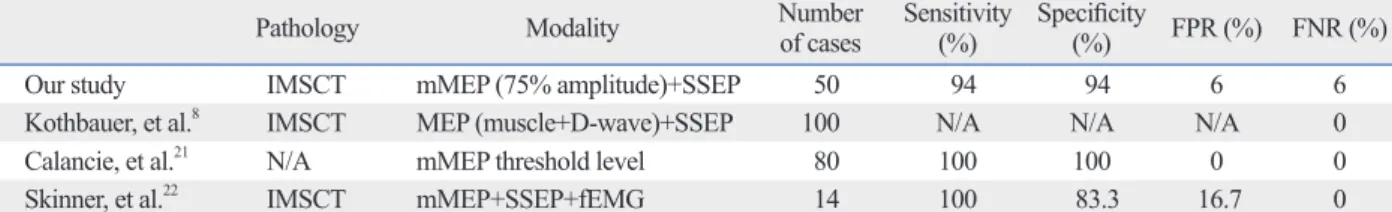
Thus, it is important to determine a reasonable compro- mise. No report has shown whether mMEP amplitude crite- ria are reliable. We suggest that a 75% criterion is the most reliable criterion. Thus, we verified the specificity, sensitivi- ty, positive predictive value, negative predictive value, false positive rate, and false negative rate (Table 6).
There have been various reports discussing IONM with gross total tumour excision. The majority reported a negative effect of IONM on gross total tumour mass removal. Surger- ies were frequently stopped too early because the IONM was too sensitive.9,22,26 In our study, gross total resection (GTR) was superior to not monitoring cases. Although it failed to exert a statistically significant effect in the final adjusted model, IONM tended to enable complete tumour mass re- cline in amplitude to be too sensitive.24-27 If the monitoring
alarm is too sensitive, the surgery is stopped prematurely. If the alarm is too specific, there is risk of damaging the nor- mal spinal cord.
Combining mMEP and D-wave monitoring may be the gold standard for IMSCT surgery. We could not use D-wave monitoring due to a holding provision with our local insur- ance. Our protocol choice was a combination of mMEP and SEP monitoring without D-wave, a unique IONM protocol for IMSCT.
The effect of various modalities for IMSCT surgery re- mains unclear.21,27-29 In our study, all surgical intervention was performed if the mMEP amplitude was less than 75%
of baseline MEP amplitude. If the surgery was abandoned due to a change in amplitude of 50%, such termination of surgery precludes a complete resection of the tumour. In contrast, if the surgery is stopped based on an ‘absence- presence criterion’, a high rate of “false positives” may oc-
0
0
0
0 1
1
1
1 2
2
2
2 3
3
3
3 4
4
4
4
McCormick scale scoreMcCormick scale score McCormick scale scoreMcCormick scale score
Preoperative period
Preoperative period
Preoperative period
Preoperative period POD 1 week
POD 1 week
POD 1 week
POD 1 week Out-patient clinic
Out-patient clinic
Out-patient clinic
Out-patient clinic Ependymoma, monitored (n=22), mean follow-up: 21 months
Ependymoma, no-monitored (n=11), mean follow-up: 45 months Astrocytoma, monitored (n=7), mean follow-up: 16 months Astrocytoma, no-monitored (n=7), mean follow-up: 6 months Hemangioblastoma, monitored (n=13), mean follow-up: 17 months Hemangioblastoma, no-monitored (n=4), mean follow-up: 35 months
Astrocytoma, monitored (n=7), mean follow-up: 16 months Astrocytoma, no-monitored (n=7), mean follow-up: 6 months
Ependymoma, monitored (n=22), mean follow-up: 21 months
Ependymoma, no-monitored (n=11), mean follow-up: 45 months
Hemangioblastoma, monitored (n=13), mean follow-up: 17 months
Hemangioblastoma, no-monitored (n=4), mean follow-up: 35 months
3.14
1.73
1.5 1.43
1.45
1.46 3.57
2.41
2 1.5
2.45
1.69 3.29
2
2.29 2
1.55
p=0.01 1.31
p=0.60
p=0.59 p=0.03
p=0.94
p=0.99 p=0.15
p=0.19
p=0.26
Fig. 2. Repeated measured ANOVA comparing effect of monitoring according IMSCT histology. POD, post operative day; IMSCT, intramedullary spinal cord tumour; ANOVA, analysis of variance.
ACKNOWLEDGEMENTS
We appreciate critical advice from Dr. Seok-Ho Hong and correction of the formatting by YM Jeong.
REFERENCES
1. Cristante L, Herrmann HD. Surgical management of intramedul- lary spinal cord tumors: functional outcome and sources of mor- bidity. Neurosurgery 1994;35:69-74.
2. McCormick PC, Torres R, Post KD, Stein BM. Intramedullary ep- endymoma of the spinal cord. J Neurosurg 1990;72:523-32.
3. Hoshimaru M, Koyama T, Hashimoto N, Kikuchi H. Results of microsurgical treatment for intramedullary spinal cord ependymo- mas: analysis of 36 cases. Neurosurgery 1999;44:264-9.
4. Constantini S, Miller DC, Allen JC, Rorke LB, Freed D, Epstein FJ. Radical excision of intramedullary spinal cord tumors: surgical morbidity and long-term follow-up evaluation in 164 children and young adults. J Neurosurg 2000;93(2 Suppl):183-93.
5. Epstein FJ, Farmer JP, Freed D. Adult intramedullary spinal cord ependymomas: the result of surgery in 38 patients. J Neurosurg 1993;79:204-9.
6. Guidetti B, Mercuri S, Vagnozzi R. Long-term results of the surgi- cal treatment of 129 intramedullary spinal gliomas. J Neurosurg 1981;54:323-30.
7. Ney JP, van der Goes DN. Evidence-based guideline update: In- traoperative spinal monitoring with somatosensory and transcrani- al electrical motor evoked potentials. Report of the Therapeutics and Technology Assessment Subcommittee of the American Academy of Neurology and the American Clinical Neurophysiol- ogy Society. Neurology 2012;79:292.
8. Kothbauer KF, Deletis V, Epstein FJ. Motor-evoked potential monitoring for intramedullary spinal cord tumor surgery: correla- tion of clinical and neurophysiological data in a series of 100 con- secutive procedures. Neurosurg Focus 1998;4:e1.
9. Sala F, Palandri G, Basso E, Lanteri P, Deletis V, Faccioli F, et al.
Motor evoked potential monitoring improves outcome after sur- gery for intramedullary spinal cord tumors: a historical control study. Neurosurgery 2006;58:1129-43.
10. Nuwer MR, Emerson RG, Galloway G, Legatt AD, Lopez J, Mi- nahan R, et al. Evidence-based guideline update: intraoperative spinal monitoring with somatosensory and transcranial electrical motor evoked potentials: report of the Therapeutics and Technolo- gy Assessment Subcommittee of the American Academy of Neu-
moval. We believe that there are several reasons that may underlie the slight increase in total tumour mass removal in the monitored group. 1) The surgeon believed that IONM enabled a safer surgery, a vitally important point for the sur- geon. A stable IONM enabled the surgeon to be more ag- gressive during tumour mass resection. IONM encouraged the surgeon to proceed with tumour mass resection. 2) IONM not only prevented neural injury but also provided opportunities to correct reversible injury by intervention. 3) Although not reaching statistical significance in the moni- tored group, the number of haemangioblastoma patients was slightly greater than the number of patients with astro- cytoma. Haemangioblastoma can be more easily removed than astrocytoma. This last point is very difficult to assess due to the surgeon’s learning curve. Thus, we considered the learning curve and excluded 10 years of operations pri- or to 1998. Nevertheless, this point may cause some bias.
Our study had several important limitations. This study was a retrospective analysis; we could not design prospec- tive or randomised controlled studies due to ethics. Without prospective studies that are powered to detect differences, it may not be possible to determine the potential value of neu- rological monitoring in these cases. The follow-up time was different between the two groups. Although we includ- ed cases that were observed for at least 1 year, the follow- up time differed by 13 months. The number of patients in the control group was relatively smaller than in the moni- tored group. A greater number in the control group will pro- vide power to detect the mean difference between groups.
Our data demonstrates the use of combined IONM using Tce-mMEP and SEP for IMSCT. During IMSCT surgery, combined Tce-mMEP and SEP, using a 75% muscle ampli- tude weaning criterion, did not cause a significant improve- ment in the rate of gross total excision of the tumour or neurologic outcome. IONM for IMSCT does not reduce the total tumour excision rate, although there had been con- cerns that IONM during IMSCT blocked the surgical pro- cedure and resulted in incomplete tumour mass removal.
Table 6. Verification of the 75% Amplitude Wearing Criteria Regarding Variable Methods
Pathology Modality Number
of cases Sensitivity
(%) Specificity
(%) FPR (%) FNR (%)
Our study IMSCT mMEP (75% amplitude)+SSEP 50 94 94 6 6
Kothbauer, et al.8 IMSCT MEP (muscle+D-wave)+SSEP 100 N/A N/A N/A 0
Calancie, et al.21 N/A mMEP threshold level 80 100 100 0 0
Skinner, et al.22 IMSCT mMEP+SSEP+fEMG 14 100 83.3 16.7 0
IMSCT, intramedullary spinal cord tumour; FPR, false positive rate; FNR, false negative rate; EMG, electromyography; mMEP, muscle motor evoked poten- tial; SSEP, somatosensory evoked potential; N/A, not available.
postoperative motor function. Neurosurgery 2005;56:982-93.
21. Calancie B, Harris W, Brindle GF, Green BA, Landy HJ. Thresh- old-level repetitive transcranial electrical stimulation for intraop- erative monitoring of central motor conduction. J Neurosurg 2001;
95(2 Suppl):161-8.
22. Skinner SA, Nagib M, Bergman TA, Maxwell RE, Msangi G. The initial use of free-running electromyography to detect early motor tract injury during resection of intramedullary spinal cord lesions.
Neurosurgery 2005;56(2 Suppl):299-314.
23. Hyun SJ, Rhim SC, Kang JK, Hong SH, Park BR. Combined mo- tor- and somatosensory-evoked potential monitoring for spine and spinal cord surgery: correlation of clinical and neurophysiological data in 85 consecutive procedures. Spinal Cord 2009;47:616-22.
24. Jones SJ, Harrison R, Koh KF, Mendoza N, Crockard HA. Motor evoked potential monitoring during spinal surgery: responses of distal limb muscles to transcranial cortical stimulation with pulse trains. Electroencephalogr Clin Neurophysiol 1996;100:375-83.
25. Sutter M, Eggspuehler A, Grob D, Jeszenszky D, Benini A, Por- chet F, et al. The validity of multimodal intraoperative monitoring (MIOM) in surgery of 109 spine and spinal cord tumors. Eur Spine J 2007;16 Suppl 2:S197-208.
26. Albright AL. Intraoperative spinal cord monitoring for intramedul- lary surgery: an essential adjunct? Pediatr Neurosurg 1998;29:112.
27. Calancie B, Molano MR. Alarm criteria for motor-evoked poten- tials: what’s wrong with the “presence-or-absence” approach?
Spine (Phila Pa 1976) 2008;33:406-14.
28. Fehlings MG, Brodke DS, Norvell DC, Dettori JR. The evidence for intraoperative neurophysiological monitoring in spine surgery:
does it make a difference? Spine (Phila Pa 1976) 2010;35(9 Suppl):
S37-46.
29. Hsu W, Bettegowda C, Jallo GI. Intramedullary spinal cord tumor surgery: can we do it without intraoperative neurophysiological monitoring? Childs Nerv Syst 2010;26:241-5.
rology and the American Clinical Neurophysiology Society. Neu- rology 2012;78:585-9.
11. Nash CL Jr, Lorig RA, Schatzinger LA, Brown RH. Spinal cord monitoring during operative treatment of the spine. Clin Orthop Relat Res 1977:100-5.
12. Calancie B, Madsen P, Lebwohl N. Stimulus-evoked EMG moni- toring during transpedicular lumbosacral spine instrumentation.
Initial clinical results. Spine (Phila Pa 1976) 1994;19:2780-6.
13. Danesh-Clough T, Taylor P, Hodgson B, Walton M. The use of evoked EMG in detecting misplaced thoracolumbar pedicle screws. Spine (Phila Pa 1976) 2001;26:1313-6.
14. Morota N, Deletis V, Constantini S, Kofler M, Cohen H, Epstein FJ. The role of motor evoked potentials during surgery for intra- medullary spinal cord tumors. Neurosurgery 1997;41:1327-36.
15. Sala F, Beltramello A, Gerosa M. Neuroprotective role of neuro- physiological monitoring during endovascular procedures in the brain and spinal cord. Neurophysiol Clin 2007;37:415-21.
16. Deletis V. Intraoperative neurophysiology of the corticospinal tract of the spinal cord. Suppl Clin Neurophysiol 2006;59:107-12.
17. Hyun SJ, Rhim SC. Combined motor and somatosensory evoked potential monitoring for intramedullary spinal cord tumor surgery:
correlation of clinical and neurophysiological data in 17 consecu- tive procedures. Br J Neurosurg 2009;23:393-400.
18. Kearse LA Jr, Lopez-Bresnahan M, McPeck K, Tambe V. Loss of somatosensory evoked potentials during intramedullary spinal cord surgery predicts postoperative neurologic deficits in motor function [corrected]. J Clin Anesth 1993;5:392-8.
19. Ginsburg HH, Shetter AG, Raudzens PA. Postoperative paraplegia with preserved intraoperative somatosensory evoked potentials.
Case report. J Neurosurg 1985;63:296-300.
20. Quiñones-Hinojosa A, Lyon R, Zada G, Lamborn KR, Gupta N, Parsa AT, et al. Changes in transcranial motor evoked potentials during intramedullary spinal cord tumor resection correlate with