Right Gastric Venous Drainage: Angiographic Analysis in 100 Patients
8
0
0
전체 글
(2) Seong et al.. ARGV can play an important role in cavernous transformation due to thrombosis in the portal vein trunk (2, 9, 10). However, the incidence of ARGV is also unclear because there are large variations, ranging from 0% to 34% in the literature including an unpublished cadaveric study (2, 3, 6, 9, 13). The left gastric vein has been well recognized as a preferential route of the portosystemic shunt in patients with portal hypertension, and is easily traced on an enhanced CT of the upper abdomen. Therefore, radiologists are familiar with its anatomy and pathologies. However, the right gastric vein or right gastric venous drainage is difficult to trace on routine enhanced CT of the upper abdomen and the pattern of right gastric venous drainage has not been systematically investigated. The purpose of this study was to investigate the pattern of right gastric venous drainage by use of digital subtraction angiography in a large study population.. MATERIALS AND METHODS This study was approved by the Institutional Review Board, and informed consent was waived. Patients This study included consecutive 100 patients who underwent right gastric arteriography during transcatheter arterial chemoembolization of hepatocellular carcinoma over the past six month period at the Seoul National University Hospital. During this period, 685 patients underwent transcatheter arterial chemoembolization. Patients with tumors in the left hepatic lobe or in the caudate lobe, portal vein thrombosis, an arterioportal shunt, and severe liver cirrhosis with reversed portal flow were excluded from the study. Also we excluded patients who did not undergo right gastric angiography because it was not necessary for their particular tumor treatment. The patient population consisted of 73 men and 27 women ranging in age from 15 to 80 years of age (mean age, 56.2 years). Angiography To obtain indirect gastric venograms, celiac arteriography and selective arteriography of the right and left gastric arteries were performed with digital subtraction angiographic equipment (Angiostar; Siemens, Erlangen, Germany or V-3000; Philips Medical Systems, Einthoven, The Netherlands). A celiac arteriography was performed 54. with a 6.5-Fr or 5-Fr Rösch hepatic catheter (Cook, Bloomington, IN). Selective arteriography of the right gastric and left gastric arteries was performed with use of a 3-Fr microcatheter (Microferret; Cook). An experienced interventional radiologist performed all of the angiographic procedures. A nonionic contrast agent (iopromide, Ultravist 370; Schering, Berlin, Germany) was used for the angiography. The injection rate and total volume of the contrast medium used were 6-7 mL/sec and 42-49 mL, respectively for celiac arteriography, 1.0-2.0 mL/sec and 10-14 mL, respectively for selective arteriography of the right or left gastric arteries Imaging Interpretation Angiographic findings for the 100 patients were retrospectively analyzed in consensus by two radiologists with respect to the presence or absence of the right gastric vein and aberrant gastric veins, multiplicity of draining veins, aberrant right gastric venous drainage sites, and the termination pattern of ARGVs. When it was difficult to determine the drainage site, CT images were referenced. We also compared the relative size of the right and left gastric veins. The right gastric vein was defined as a vein which runs rightward along the lesser curvature of the stomach in parallel with the right gastric artery, and drains into the main portal vein or the left portal vein trunk. The ARGV was defined as a vein which takes off from the right side of the lesser curvature of the stomach and drains into the superficial liver parenchyma or peripheral portal branches. Aberrant right gastric venous drainage sites were classified by hepatic segments based on the Couinaud classification (Couinaud C. Le foie: etudes anatomiques et chirurgicales. Paris, France: Masson, 1957).. RESULTS Presence or Absence of the Right Gastric Vein and Aberrant Gastric Veins Of the 100 patients evaluated, only forty eight patients had the right gastric vein draining into the main portal vein (n = 43) or into the left portal vein trunk (n = 5) without aberrant right gastric venous drainage (Fig. 1). Eleven patients had aberrant right gastric venous drainage in addition to the right gastric vein draining into the main portal vein. Thirty eight patients had aberrant right gastric venous drainage without the right gastric vein (Fig. 2). Korean J Radiol 13(1), Jan/Feb 2012. kjronline.org.
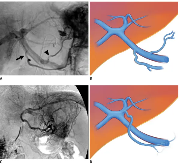
(3) Right Gastric Venous Drainage Patterns on Angiography. In the remaining three patients, the right gastric vein or aberrant gastric venous drainage was not demonstrated at all. In six of the fifty nine patients with right gastric vein drainage, the left gastric vein was not identified. The conjunction of the right and left gastric vein just before entering the main portal vein trunk was noted in one patient (Fig. 3).. which had aberrant gastric venous drainage in addition to the right gastric vein draining into the main portal vein. The aberrant left gastric vein was drained into the portal vein of the hepatic segment II. Aberrant Right Gastric Venous Drainage Sites The most common drainage site of the 66 ARGVs is hepatic segment IV (n = 35) followed by segment I (n = 15). The 12 ARGVs were drained into the umbilical segment of the left portal vein or adjacent liver parenchyma. The uncommon drainage sites included the hepatic segment II (n = 3) and hepatic segment III (n = 1).. Multiplicity of Aberrant Gastric Veins Among the forty nine patients with aberrant gastric venous drainage, thirteen patients had two ARGVs and two patients had three. Therefore, a total of 66 ARGVs were demonstrated in forty nine patients. Aberrant left gastric venous drainage was found in four patients; three of which had aberrant right gastric venous drainage without the right gastric vein (Fig. 2) and one. Termination Pattern of Aberrant Right Gastric Veins The termination pattern of ARGV could be classified into 4 different types which were illustrated on Figure 4. Type I. A. B. C. D. Fig. 1. Normal venous drainage of stomach.. A. Right gastric vein draining into main portal vein in 72-year-old man. Right gastric vein drains into main portal vein (arrow), while left gastric vein drains into splenic vein (arrowhead). B. Schematic diagram of right gastric vein draining into main portal vein. Right gastric vein drainage site is more distal to left gastric vein drainage site and on right side of main portal vein. C. Right gastric vein draining into left portal vein trunk in 43-year-old woman. Right gastric vein runs parallel to main portal vein and drains into left portal vein trunk. D. Schematic diagram of right gastric vein draining into left portal vein trunk.. kjronline.org. Korean J Radiol 13(1), Jan/Feb 2012. 55.
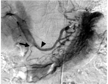
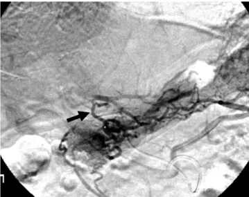
(4) Seong et al.. Fig. 2. Coexisting aberrant right and left gastric venous drainage in 46-year-old man. Aberrant right gastric vein drains into. Fig. 3. Conjunction type of gastric venous drainage in 60-yearold woman. Right gastric vein (arrow) anastomose with left gastric. superficial areas of hepatic segment IV (black arrows) and aberrant left gastric vein drains into segment II portal branches (white arrow).. vein (arrowhead) just before entering main portal vein.. A. B. D E Fig. 4. Termination patterns of aberrant right gastric vein.. C. F. A. Type I. Aberrant right gastric vein continues smoothly into peripheral portal vein as single channel, hence sequestering territory supplied by aberrant right gastric vein from normal portal supply. B. Type IIa. Aberrant right gastric vein is connected to peripheral portal vein in end-to-end or end-to-side (dotted line) fashion via single collateral channel. C. Type IIb. Aberrant right gastric vein is connected to peripheral portal vein in end-to-end or end-to-side (dotted line) fashion via multiple collateral channels. D. Type IIIa. Aberrant right gastric vein is terminated as small superficial parenchymal blush formation without demonstrable portal branches. E. Type IIIb. Aberrant right gastric vein branches in extrahepatic location and branches are terminated as multifocal small superficial parenchymal blush formation without demonstrable portal branches. F. Type IV. Aberrant right gastric vein forms network around sectional or segmental portal vein, and subsequently drains into it.. 56. Korean J Radiol 13(1), Jan/Feb 2012. kjronline.org.
(5) Right Gastric Venous Drainage Patterns on Angiography. is the smooth continuation of ARGV as a single channel into the peripheral portal vein (Fig. 5). Type II is the collateral connection of ARGV into the peripheral portal vein (Fig. 6). Type III is the superficial parenchymal blush formation in a small area without demonstrable portal branches. Type IV is the network connection to the sectional or segmental portal vein (Fig. 7). For type II and III, subclassifications are made according to the multiplicity of channels. The most common termination pattern of ARGV was type III (n = 38, 58%) (Table 1). Sixteen of the 38 type III ARGVs branched into the extrahepatic location, and the branches terminated as multifocal small superficial parenchymal blush without demonstrable portal branches (Fig. 7). Twelve ARGVs showed a type II termination pattern. They were connected to the peripheral portal vein in an end-to-end or end-to-side (dotted line, Fig. 4) fashion via single or multiple collateral channels (Fig. 6). Nine. Fig. 6. Type IIb aberrant right gastric vein in 69-year-old man. Aberrant right gastric vein (arrow) is connected to segment I portal vein in end-to-side fashion via multiple collateral channels.. Fig. 5. Type I aberrant right gastric vein in 58-year-old woman.. Fig. 7. Type IV aberrant right gastric vein in 53-year-old man. Three aberrant right gastric veins are seen; one in type IV. Venous phase image of selective right gastric arteriography shows two aberrant right gastric veins, one in type I (arrow) and other in type IIIb (arrowhead).. (white arrow) and two in type III (black arrows). Network formation around umbilical segment of left portal vein is clearly demonstrated (arrowheads).. Table 1. Termination Pattern of Aberrant Gastric Vein Type Termination Pattern I Smooth continuation as single channel into peripheral portal vein II Collateral connection into peripheral portal vein IIa Single collateral channel IIb Multiple collateral channels III Superficial parenchymal blush formation in small areas without demonstrable portal branches IIIa Unifocal IIIb Multifocal IV Network formation around sectional or segmental portal vein. Subtotal. Total 9 12. 3 9 (6*) 38 22 16 7. Note.— *In six patients, connection between peripheral portal vein and aberrant right gastric vein was proven to be in end-toside fashion.. kjronline.org. Korean J Radiol 13(1), Jan/Feb 2012. 57.
(6) Seong et al.. Table 2. Relative Size of Right and Left Gastric Veins Depending on Presence or Absence of Aberrant Right Gastric Vein Aberrant Right Gastric Vein + RGV ≥ LGV 7 26 RGV < LGV 42 25 Note.— *P value is 0.001. LGV = left gastric vein, RGV = right gastric vein. ARGVs showed a type I termination pattern. The territory supplied by the type I aberrant right gastric vein was considered to be sequestered from the normal portal supply. The seven remaining ARGVs showed a type IV termination pattern. Relative Size of the Right and Left Gastric Veins In the group of patients with ARGV (n = 49), the right gastric vein was equal to (n = 5) or larger than (n = 2) the left gastric artery in seven patients (7 of 49, 14%) (Table 2). However, in the group of patients without ARGV (n = 51), the right gastric vein was equal to (n = 9) or larger than (n = 17) the left gastric vein in 26 patients (26 of 51, 51%). A statistically significant difference was found for the relative size of the right and left gastric veins (dominance of the right gastric vein in gastric venous drainage) between the two groups (p < 0.05, Fisher’s exact test).. DISCUSSION Based on previous studies, the incidence of an ARGV variable from 2% to 14%; angiographic studies have reported ARGV incidence as 2% and the assumption from the lesion study at hepatic segment IV was 14% with the CT during arterial portography (3, 14). An unpublished cadaver study showed an incidence as high as 34% for ARGV prevalence (2, 9, 14). An aberrant left gastric vein (ALGV) is a very rare variation, having an incidence of 0.8% (2 of 245 cadavers) (15). However, in our study, the prevalence of an ARGV was 49% and the prevalence of an ALGV was 4%; both frequencies being higher than the frequencies in autopsy reports or angiographic reports previously published. Usually, aberrant gastric veins are slender and may be missed on an autopsy or radiological imaging such as CT, MRI or US. However, with angiography, the presence of aberrant gastric veins is more readily visualized. We performed a selective arteriography of the right and left gastric artery, but a previous angiographic study was 58. performed at the celiac artery. This is the one reason for a large discrepancy between previous studies and our study. The most common drainage site was hepatic segment IV (35 out 66 ARGVs) and hepatic segment I (15 of 66 ARGVs). Our results support the previous study about aberrant gastric venous drainage on the basis of CT arterial portography (CTAP) (9). When the incidence (12 out of 66 ARGVs) of drainage to the left portal vein or around the portal vein is included, almost all ARGVs drained into or adjacent to the medial segment of the left hepatic lobe, including the caudate lobe (62 out of 66 ARGVs, 94%). The reason for the difference between hepatic segments IV/I and the other hepatic segments is the following. Although the major portion of the liver and portal venous system develops at approximately days 26-28 of gestation, the bile ducts, parabiliary venous system, hepatic artery, and segment I and IV of the liver develop later, at approximately days 3234 of gestation. The parabiliary venous system extends along the hepatic artery and bile duct, and finally directly supply the liver in the later stage, after the intrahepatic distribution of the portal veins is established (16). This is the first report to describe the termination pattern of ARGVs. We classified the ARGVs based on the extrahepatic ramification and portal vein connection in the liver. Type I is the smooth continuation of ARGV into the intrahepatic portal vein as a single channel (Fig. 4A), which means that type I ARGV is the only source of portal venous supplies in the drainage territory. Type II is the single or multiple collateral connection of the ARGV to the peripheral portal vein in an end-to-end or end-to-side fashion (Fig. 4B, C), which suggests that type II ARGV may not be the only source of portal venous supply to the drainage territory because normal portal venous connection can be patent. Type III is the superficial parenchymal blush formation in a small area without demonstrable portal branches, which suggests that type III ARGV supplies only the superficial capsular or subcapsular areas without penetrating into the deeper portion of the liver (Fig. 4D, E). Type IV is the network connection to the sectional or segmental portal vein, which suggests that type IV ARGV contributes little to the portal venous supply of the section or segment. Therefore, we theorize that a relatively large type I and type II ARGV without normal portal venous connection can result in a pseudo lesion on imaging studies. In this study, the incidence of type I and II ARGVs without a normal portal venous connection (10 of 100, 10%) is very similar to the incidence of a pseudo lesion of the liver as reported Korean J Radiol 13(1), Jan/Feb 2012. kjronline.org.
(7) Right Gastric Venous Drainage Patterns on Angiography. in 6-14% of cases (14, 17, 18). The termination pattern or ARGVs explain why the incidence of pseudo lesions in the clinical setting is much lower than the true incidence of ARGVs. Of the 94 patients evaluated for left gastric veins, 47 left gastric veins drained into the splenic vein, 30 drained into the main portal vein and 13 drained into the portal confluence. Only four patients had an ALGV which drained into hepatic segment II and all these patients (n = 4) also had an ARGV. We found only one case of conjunction between the right and left gastric vein drained into the main portal vein (Fig. 3). To our knowledge, this is a unique report of the conjunction type involving the right and left gastric vein. Aberrant gastric venous drainage is important to both the radiologist and clinician because of the pseudo lesion formation in the portal phase of CT angiography or CTAP (9, 14), one route of cavernous transformation in the main portal thrombosis, and unexpected hemorrhage during hepatobiliary surgery due to a missed aberrant gastric vein by the surgeon (19). It is also probable that the ARGV provides a direct metastatic pathway for gastric cancer in the lesser curvature and a potential route of hepatofugal arterioportal shunt in a case of main portal vein tumor thrombosis (19-21). It can be used as the alternative route for placing a stent in a transjugular intrahepatic portosystemic shunt with main portal thrombosis (20). First a limitation of our study is the absence of secondary confirmative modality for the exact frequency of aberrant gastric venous drainage. Even an autopsy could not confirm the exact frequency (2). Secondly, most of the patients in this study had liver cirrhosis, which elevates portal venous pressure. In turn, liver cirrhosis and portal hypertension may alter hemodynamics in the portal system, which may affect angiographic visualization of aberrant gastric venous drainage. Further study is necessary in the population with a non-cirrhotic normal liver. In conclusion, aberrant right gastric venous drainage was found in almost half of the patients. The main venous drainage site was hepatic segment IV, and I around the umbilical segment of the left portal vein in descending order. The termination pattern of ARGV could be divided into four different types.. REFERENCES 1. Gilfillan RS, Hills HL. Anatomic study of the portal vein and. kjronline.org. Korean J Radiol 13(1), Jan/Feb 2012. its main branches. Arch Surg 1950;61:449-461 2. Deneve E, Caty L, Fontaine C, Guillem P. Simultaneous aberrant left and right gastric veins draining directly into the liver. Ann Anat 2003;185:263-266 3. Takayasu K, Aoki K, Ichikawa T, Ohmura T, Sekiguchi R, Terauchi T, et al. Aberrant right gastric vein directly communicating with left portal vein system. Incidence and implications. Acta Radiol 1990;31:575-577 4. Yoon KH, Matsui O, Kadoya M, Yoshigawa J, Gabata T, Arai K. Pseudolesion in segments II and III of the liver on CT during arterial portography caused by aberrant right gastric venous drainage. J Comput Assist Tomogr 1999;23:306-309 5. Caty L, Deneve E, Fontaine C, Guillem P. Concurrent aberrant right gastric vein directly draining into the liver and variations of the hepatic artery. Surg Radiol Anat 2004;26:7073 6. Gabata T, Matsui O, Kadoya M, Ueda K, Kawamori Y, Yoshikawa J, et al. Aberrant gastric venous drainage in a focal spared area of segment IV in fatty liver: demonstration with color Doppler sonography. Radiology 1997;203:461-463 7. Kurosaki Y, Tanaka YO, Itai Y. Aberrant gastric venous drainage in focal fatty liver of segment IV: demonstration with sonography. AJR Am J Roentgenol 1998;171:897-898 8. Maeda H, Sato M, Kimura M, Kawai N, Sonomura T, Kishi K, et al. Focal fatty infiltration in the quadrate lobe of the liver accompanied by aberrant right gastric vein. Radiat Med 1998;16:61-64 9. Matsui O, Takahashi S, Kadoya M, Yoshikawa J, Gabata T, Takashima T, et al. Pseudolesion in segment IV of the liver at CT during arterial portography: correlation with aberrant gastric venous drainage. Radiology 1994;193:31-35 10. Tajima H, Murakami R, Tajima N, Kumazaki T. Aberrant right gastric vein draining directly into the quadrate lobe of the liver. A case report. Acta Radiol 1995;36:270-272 11. Kawamori Y, Matsui O, Takahashi S, Kadoya M, Takashima T, Miyayama S. Focal hepatic fatty infiltration in the posterior edge of the medial segment associated with aberrant gastric venous drainage: CT, US, and MR findings. J Comput Assist Tomogr 1996;20:356-359 12. Yamagami T, Nakamura T, Maeda T. Aberrant gastric venous inflow to the left lobe of the liver parenchyma adjacent to the falciform ligament. Br J Radiol 1999;72:903-905 13. Zhang J, Rath AM, Chevrel JP. Anatomic basis of venous drainage in gastric tubular esophagoplasty. Surg Radiol Anat 1994;16:221-228 14. del Pilar Fernandez M, Bernardino ME. Hepatic pseudolesion: appearance of focal low attenuation in the medial segment of the left lobe at CT arterial portography. Radiology 1991;181:809-812 15. Miyaki T, Yamada M, Kumaki K. Aberrant course of the left gastric vein in the human. Possibility of a persistent left portal vein. Acta Anat (Basel) 1987;130:275-279 16. Couinaud C. The parabiliary venous system. Surg Radiol Anat 1988;10:311-316 17. Nelson RC, Thompson GH, Chezmar JL, Harned RK 2nd,. 59.
(8) Seong et al.. Fernandez MP. CT during arterial portography: diagnostic pitfalls. Radiographics 1992;12:705-718; discussion 719-720 18. Ohashi I, Ina H, Gomi N, Himeno Y, Okada Y, Hanafusa K, et al. Hepatic pseudolesion in the left lobe around the falciform ligament at helical CT. Radiology 1995;196:245-249 19. Ohnishi K, Okuda K, Ohtsuki T, Nakayama T, Hiyama Y, Iwama S, et al. Formation of hilar collaterals or cavernous transformation after portal vein obstruction by hepatocellular. 60. carcinoma. Observations in ten patients. Gastroenterology 1984;87:1150-1153 20. Bezzi M, Broglia L, Lemos AA, Rossi P. Transjugular intrahepatic portosystemic shunt in portal vein thrombosis: role of the right gastric vein with anomalous insertion. Cardiovasc Intervent Radiol 1995;18:102-105 21. Parker RA, Seal RM. Cavernous transformation of the portal vein. J Pathol Bacteriol 1955;70:97-103. Korean J Radiol 13(1), Jan/Feb 2012. kjronline.org.
(9)
수치
관련 문서
The cucumber sprouts pre-treated with all seven rhizobacterial strains at concentration of 5.0×10 6 cfu/ml were significantly increased plant height and
학생들이 구조적으로 완벽하게 만들지... http://www.nasa.gov/multimedia/imagegallery/index.ht ml
캐나다정부간행물목록(Weekly checklist of Canadian government publications) 에 수록된 자료 중 Folder자료, 인구센서스, 전화번호 자료 등을 제외한
이에 전남지역 중학생들 대상의 설문조사를 통해서 체벌의 실태와 중학교 교사와 학생들의 체벌에 관한 인식 및 체벌의 교육적 효과 등을 파악하여 체벌이 진정
상기 신입생 장학금 외에도 본교는 신입생장학금-재학생장학금-해외연수장학금-대학원진학장학금에 이르는 전주기 장학제도를 운영하고 있으며, 다양한 교외장학금
The 4-digit display shows the pump’s maximum flow rate (mL/min), pressure setting (PSI), the set upper or lower pressure limit (PSI), the actual pressure (PSI), or actual
Basic aspects of AUTOSAR architecture and methodology Safety mechanisms supported by AUTOSAR.. Technical safety concepts supported by AUTOSAR Relationship to ISO
v i 는 측정하려는 단자간 전압이고, Probe는 RC 병렬 회로이고 오실로스코프에서는 내부에 RC 병렬회로가 우선적
