Correction of Angle Class II division 1 malocclusion with a mandibular protraction appliances and multiloop edgewise archwire technique
10
0
0
전체 글
(2) Freitas et al • Treatment of Class II malocclusion. INTRODUCTION According to Angle,1 a Class II malocclusion is charac terized by the distal occlusion of the mandibular first molar in relation to the maxillary first molar; in a Class II division 1 malocclusion, the maxillary incisors addi tionally exhibit proclination. This malocclusion is also characterized by an anteroposterior dental discrepancy, which may be associated with skeletal changes. The overjet may be excessive and the overbite is most likely deep. The retrognathic profile and excessive overjet result in abnormal contraction patterns of the facial muscles and tongue. Typically, the mentalis becomes hyperactive, to elevate the orbicularis oris and achieve lip sealing. 2 The marked overjet also increases the patient's susceptibility to dental trauma. Additionally, the unaesthetic facial appearance often has psychosocial consequences.3 Class II division 1 malocclusions have a multifactorial. origin and are mainly attributable to evolutionary changes in craniofacial growth, dietary and social habits, and ethnic admixture. Therefore, orthodontic treatment planning depends on several factors, including the na ture of the malocclusion, patient characteristics, and family history.4 One treatment option is the combined use of a mandibular protraction appliance (MPA) and multiloop edgewise archwires (MEAWs). The MPA is a fixed orthopedic appliance used for treating Class II malocclusions. Its advantages include ease of fabrication by the dentist or assistant, easy placement, and the possibility of concomitant use with other appliances, thus reducing the total treatment time and increasing post-treatment stability. 5 The MEAW technique was developed in 1967 to treat severe open bites and was found to be extremely effective. Since then, it has been applied in various malocclusions, especially at the final treatment stage, to achieve better intercuspation.6 This paper reports a case of severe Angle Class II. Figure 1. Pretreatment facial and intraoral photographs. www.e-kjo.org. http://dx.doi.org/10.4041/kjod.2014.44.5.268. 269.
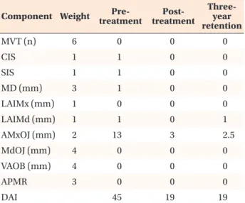
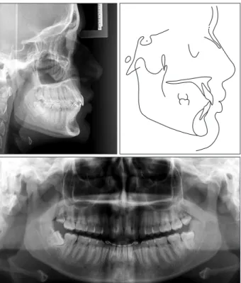
(3) Freitas et al • Treatment of Class II malocclusion. division 1 malocclusion in a female patient outside the maximum pubertal growth peak who was treated by orthodontic camouflage using the MPA and MEAW technique.. DIAGNOSIS AND ETIOLOGY A Brazilian girl aged 14 years and 9 months presented with a chief complaint of protrusive teeth. She had a convex facial profile, deep bite, lack of passive lip seal, acute nasolabial angle, retrognathic mandible, and no midline deviation. No signs of temporomandibular dysfunction such as clicks, cracks, and crepitation were noted. She also did not report systemic problems or a family history of the same malocclusion. Intraoral examination revealed good oral hygiene, maxillary dia stemas, slight crowding of the mandibular incisors, a small maxillary arch, overjet of 13 mm, and overbite of 4 mm (Figure 1). Her occlusion was assessed using the Dental Aesthetic Index (DAI), as recommended by the World Health Organization.7 The assessment revealed a very severe or disfiguring malocclusion, necessitating orthodontic treatment (Table 1). Table 1. Dental Aesthetic Index (DAI) values before and after treatment and at the 3-years follow-up examination PreComponent Weight treatment 6. 0. 0. 0. CIS. 1. 1. 0. 0. SIS. 1. 1. 0. 0. MD (mm). 3. 1. 0. 0. LAIMx (mm). 1. 0. 0. 0. LAIMd (mm). 1. 1. 0. 1. AMxOJ (mm). 2. 13. 3. 2.5. MdOJ (mm). 4. 0. 0. 0. VAOB (mm). 4. 0. 0. 0. APMR. 3. 0. 0. 0. 45. 19. 19. MVT, Missing visible teeth (incisors, canines, and premolars in the maxillary and mandibular dentitions); CIS, crowding in the incisal segments (0 = no crowding; 1 = one segment crowded; 2 = two segments crowded); SIS, spacing in the incisal segments (0 = no spacing; 1 = one segment spaced; 2 = two segments spaced); MD, midline diastema; LAIMx, largest anterior irregularity in the maxilla; LAIMd, largest anterior irregularity in the mandible; AMxOJ, anterior maxillary overjet; AMdOJ, anterior mandibular overjet; VAOB, vertical anterior open bite; APMR, anteroposterior molar relationship (0 = normal; 1 = half cusp; 2 = one cusp).. 270. TREATMENT OBJECTIVES The treatment objectives were to improve the facial aesthetics, balance the lip musculature, achieve stable occlusion, correct the maxillary dental protrusion and canine relationship, reduce the overjet and overbite, and correct the mandibular incisor crowding. MPA fabrication The MPA consisted of three parts: the maxillary and mandibular parts and the bootstrap. To construct the maxillary portion, a short piece of stainless steel tubing was joined transversely to one end of a telescopic stainless steel tube (outer diameter = 1.0 mm; inner diameter = 0.9 mm; length = 35 mm) by point welding (fusion welding held the tubes together while silver soldering them with flux and a blowtorch). After the tubing was cut flush with the telescopic tube, a 0.9-mm-diameter stainless steel wire clip was inserted into the telescopic tube and maxillary first molar tube. ThreePostyear treatment retention. MVT (n). DAI. The initial panoramic radiograph (Figure 2) revealed the presence of well-positioned third molars and the absence of morphologic changes to the condyles. The initial lateral cephalogram showed a horizontal growth pattern (FMA = 22°), well-positioned maxilla (SNA = 79°), retrognathic mandible (SNB = 76°), and marked incisor proclination (1.NA = 44°) (Figure 2 and Table 2).. Figure 2. Pretreatment panoramic and cephalometric radiographs and tracing.. http://dx.doi.org/10.4041/kjod.2014.44.5.268. www.e-kjo.org.
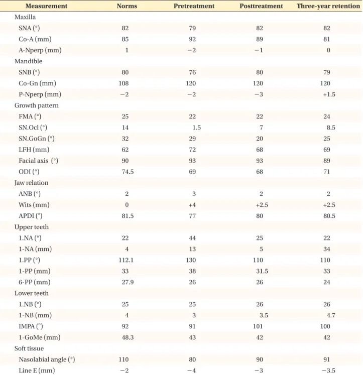
(4) Freitas et al • Treatment of Class II malocclusion. Table 2. Cephalometric measurements Measurement. Norms. Pretreatment. Posttreatment. Three-year retention. SNA (°). 82. 79. 82. 82. Co-A (mm). 85. 92. 89. 81. 1. −2. −1. 0. 80. 76. 80. 79. Co-Gn (mm). 108. 120. 120. 120. P-Nperp (mm). −2. −2. −3. FMA (°). 25. 22. 22. SN.Ocl (°). 14. SN.GoGn (°). 32. 29. 20. 25. LFH (mm). 62. 72. 68. 69. Facial axis (°). 90. 93. 93. 89. ODI (°). 74.5. 69. 68. 71. Maxilla. A-Nperp (mm) Mandible SNB (°). +1.5. Growth pattern 1.5. 7. 24 8.5. Jaw relation ANB (°). 2. 3. 2. 2. Wits (mm). 0. +4. +2.5. +2.5. 81.5. 77. 80. 80.5. 22. 44. 25. 22. 4. 13. 5. 34. 110. 110. APDI (o) Upper teeth 1.NA (°) 1-NA (mm) 1.PP (°). 112.1. 130. 1-PP (mm). 33. 38. 31.5. 33. 6-PP (mm). 27.9. 26. 26. 24. 25. 25. 26. 26. 4. 3. IMPA ( ). 92. 91. 101. 100. 1-GoMe (mm). 48.3. 43. 42. 42. Lower teeth 1.NB (°) 1-NB (mm) o. 3.5. 4.7. Soft tissue Nasolabial angle (°). 110. 80. 90. 91. Line E (mm). −2. −4. −3. −3.5. SNA, Sella-nasion-A point; Co-A, distance from condylion to A point; A-Nperp, distance from A point to nasion perpendicular line; SNB, sella-nasion-B point; Co-Gn, distance from condylion to gnathion; P-Nperp, distance from pogonion to nasion perpendicular line; FMA, Frankfurt-mandibular plane angle; FH, Frankfurt horizontal plane; SN.Ocl, sella-nasion-occlusal plane angle; SN.GoGn, sella-nasion line to gonion-menton line angle; LFH, lower facial height; facial axis, basion-nasion line to pterygoid-gnathion line angle; ODI, overbite depth indicator; ANB, A point-nasion-B point; Wits, distance from A point to B point at the occlusal plane; APDI, anteroposterior dysplasia indicator; 1.NA, angle between the maxillary central incisor axis and nasion-A point line; 1-NA, distance from the maxillary central incisor to nasion-A point line; 1.PP, angle between the maxillary central incisor axis and the palatal plane; 1-PP, distance from the edge of the maxillary central incisor to the palatal plane; 6-PP, distance from the occlusal surface of the maxillary first molar to the palatal plane; 1.NB, angle between the mandibular central incisor and nasion-B point line; 1-NB, distance from the edge of the mandibular central incisor to nasion-B point; IMPA, incisor axis-mandibular plane angle; 1-GoMe, distance from the mandibular central incisor edge to the mandibular plane; Nasolabial angle, angle between the line drawn through the midpoint of the nasal aperture and the line drawn perpendicular to the Frankfurt horizontal plane while intersecting subnasale; Line E, distance from the lower lip connecting the tip of nose and soft tissue pogonion.. www.e-kjo.org. http://dx.doi.org/10.4041/kjod.2014.44.5.268. 271.
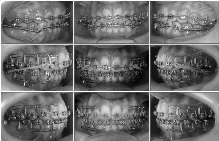
(5) Freitas et al • Treatment of Class II malocclusion. Figure 3. The mandibular protraction appliances. A, Right side; B, left.. on each side (Figure 3). For the bootstrap, a piece of 0.9-mm-diameter stain less steel wire was bent at 90° on one end and inserted into the maxillary telescopic tube to prevent subsequent deformation of the tube. Then, the straight end of the wire was inserted into the maxillary tubing, and the wire was bent until it was parallel to the maxillary telescopic tube. The wire was cut so that its total length was ap proximately twice the length of the maxillary first molar tube. The wire was annealed to allow for easy ben ding around the maxillary first molar tube during its placement and to prevent dislodgement of the appliance (Figure 3). The mandibular part was fabricated from a 0.9-mmdiameter stainless steel rod and 0.019 × 0.025-inch stainless steel archwire with a helix between the canine and the first premolar on each side. The rod had a U-shaped bend at one end; the bend was threaded through each helix from the lingual side and turned parallel to the archwire. During MPA placement, the rods were inserted into the maxillary tubes, which were shortened to match the helices when the mandible protruded to the point where the optimal overbite, overjet, and midline were achieved. The rods extended. Figure 4. Intraoral progress photographs showing the mandibular protraction appliance used in conjunction with Class II elastics in the multiloop edgewise archwire technique. 272. http://dx.doi.org/10.4041/kjod.2014.44.5.268. www.e-kjo.org.
(6) Freitas et al • Treatment of Class II malocclusion. less than a millimeter distally from the maxillary tubes (Figure 3).. TREATMENT ALTERNATIVES Two treatment options were presented to the patient. The first option was orthognathic surgery including mandibular advancement and genioplasty. The second option was nonsurgical treatment by dentoalveolar compensation without extraction (orthodontic camou flage). The patient rejected the first option, so nonsurgical treatment comprising mandibular advancement with the MPA and orthodontic finishing with the MEAW technique (Figure 4) was planned.. TREATMENT PROGRESS Treatment was initiated by banding the maxillary and mandibular first molars and bonding pre-adjusted. edgewise brackets (0.022 × 0.025-inch slot, Roth prescription). Leveling was performed with 0.014-inch nickel titanium (NiTi), 0.016-inch NiTi, 0.018-inch stainless steel, 0.020-inch stainless steel, and 0.019 × 0.025-inch stainless steel archwires. During leveling, in addition to the 0.016-inch NiTi archwire, a continuous ligature was tied from molar to molar to reduce the maxillary diastemas and to prevent labial tipping of the mandibular incisors. Interproximal enamel reduction of 2 mm was performed on the mandibular lateral incisors to relieve the mandibular crowding. After the 0.020inch stainless steel archwire was placed, the ligature was replaced with an elastic chain. In the mandibular 0.019 × 0.025-inch stainless steel archwire, a helix was included between canines and premolars for placement of the MPA. The MPA was maintained for 10 months in total. The initial mandibular advancement was 6 mm. After 4 months, further advancement was performed to achieve an edge-to-edge relationship. The MPA was. Figure 5. Posttreatment facial and intraoral photographs. www.e-kjo.org. http://dx.doi.org/10.4041/kjod.2014.44.5.268. 273.
(7) Freitas et al • Treatment of Class II malocclusion. removed after an additional 6 months of use. Although correction of the molar relationship was observed, a mild Class II malocclusion remained in the canine and premolar regions. MEAWs (0.019 × 0.025-inch stainless steel archwires) were placed in the dental arches. Intermaxillary (5/16 inch) elastics were used from the first “L” loops on the maxillary lateral incisors to the mandibular first molar tubes. The MEAWs were maintained for 2 months to avoid possible relapse of the Class II relationship (Figure 4). The patient showed excellent compliance during the treatment. After 26 months of active treatment, the appliances were removed and impressions were taken to fabricate retainers. A modified Hawley plate and 3 by 3 fixed retainer were used in the maxillary and mandibular arches, respectively.. RESULTS The post-treatment photographs revealed an improved facial profile (Figure 5). The intraoral photographs exhi bited bilateral Class I molar and canine relationships and an occlusion with a normal overjet and overbite (Figures 5 and 6). Good intercuspation, proximal contacts, and root parallelism were achieved (Figure 6). The decreased DAI value suggested normal occlusion at the completion of orthodontic treatment (DAI = 19) and 3 years. Figure 6. Posttreatment panoramic and cephalometric radiographs and tracing. 274. thereafter (DAI = 19) (Table 1). The final lateral cephalogram demonstrated proper inc lination of the maxillary incisors (Figure 6). The mandibular incisors were facially inclined and the upper lip projection was reduced. The patient was satisfied with her dental and facial appearance. Dentoalveolar stability was maintained even after 3 years (Figures 7, 8, and 9).. DISCUSSION Angle Class II malocclusions, commonly characterized by an anteroposterior dental discrepancy, are more severe when combined with skeletal disharmony, which may be caused by mandibular deficiency, maxillary protrusion, or a combination of both.8 Mandibular re trusion is the most common characteristic in children with Class II malocclusions9 and shows no tendency for self-correction with growth. Furthermore, mandibular retrusion worsens during the pubertal growth spurt,10 and maintains the same standard after this period until young adulthood.11 For patients with skeletal Class II malocclusions who have completed growth, the fol lowing treatment options are possible: (1) orthodontic camouflage, which may be combined with extraction, based on retraction of the facially inclined maxillary incisors and facial inclination of the mandibular inci sors, to improve occlusion and facial aesthetics without correcting the underlying skeletal problem; or (2) or thog n athic surgery to reposition the mandible or maxilla, depending on the skeletal Class II problems associated with mandibular deficiency and downward and backward mandibular rotation caused by excessive maxillary vertical growth. Another option would have been orthodontic treatment with first premolar extraction; however, given the horizontal growth pattern of the patient, this alternative was not considered be cause it would impair deep bite correction and affect facial aesthetics. Surgical treatment includes mandibular advancement, superior maxillary repositioning, or a combination of both. Mandibular deficiency is a problem existing in nearly two thirds of surgical patients, and one third of surgical patients require maxillary surgery alone (15%) or in combination with mandibular surgery (20%). In the present case, orthognathic surgery was considered for anterior mandibular repositioning and genioplasty after the growth period, but the patient did not accept this option. Although surgical patients achieve an ideal skeletal relationship, with the mandible positioned anteriorly and the mandibular incisors in an ideal relationship with the basal bone, patients treated by orthodontic camouflage usually present less problems than those who are surgically treated.12 Orthognathic. http://dx.doi.org/10.4041/kjod.2014.44.5.268. www.e-kjo.org.
(8) Freitas et al • Treatment of Class II malocclusion. Figure 7. Three-years follow-up facial and intraoral photographs. surgery may cause complete condylar resorption in 10% of surgical cases. 13 Patients treated with orthodontic camouflage also report less functional problems in the temporomandibular joint than those treated by orthog nathic surgery. Finally, with regard to the cost-bene fit relationship for patients outside the growth period, similar results have been observed between the treat ment options, although orthodontic camouflage may yield a slightly greater overjet one year after treatment.8 In the present case, both the overjet and the overbite were in the normal range even at 3 years post-treatment (Figure 9). Treatment with extraction of the two first premolars, which is often indicated in comparison to treatment without extraction, is reportedly the most effective protocol when assessed by a normative index.14 This protocol was considered for the present patient; how. www.e-kjo.org. http://dx.doi.org/10.4041/kjod.2014.44.5.268. ever, extractions may have led to a marked facial con cavity and worsened the facial profile.15 Furthermore, the normative index used showed a dramatic change in the severity of malocclusion, with reduction to a level considered to require little or no orthodontic treatment after treatment without extraction. More recently, several approaches to orthopedic treatm ent of Class II malocclusions in young adults have been indicated with mandibular advancement appliances.5,16-25 Some studies have indicated associated problems, such as increased treatment time26 or partial loss of outcomes after use of Class II elastics. 27 The present patient underwent orthodontic treatment after her maximum growth peak. Orthodontic camouflage with the MPA16 was used in addition to Class II elastics in the MEAW technique within a relatively normal treatment time. However, one should also consider the. 275.
(9) Freitas et al • Treatment of Class II malocclusion. Figure 10. Superimposition of the pretreatment (black line) and post-treatment (gray line) tracings.. Figure 8. Three-years follow-up panoramic and cepha lometric radiographs and tracing.. stages 5 and 6, when treatment was started (Figure 3). Some growth could still occur, but not enough to correct the Class II malocclusion by mandibular growth (Figure 10).. CONCLUSION Orthodontic camouflage using the MPA and MEAW technique is an effective option for correcting Class II malocclusions in patients who refuse orthognathic surgery. In the present case, this treatment significantly improved the facial profile, achieved a satisfactory occlusion and pleasant aesthetics, and ensured good dentoalveolar stability even at 3 years after treatment was completed.. REFERENCES. Figure 9. Superimposition of the post-treatment (solid line) and three years follow-up (dotted line) tracings. greater success of treatment of a bilateral half cusp Class II malocclusion, as in the present case, than treatment with extraction resulting in a bilateral full cusp Class II malocclusion.15 According to Franchi et al.,28 the peak in skeletal growth occurs between stages 3 and 4 of cervical vertebral maturation in 93.5% of individuals. The present patient was past her growth peak, between 276. 1. Angle EH. Classification of malocclusion. Dental cosmos 1899;41:248-64. 2. Thüer U, Ingervall B. Pressure from the lips on the teeth and malocclusion. Am J Orthod Dentofacial Orthop 1986;90:234-42. 3. Jenny J, Cons NC. Comparing and contrasting two orthodontic indices, the Index of Orthodontic Treatment need and the Dental Aesthetic Index. Am J Orthod Dentofacial Orthop 1996;110:410-6. 4. Dolce C, Mansour DA, McGorray SP, Wheeler TT. Intrarater agreement about the etiology of Class II malocclusion and treatment approach. Am J Orthod Dentofacial Orthop 2012;141:17-23. 5. Coelho Filho CM. Mandibular protraction appliances for Class II treatment. J Clin Orthod 1995;29:31936.. http://dx.doi.org/10.4041/kjod.2014.44.5.268. www.e-kjo.org.
(10) Freitas et al • Treatment of Class II malocclusion. 6. Kim YH. Tratamiento de maloclusiones severas mediante la técnica de alambre Edgewise Multiloop (Multiloop Edgewise Arch-Wire, MEAW). Ortodoncia Clínica 2004;7:22-34. 7. WHO. Oral Health Surveys: Basic Methods. Geneva, Switzerland: World Health Organization; 1997. 8. Ghafari J, Shofer FS, Jacobsson-Hunt U, Markowitz DL, Laster LL. Headgear versus function regulator in the early treatment of Class II, Division 1 maloc clusion: a randomized clinical trial. Am J Orthod Dentofacial Orthop 1998;113:51-61. 9. Wong L, Hägg U, Wong G. Correction of extreme overjet in 2 phases. Am J Orthod Dentofacial Orthop 2006;130:540-8. 10. Stahl F, Baccetti T, Franchi L, McNamara JA Jr. Lon gitudinal growth changes in untreated subjects with Class II Division 1 malocclusion. Am J Orthod Den tofacial Orthop 2008;134:125-37. 11. Baccetti T, Stahl F, McNamara JA Jr. Dentofacial growth changes in subjects with untreated Class II malocclusion from late puberty through young adulthood. Am J Orthod Dentofacial Orthop 2009; 135:148-54. 12. Mihalik CA, Proffit WR, Phillips C. Long-term followup of Class II adults treated with orthodontic camouflage: a comparison with orthognathic sur gery outcomes. Am J Orthod Dentofacial Orthop 2003;123:266-78. 13. Ruf S, Pancherz H. Orthognathic surgery and dento facial orthopedics in adult Class II Division 1 treat ment: mandibular sagittal split osteotomy versus Herbst appliance. Am J Orthod Dentofacial Orthop 2004;126:140-52. 14. Bock NC, von Bremen J, Ruf S. Occlusal stability of adult Class II Division 1 treatment with the Herbst appliance. Am J Orthod Dentofacial Orthop 2010;138:146-51. 15. Janson G, Valarelli FP, Cançado RH, de Freitas MR, Pinzan A. Relationship between malocclusion severity and treatment success rate in Class II non extraction therapy. Am J Orthod Dentofacial Orthop 2009;135:274.e1-8. 16. Johnston LE Jr. A comparative analysis of Class II treatments. In: Vig PS, Ribbens KA, editors. Science and clinical judgment in orthodontics. Monograph 19, Craniofacial Growth Series. Ann Arbor: Center for Human Growth and Development, The University of Michigan; 1986. 17. Janson I. Skeletal and dentoalveolar changes in. www.e-kjo.org. http://dx.doi.org/10.4041/kjod.2014.44.5.268. patients treated with a bionator during prepubertal and pubertal growth. In: McNamara JA Jr, Ribbens KA, Howe RP, editors. Clinical alteration of the growing face. Monograph 14, Craniofacial Growth Series. Ann Arbor: Center for Human Growth and Development, The University of Michigan; 1983. 18. Janson G, Barros SEC, de Freitas MR, Henriques JFC, Pinzan A. Class II treatment efficiency in maxillary premolar extraction and nonextraction protocols. Am J Orthod Dentofacial Orthop 2007;132:490-8. 19. Xu TM, Liu Y, Yang MZ, Huang W. Comparison of extraction versus nonextraction orthodontic treatment outcomes for borderline Chinese patients. Am J Orthod Dentofacial Orthop 2006;129:672-7. 20. Coelho Filho CM. Mandibular protraction appliance IV. J Clin Orthod 2001;35:18-24. 21. Ruf S, Pancherz H. Dentoskeletal effects and facial profile changes in young adults treated with the Herbst appliance. Angle Orthod 1999;69:239-46. 22. Ruf S, Pancherz H. Temporomandibular joint remodeling in adolescents and young adults during Herbst treatment: A prospective longitudinal mag netic resonance imaging and cephalometric radio graphic investigation. Am J Orthod Dentofacial Orthop 1999;115:607-18. 23. Kinzinger G, Diedrich P. Skeletal effects in class II treatment with the functional mandibular advancer (FMA)? J Orofac Orthop 2005;66:469-90. 24. Nalbantgil D, Arun T, Sayinsu K, Fulya I. Skeletal, dental and soft-tissue changes induced by the Jasper Jumper appliance in late adolescence. Angle Orthod 2005;75:426-36. 25. Ruf S, Pancherz H. Herbst/multibracket appliance treatment of Class II division 1 malocclusions in early and late adulthood. a prospective cephalometric study of consecutively treated subjects. Eur J Orthod 2006;28:352-60. 26. Popowich K, Nebbe B, Heo G, Glover KE, Major PW. Predictors for Class II treatment duration. Am J Orthod Dentofacial Orthop 2005;127:293-300. 27. Herrera FS, Henriques JF, Janson G, Francisconi MF, de Freitas KM. Cephalometric evaluation in different phases of Jasper jumper therapy. Am J Orthod Dentofacial Orthop 2011;140:e77-84. 28. Franchi L, Baccetti T, McNamara JA Jr. Mandibular growth as related to cervical vertebral maturation and body height. Am J Orthod Dentofacial Orthop 2000;118:335-40.. 277.
(11)
수치

+3

관련 문서
- 각종 지능정보기술은 그 자체로 의미가 있는 것이 아니라, 교육에 대한 방향성과 기술에 대한 이해를 바탕으로 학습자 요구와 수업 맥락 등 학습 환경에 맞게
Quality life assessment of bone-anchored fixed partial denture patients with unilateral mandibular distal-extension edentulism.. Kent G, Johns R..Effects
success rates of dental implants placed at the time of or after alveolar ridge augmentation with an autogenous mandibular bone graft and titanium mesh: a 3-to
The purpose of this study was to identify the frequency and related factors of advanced airway management for patients with cardiac arrest by the
This is because compared with the free-year English classes, the general semester English classes consisted of teacher oriented grammar class and reading
The purpose of this study was to analyze the impaction pattern of the impacted mandibular third molar and the relationship with the inferior alveolar nerve
For class flow, autotelic experience of the first and second graders was higher than that of the third graders, and matching of behavior with consciousness
The purpose of this study was to evaluate the curvature of Vertucci's type II mesial canals of mandibular molar using new method; The radius and angle
