INTRODUCTION
Parkinson’s disease (PD) is an neurodegenerative disease characterized by the loss of dopaminergic neurons and mus- cular rigidity, affecting several million patients globally, espe-
cially older adults.1 Due to its complex causes and mechanisms, PD has attracted increasing attention from scientists and clin- ical physicians.2 Occurrence of inflammatory response, over- production of oxidative stress, and activation of apoptotic cas- cade have been reported to be associated with the development of PD.3 Although many disease-modifying therapies for PD have been discovered and developed,4 it is still necessary to ex- plore novel molecular mechanisms involved in PD for further breakthroughs in PD treatment.
MicroRNAs (miRNAs), a class of endogenous single-stranded noncoding transcript with 18−23 nucleotides (nt), play vital roles in a series of physiological and pathological processes.5 MiRNAs could regulate gene expression at the post-transcrip- tional level by targeting the 3’-untranslated region (3’-UTR) of messenger RNAs (mRNAs), leading to mRNA degradation or translational repression. Increasing evidence has described Received: November 20, 2017 Accepted: February 12, 2018
Corresponding author: Dr. Xiaowei Chen, Department of Internal Medicine-Neu- rology, Hua Mei Branch of the Second People’s Hospital of Liaocheng, South End of Xinhua Road, Linqing 252600, China.
Tel: 86-635-2320346, Fax: 86-635-2341280 E-mail: [email protected]
•The authors have no financial conflicts of interest.
© Copyright: Yonsei University College of Medicine 2018
This is an Open Access article distributed under the terms of the Creative Com- mons Attribution Non-Commercial License (http://creativecommons.org/licenses/
by-nc/4.0) which permits unrestricted non-commercial use, distribution, and repro- duction in any medium, provided the original work is properly cited.
MiR-212 Attenuates MPP + -Induced Neuronal Damage by Targeting KLF4 in SH-SY5Y Cells
Yanfeng Song, Ying Liu, and Xiaowei Chen
Department of Internal Medicine-Neurology, Hua Mei Branch of the Second People’s Hospital of Liaocheng, Linqing, China.
Purpose: Parkinson’s disease (PD) is a common age-dependent neurodegenerative disease. MiR-212 has been demonstrated to exert protective effects in several neurological disorders. The present study aimed to investigate the role and underlying molecu- lar mechanism of miR-212 in PD.
Materials and Methods: 1-methyl-4-phenylpyridinium (MPP+)-induced SH-SY5Y cells were applied as a PD model in vitro. RT- qPCR was used to measure the expression of miR-212 and Kruppel-like factor 4 (KLF4) mRNA. Western blot analysis was per- formed to detect the protein levels of KLF4, Notch1 and Jagged1. Cell viability and apoptosis were determined by the Cell Count- ing Kit-8 and flow cytometry, respectively. Quantitative analysis of caspase-3 activity, lactate dehydrogenase (LDH), reactive oxygen species (ROS), superoxide dismutase (SOD), tumor necrosis factor-α (TNF-α), and interleukin-1 beta (IL-1β) was con- ducted with corresponding ELISA kits. Dual-luciferase reporter assay was employed to evaluate the relationship between miR- 212 and KLF4.
Results: MiR-212 was downregulated in MPP+-induced SH-SY5Y cells. Also, miR-212 alleviated MPP+-induced SH-SY5Y cell dam- age, embodied by increased cell viability, decreased caspase-3 activity, LDH release, ROS production, TNF-α, and IL-1β expres- sion, as well as elevated SOD levels. KLF4 was a direct target of miR-212, and miR-212 repressed KLF4 expression in a post-tran- scriptional manner. Moreover, miR-212-mediated protection effects were abated following KLF4 expression restoration in MPP+- induced SH-SY5Y cells, represented as lowered cell viability and enhanced apoptotic rate. Furthermore, Notch signaling was involved in the regulation of miR-212/KLF4 axis in MPP+-induced SH-SY5Y cells.
Conclusion: miR-212 might attenuate MPP+-induced neuronal damage by regulating KLF4/Notch signaling pathway in SH-SY5Y cells, a promising target for PD therapy.
Key Words: Parkinson’s disease, miR-212, KLF4, Notch signaling pathway, SH-SY5Y cells
pISSN: 0513-5796 · eISSN: 1976-2437 Yonsei Med J 2018 May;59(3):416-424
https://doi.org/10.3349/ymj.2018.59.3.416
the participation of miRNA dysregulation in the pathogenesis of neurodegenerative disorders, including PD.6,7 For instance, Yang, et al.8 demonstrated that miR-22 overexpression promot- ed cell survival in 6-hydroxydopamine-induced PC12 cells via regulating transient receptor potential melastatin 7 (TRPM7) expression. MiR-590-3p suppressed mitochondrial dysfunc- tion and oxidative stress through modulating its target gene JMJD1C.9 MiR-7 repressed 1-methyl-4-phenylpyridinium (MPP+)- triggered neuronal apoptosis by targeting Bax and sirtuin2 (Sirt2).10 MiR-212, necessary for the proper development, maturation, and function of neurons,11 has been implicated in a number of neurodegenerative and neurocognitive disorders, such as Alzheimer’s disease, Huntington’s disease, autism, Rett syndrome, and schizophrenia.12 Moreover, a previous study re- ported that miR-212 was downregulated in cerebrospinal fluid of PD patients.13 Nevertheless, little is known about the detailed function and molecular basis of miR-212 in PD progression.
In this study, we used MPP+-induced SH-SY5Y cells as an in vitro PD model to investigate the possible role and mechanism of miR-212. We found that miR-212 was downregulated in MPP+-induced SH-SY5Y cells. Moreover, miR-212 overexpres- sion alleviated MPP+-induced damage in SH-SY5Y cells. Fur- thermore, the neuroprotective effect of miR-212 might be me- diated by Kruppel-like factor 4 (KLF4)/Notch pathway in SH- SY5Y cells.
MATERIALS AND METHODS
Cell culture and treatment
Human neuroblastoma SH-SY5Y cells were obtained from American Type Culture Collection (ATCC, Manassas, VA, USA).
Cells were maintained in Dulbecco’s modified Eagle’s medium (DMEM, Gibco, Rockville, MD, USA) supplemented with 10%
fetal bovine serum (Gibco) and 1% penicillin/streptomycin (Gibco) at 37°C in a humidified incubator with 5% CO2.
MPP+ iodide was purchased from Sigma-Aldrich (St. Louis, MO, USA). To establish the PD model in vitro, SH-SY5Y cells were treated with various concentrations (0, 1, 2, and 4 mM) of MPP+ for 24 h or 2 mM of MPP+ for different treated times (0, 6, 12, and 24 h).
Cell transfection
The KLF4-overexpression vector (pcDNA-KLF4) and pcDNA empty vector were obtained from Invitrogen (Waltham, MA, USA). MiR-212 mimics, miR-212 inhibitor (anti-miR-212), siR- NA specifically against KLF4 (si-KLF4), and all control oligonu- cleotides (NC and si-NC) were synthesized by Sangon Biotech (Shanghai, China). All plasmids and oligonucleotides were transfected into SH-SY5Y cells with LipofectamineTM 2000 transfection reagent (Invitrogen) referring to the manufacturer’s instructions.
Cell viability assay
The Cell Counting Kit-8 (CCK-8, Dojindo Laboratories, Kuma- moto, Japan) was applied to measure cell viability according to the manufacturer’s instructions. Briefly, at indicated time points, CCK-8 solution (10 µL) was added into each cell culture medium for 2 h at 37°C, followed by the detection of absor- bance at 450 nm with a microplate reader (Bio-Rad Laborato- ries, Hercules, CA, USA).
Apoptosis analysis
The apoptotic rate of SH-SY5Y cells was analyzed by the Annex- in V apoptosis detection kit I (BD Biosciences, San Jose, CA, USA) and flow cytometry (BD Biosciences).
ELISA detection
The Caspase-3 Human Instant ELISATM Kit (Invitrogen) was used to measure the caspase-3 activity according to the in- structions. Briefly, cell lysates were incubated with 100 µM of enzyme-specific substrates at 37°C for 4 h, followed by the measurement of absorbance at 450 nm with a microplate reader (Bio-Rad Laboratories). Additionally, lactate dehydrogenase (LDH) release, superoxide dismutase (SOD) level, reactive ox- ygen species (ROS) production, and tumor necrosis factor-α (TNF-α), and interleukin-1 beta (IL-1β) expression were mea- sured using corresponding commercially available ELISA kits (Invitrogen), according to the provided instructions.
Dual-luciferase reporter assay
Online software algorithms (TargetScan, http://www.targetscan.
org; MiRanda, http://www.microrna.org) were applied to search for the potential targets of miR-212. The 3’-UTR sequences of KLF4 containing the putative binding sites of miR-212 were amplified from human genomic DNA and then inserted into the pmirGLO Dual-Luciferase miRNA Target Expression Vec- tor (Promega, Madison, WI, USA) to generate wild-type KLF4 reporter vector (KLF4-WT). Site-directed mutagenesis of the miR-212 binding sites in the KLF4 3’-UTR was carried out us- ing GeneArtTM Site-Directed Mutagenesis System (Invitro- gen), and then inserted into pmirGLO vector to generate mu- tant-type KLF4 reporter vector (KLF4-Mu). For reporter assays, SH-SY5Y cells were transfected with KLF4-WT or KLF4- Mu together with miR-212 mimics or anti-miR-212 using Li- pofectamineTM 2000 (Invitrogen). The Dual-Luciferase Reporter Assay system (Promega) was used to measure the luciferase activity in cell lysates.
RT-qPCR
Total RNA was extracted from treated cells using TRIzol re- agent (Invitrogen), and RNA (1 µg) was converted into cDNA using the cDNA Reverse Transcription Kit (Invitrogen) accord- ing to the manufacturer’s instructions. qPCR was performed using HotStart-IT SYBR Green qPCR Master Mix (TOYOBO, Osaka, Japan) in the ABI PRISM® 7300 real-time PCR system
(Applied Biosystems, Foster City, CA, USA). 2−ΔΔCt method was used to calculate the relative gene expression levels with GAP- DH and U6 as internal controls. The primer sequences used for PCR amplification were as follows: miR-212, 5’-CCCTCTGG GACATCTTTGACG-3’ (forward) and 5’-TGCTCCGCC TCCCCTGCGTCTC-3’ (reverse); KLF4, 5’-CAAGTCCCGCCG CTCCATTACCAA-3’ (forward) and 5’-CCACAGCCGTCCC AGTCACAGTGG-3’ (reverse); Notch1, 5’-CTGGACCCCATG GACATC-3’ (forward) and 5’-AGGATGACTGCACACATT GC -3’ (reverse); Jagged1, 5’-GAATGGCAACAAAACTTGCAT-3’
(forward) and 5’-AGCCTTGTCGGCAAATAGC-3’ (reverse);
GAPDH, 5’-AAGGTGAAGGTCGGAGTCAAC-3’ (forward) and 5’-GGGGTCATTGATGGCAACAATA-3’ (reverse); and U6, 5’-GCTTCGGCAGCACATATACTAAAAT-3’ (forward) and 5’-CGCTTCACGAATTTGCGTGTCAT-3’ (reverse).
Western blot assay
Total proteins were extracted from treated cells with cell lysis buffer (RIPA, Beyotime, Shanghai, China). Then, proteins were subjected to 10% SDS-PAGE and transferred to PVDF mem- branes (Sigma-Aldrich). The PVDF membranes were blocked in TBST buffer (TBS supplemented with 0.1% Tween-20) con- taining 5% (wt/vol) skimmed milk powder for 2 h at room tem- perature. Followed by three washes in TBST, the PVDF mem- branes were then incubated with primary antibodies, including anti-β-actin (Santa Cruz Biotechnology, Santa Cruz, CA, USA), anti-KLF4 (Santa Cruz Biotechnology), anti-Notch1 (Santa Cruz Biotechnology), and anti-Jagged1 (Santa Cruz Biotechnol- ogy) respectively overnight at 4°C. After being washed three times in TBST for 10 min, the PVDF membranes were further probed with HRP-conjugated secondary antibodies (Abcam, Cambridge, UK). Lastly, the membranes were exposed to Hy- perfilm-ECL (Thermo Fisher Scientific, Waltham, MA, USA) for 5 min, and visualized using a Fluor S Multimager and Quantity One 4.1 (Bio-Rad Laboratories).
Statistical analysis
Two-tailed Student’s t-test and one-way ANOVA were used to detect significant differences between groups using SPSS 16.0 software (SPSS Inc., Chicago, IL, USA). All data are presented as means±standard deviations. p<0.05 was considered statis- tically significant.
RESULTS
MiR-212 relieves MPP
+-induced suppression of SH-SY5Y cell viability
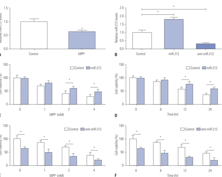
In order to explore the effect of miR-212 on PD, RT-qPCR was first performed to detect the expression pattern of miR-212 in MPP+-induced SH-SY5Y cells. The results revealed that, com- pared with the control group, miR-212 expression was mark- edly lower in SH-SY5Y cells following MPP+ treatment (Fig. 1A).
Then, miR-212 mimics and anti-miR-212 were synthesized and transfected into SH-SY5Y cells to examine their efficiency.
As displayed in Fig. 1B, the expression level of miR-212 was significantly promoted by introduction with miR-212 mimics, while miR-212 expression was greatly inhibited after transfec- tion with anti-miR-212 in SH-SY5Y cells, compared with the negative control. Subsequently, we further observed the effect of miR-212 on viability by gain- and loss-of-function analysis in MPP+-treated SH-SY5Y cells. SH-SY5Y cells were transfect- ed with miR-212 mimics or anti-miR-212, followed by treat- ment with various concentrations of MPP+ (0−4 mM) for 24 h or with 2 mM for different times (0−24 h). As expected, MPP+ treat- ment led to an obvious viability suppression of SH-SY5Y cells in a dose- or time-dependent manner (Fig. 1C-F). However, MPP+-induced vitality diminishment at 2 mM and 4 mM for 24 h (Fig. 1C) or 2 mM for 12 h and 24 h (Fig. 1D) was markedly ameliorated by the restoration of miR-212 expression. Con- versely, miR-212 downregulation aggravated MPP+-induced viability suppression in SH-SY5Y cells, compared with corre- sponding control (Fig. 1E and F). All these results implied that miR-212 weakened MPP+-induced decreases in the viability of SH-SY5Y cells.
MiR-212 alleviates MPP
+-induced SH-SY5Y cell damage
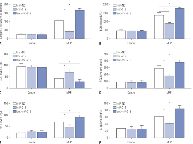
Oxidative stress, activation of apoptotic cascade, and neuroin- flammation have been confirmed to play central roles in the pathogenesis of PD.14 Hence, to further investigate the potential effect of miR-212 on MPP+-induced SH-SY5Y cells injury, SH- SY5Y cells transfected with miR-212 mimics or anti-miR-212 were stimulated with 2 mM MPP+ for 24 h, followed by detec- tion of caspase-3 activity, SOD generation, ROS production, IL- 1β expression, TNF-α levels, and LDH release. These experi- mental data demonstrated that MPP+ treatment results in a significant enhancement in caspase-3 activity (Fig. 2A), LDH release (Fig. 2B), ROS production (Fig. 2D), and TNF-α (Fig. 2E) and IL-1β (Fig. 2F) secretions, as well as a dramatic decrease in SOD level (Fig. 2C). However, these MPP+-elicited effects were attenuated by miR-212 overexpression and were exacerbated following miR-212 downregulation (Fig. 2). Taken together, these results suggested that miR-212 might alleviate MPP+-in- duced damage in SH-SY5Y cells.KLF4 is a direct target of miR-212
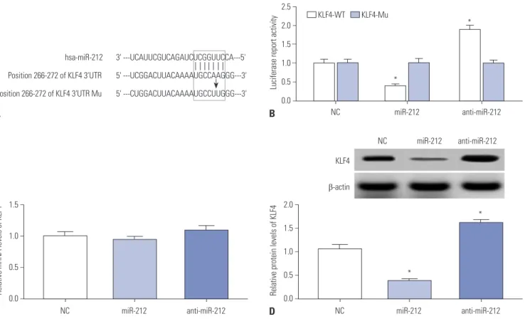
To further investigate the underlying molecular mechanism of miR-212 in protecting against MPP+-induced injury in SH- SY5Y cells, TargetScan software was used to search for the en- dogenous targets of miR-212. Interestingly, the 3’-UTR of KLF4 was predicted to have a higher probability of binding to miR- 212 (Fig. 3A). Thus, dual-luciferase reporter assay was per- formed to test whether KLF4 was a direct target of miR-212 in SH-SY5Y cells. As displayed in Fig. 3B, the luciferase activities of KLF4-WT reporter were repressed by upregulation of miR- 212, while it was enhanced after suppression of miR-212 (Fig.
3B). Nevertheless, no difference was found in the luciferase ac- tivities of KLF4-Mu reporter following a change of miR-212 ex- pression (Fig. 3B). Furthermore, miR-212 mimics or anti-miR-212 were transfected into SH-SY5Y cells to confirm the regulation role of miR-212 on KLF4 expression. RT-qPCR assay displayed that there was little influence on KLF4 mRNA levels when trans- fection with miR-212 mimics or anti-miR-212, compared with the control (Fig. 3C). Western blot assay presented that miR-212 upregulation repressed the expression of KLF4 protein, while miR-212 downregulation promoted KLF4 protein expression (Fig. 3D). These results indicated that miR-212 may modulate KLF4 expression in a post-transcriptional manner. Taken to- gether, these data suggested that KLF4 is a direct target of miR- 212 and that miR-212 inhibits the expression of KLF4 by bind- ing to its 3’UTR.
MiR-212-mediated protection effect is abated following restoration of KLF4 expression in MPP
+-induced SH-SY5Y cells
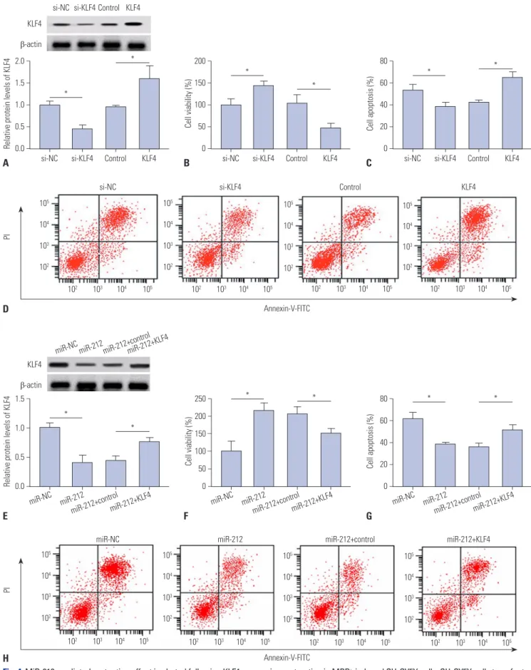
To explore the role of KLF4 on MPP+-induced damage in SH- SY5Y cells, siRNA of KLF4 (si-KLF4) and KLF4 overexpression plasmid (KLF4) were used to reduce or elevate the expression levels of KLF4. As shown in Fig. 4A, the expression of KLF4 was remarkably down-regulated by transfection of si-KLF4, while it was substantially up-regulated upon introduction with pcD- NA-KLF4, in comparison to their counterparts. Then, transfect- ed SH-SY5Y cells were treated with 2 mM MPP+ for 24 h, fol- lowed by evaluation of cell viability and apoptosis. The results revealed that cell viability was markedly enhanced after KLF4 downregulation, whereas KLF4 overexpression led to a drop of cell viability, compared with corresponding controls, in MPP+- induced SH-SY5Y cells (Fig. 4B). As expected, MPP+-induced apoptosis was decreased by KLF4 knockdown, while it was in-
Relative miR-212 levels Relative miR-212 levels
Control MPP+ Control miR-212 anti-miR-212
* 1.5
1.0
0.5
0.0
2.5 2.0 1.5 1.0 0.5 0.0
−
−
−
−
−
− – –
−
−
*
*
A B
D
F
Cell viability (%)Cell viability (%) Cell viability (%)Cell viability (%)
4
4
24
24 2
2
12
12 1
1
6
6 0
0
MPP+ (mM)
MPP+ (mM)
0
0
Time (hr)
Time (hr)
*
*
*
*
*
* 150
100
50
0
150
100
50
0
150
100
50
0
150
100
50
0
−
−
−
−
−
−
−
−
−
−
−
−
−
−
−
−
*
* *
*
*
* Control miR-212
Control anti-miR-212 Control anti-miR-212
Control miR-212
C
E
Fig. 1. MiR-212 attenuates MPP+-induced suppression of the viability of SH-SY5Y cells. (A) SH-SY5Y cells were treated with 2 mM of MPP+ for 12 h, and then RT-qPCR assay was performed to assess miR-212 expression patterns. (B) The expression levels of miR-212 were detected in SH-SY5Y cells transfected with miR-212 mimics or anti-miR-212. (C-F) SH-SY5Y transfected with miR-212 mimics or anti-miR-212 were treated with various concen- trations (0, 1, 2, and 4 mM) of MPP+ for 24 h or with 2 mM of MPP+ for different times (0, 6, 12, and 24 h), followed by the detection of cell viability using CCK-8 at optical density 450 nm. *p<0.05 vs. negative control.
creased following KLF4 upregulation (Fig. 4C and D). Subse- quently, to further investigate whether the protection effect of miR-212 was mediated by KLF4, SH-SY5Y cells were transfect- ed with miR-212 mimics alone, or in combination with KLF4, prior to treatment with MPP+. Western blot assay displayed that miR-212-triggered reduction of KLF4 expression was signifi- cantly reversed by cotransfection with KLF4-overexpressing plasmid (Fig. 4E). Further, miR-212-mediated viability im- provement was remarkably abated by the restoration of KLF4 expression in MPP+-induced SH-SY5Y cells (Fig. 4F). More- over, the inhibitory effect of miR-212 on MPP+-induced apopto- sis was greatly undermined by overexpressing KLF4 (Fig. 4G and H). All these results hinted that miR-212 exerts neuroprotec- tive effect by regulating KLF4 in MPP+-induced SH-SY5Y cells.
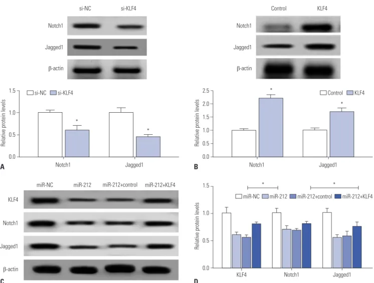
Notch signaling pathway is involved in miR-212/
KLF4-mediated regulation on MPP
+-induced damage in SH-SY5Y cells
Notch signaling, an important regulator of the nervous system,
has been shown to be able to maintain immature neurons, modulate neurite outgrowth of differentiated neurons, and con- trol synaptic plasticity and olfactory functions in the adult brain.15 Moreover, KLF4 was previously reported as an upstream regu- lator of Notch signaling in multiple biological processes.16 Thus, we further determined whether KLF4 could regulate Notch signaling in MPP+-induced SH-SY5Y cells by analyzing the ex- pression levels of Notch signaling molecules following KLF4 overexpression or downregulation. The data revealed that KLF4 depletion leads to an apparent suppression of Notch1 and Jagged1 expression (Fig. 5A). Conversely, enforced expression of KLF4 considerably elevated the expression of Notch1 and Jagged1 (Fig. 5B). These results suggested that KLF4 could stimulate Notch signaling pathway. Further, we found that miR- 212 upregulation suppressed the expression of Notch1 and Jag- ged1, whereas this inhibitory effect of miR-212 was reversed by the restoration of KLF4 expression (Fig. 5C and D). Taken together, these results suggested that miR-212 abates MPP+- induced injury of SH-SY5Y cells by blocking Notch signaling
Caspase 3 activity (% of change)SOD levels (U/mL)TNF-α levels (ng/L) LDH release (U/L)ROS levels (% count)IL-1β levels (ng/L)
Control
Control
Control
Control
Control
Control MPP+
MPP+
MPP+
MPP+
MPP+
MPP+
*
*
*
*
*
*
*
*
*
*
*
* 800
600 400 200 0
150
100
50
0
150
100
50
0
2000 1500 1000 500 0
500 400 300 200 100 0
100 80 60 40 20 0
−
−
−
−
−
−
−
−
−
−
−
−
−
−
− – –
−
−
− – – –
−
−
− – – –
− miR-NC
miR-212 anti-miR-212
miR-NC miR-212 anti-miR-212
miR-NC miR-212 anti-miR-212
miR-NC miR-212 anti-miR-212
miR-NC miR-212 anti-miR-212
miR-NC miR-212 anti-miR-212 A
C
E
B
D
F
Fig. 2. MiR-212 ameliorates MPP+-induced damage of SH-SY5Y cells. SH-SY5Y cells transfected with miR-212 mimics or anti-miR-212 were treated with or without 2 mM MPP+ for 24 h, followed by ELISA assays of caspase-3 activity (A), LDH release (B), SOD level (C), ROS production (D), TNF-α ex- pression (E), and IL-1β secretion (F). *p<0.05 vs. corresponding control. LDH, lactate dehydrogenase; SOD, superoxide dismutase; ROS, reactive oxy- gen species; TNF-α, tumor necrosis factor-α; IL-1β, interleukin-1 beta.
pathway via regulating KLF4.
DISCUSSION
PD is a chronic neurodegeneration disease in older adult in- dividuals, characterized by resting tremor, rigidity, bradykine- sia, and postural instability.1 To date, a number of documents have revealed that the pathophysiologic mechanisms under- lying neurodegeneration are related to oxidative stress and in- flammation response.3 SOD, an antioxidant enzyme, acts as a scavenger of ROS to prevent PD development.17 LDH, released by broken cells, can be measured as a surrogate for cell dam- age.18 IL-1β and TNF-α, two pivotal harmful inflammatory me- diators, play vital roles in the development of PD.19 In this study, we employed MPP+-induced SH-SY5Y cells as a PD model in vitro. Our results showed that MPP+ treatment leads to a sup- pression of SH-SY5Y cell viability in a dose- or time-dependent manner. Moreover, MPP+ treatment promoted caspase-3 activi- ty, LDH release, oxidative stress production, and inflammation response of SH-SY5Y cells. Altogether, our data implied that MPP+ treatment contributes to the damage of SH-SY5Y cells.
Up to now, several miRNAs have been implicated in MPP+-trig- gered PD model in SH-SY5Y cells. For instance, Wang, et al. 20 found that miR-124 upregulation affected apoptosis and im-
paired autophagy process in MPTP-treated mice and MPP+- induced SH-SY5Y cells by targeting a BH3-only protein (Bim).
Additionally, it was reported that miR-7 upregulation improved viability and inhibited apoptosis in MPP+-induced SH-SY5Y cells by directly targeting KLF4.21 MiR-212, expressed in prima- ry neuronal cultures, has been found to be able to exert a neu- roprotective role in a series of human neurological diseases.11 For example, Wong, et al.22 reported that miR-212 overexpres- sion protected neurons against oxidative stress and that miR- 212 depletion induced neuronal apoptosis through targeting P300, PTEN, and FOXO3a in Alzheimer’s disease. Also, sup- pression of miR-132/212 destroyed the balance of S-nitrosyl- ation and promoted tau phosphorylation in a neuronal nitric oxide synthase (NOS1)-dependent way, contributing to the progression of Alzheimer’s disease.23 Moreover, Gillardon, et al.24 stated that miR-212 may represent a potential therapeutic biomarker and target for human neurodegenerative diseases, such as PD. Our present results revealed that miR-212 is down- regulated in MPP+-induced SH-SY5Y cells. miR-212 overexpres- sion mitigated MPP+-induced decreases in SH-SY5Y cell via- bility. Moreover, miR-212 alleviated MPP+-induced SH-SY5Y cell damage, as evidenced by decreases in caspase-3 activity, LDH release, oxidative stress, and inflammation response. All these results implied that miR-212 plays a protective role against MPP+-induced cell damage of SH-SY5Y cells.
Relative mRNA levels of KLF4 Relative protein levels of KLF4
hsa-miR-212 Position 266-272 of KLF4 3'UTR Position 266-272 of KLF4 3'UTR Mu
NC miR-212 anti-miR-212 NC miR-212 anti-miR-212
1.5
1.0
0.5
0.0
2.0 1.5 1.0 0.5 0.0
−
−
−
−
−
−
−
−
− A
C D
Fig. 3. KLF4 is a direct target of miR-212 in SH-SY5Y cells. (A) Sequence alignment between miR-212 and the 3’-UTR of KLF4. (B) Luciferase reporter as- say was performed in SH-SY5Y cells after transfection with KLF4-WT or KLF4-Mu together with miR-212 mimics or anti-miR-212. SH-SY5Y cells were transfected with miR-212 mimics or anti-miR-212, followed by the measurement of KLF4 mRNA (C) and protein (D). p<0.05 vs. control. KLF4, Kruppel-like factor 4; 3’UTR, 3’-untranslated region; KLF4-WT, wild-type KLF4 reporter vector; KLF4-Mu, mutant-type KLF4 reporter vector; mRNA, messenger RNA.
3' ---UCAUUCGUCAGAUCUCGGUUCCA---5' 5' ---UCGGACUUACAAAAUGCCAAGGG---3' 5' ---CUGGACUUACAAAAUGCCUUGGG---3' Luciferase report activity
NC miR-212 anti-miR-212
*
* 2.5
2.0 1.5 1.0 0.5 0.0
−
− – –
−
−
KLF4-WT KLF4-Mu
B
*
* NC
KLF4
β-actin
miR-212 anti-miR-212
Relative protein levels of KLF4Relative protein levels of KLF4 Cell apoptosis (%)Cell viability (%)Cell viability (%) Cell apoptosis (%) 2.0
1.5 1.0 0.5 0.0
105 104 103 102
105 104 103 102
105 104 103 102
105 104 103 102
105 104 103 102 105
104 103 102 105
104 103 102 105
104 103 102 1.5
1.0
0.5
0.0
80 60 40 20 0 250
200 150 100 50 0 200 150 100 50 0
80 60 40 20 0
−
−
−
−
−
−
−
−
−
−
−
−
−
−
−
− – –
−
−
−
−
−
−
−
−
−
−
−
−
*
*
* *
*
*
*
*
* *
* *
si-NC
102 103 104 105 102 103 104 105 102 103 104 105 102 103 104 105
102 103 104 105 102 103 104 105
102 103 104 105 102 103 104 105
miR-NC miR-NC miR-NC
si-NC si-NC
Control
miR-212+control miR-212+control miR-212+control
Control Control
si-KLF4
miR-212 miR-212 miR-212
si-KLF4 si-KLF4
KLF4
miR-212+KLF4 miR-212+KLF4 miR-212+KLF4
KLF4 KLF4
A
E D
H
B
F
C
G si-NC si-KLF4Control KLF4
miR-NCmiR-212miR-212+controlmiR-212+KLF4 KLF4
KLF4 β-actin
β-actin
si-NC
miR-NC
Annexin-V-FITC
Annexin-V-FITC
PIPI
Control
miR-212+control si-KLF4
miR-212
KLF4
miR-212+KLF4
Fig. 4. MiR-212-mediated protection effect is abated following KLF4 expression restoration in MPP+-induced SH-SY5Y cells. SH-SY5Y cells transfected with si-NC, si-KLF4, control, or KLF4 were stimulated with 2 mM MPP+ for 24 h, followed by the detection of KLF4 protein (A), cell viability (B), and apoptosis (C and D). SH-SY5Y cells were transfected with miR-212 mimics alone or in combination with KLF4 prior to treatment with MPP+, followed by the analysis of KLF4 protein (E), cell viability (F), and apoptosis (G and H). *p<0.05 vs. corresponding control. KLF4, Kruppel-like factor 4.
Blocking translation initiation is one of the main mecha- nisms of miRNA in regulating gene expression.25 Therefore, Tar- getScan software was employed to search for the candidate tar- gets of miR-212. Interestingly, the data predicted that KLF4 contained a conserved sequence complementary with miR- 212 at its 3’UTR. Subsequent dual-luciferase reporter assay, RT-qPCR, and western blot assay showed that miR-212 repress- es KLF4 expression in a post-transcriptional manner. KLF4, a member of the family of zinc-finger transcription factors, was reported to be implicated in multiple pathophysiological pro- cesses, such as cell growth, proliferation, differentiation, and inflammation.26 In this study, we found that exogenous ex- pression of KLF4 lowered viability and promoted apoptosis in MPP+-stimulated SH-SY5Y cells. Moreover, KLF4 overexpres- sion effectively reversed the effect of miR-212 on vitality and apoptosis in MPP+-administrated SH-SY5Y cells. Similarly, a recent document displayed that knockdown of KLF4 inhibited SH-SY5Y cell apoptosis under MPP+ treatment, and KLF4 up-
regulation abated the protective effect of miR-7 on MPP+-elic- ited apoptosis.21 Moreover, KLF4 was demonstrated to enhance oxidative stress, block proliferation, and promote LDH release in human dopamine neuroblastoma M17 cells induced by MPP+.27
Notch signaling has been reported to be important to the normal development of neurons, oligodendrocytes and astro- cytes, and to affect neurological disorders of the central ner- vous system.28 In our current study, we found that the expres- sion of Notch1 and Jagged1 were strikingly decreased after downregulating KLF4, while Notch1 and Jagged1 expression were increased when KLF4 was upregulated. In accordance with our finding, Yu, et al.16 also presented that the expressions of Notch1, Notch2, and Jagged1 were strikingly inhibited in KLF4-depletion cells, and KLF4 was verified to be a central reg- ulator of sprouting angiogenesis by affecting Notch signaling.29 Moreover, we also found that miR-212 upregulation suppressed the expression of Notch1 and Jagged1, whereas the suppres- sion effect of miR-212 was strikingly reversed by the regaining
Relative protein levels Relative protein levels
Notch1 Notch1
* *
Jagged1 Jagged1
*
*
*
* 1.5
1.0
0.5
0.0
2.5 2.0 1.5 1.0 0.5 0.0
−
–
−
−
− – – –
−
−
si-NC si-KLF4 Control KLF4
A
C
B
D
Notch1 Notch1
Jagged1 Jagged1
β-actin β-actin
si-NC si-KLF4 Control KLF4
Relative protein levels
1.5
1.0
0.5
0.0
−
−
−
−
miR-NC miR-212 miR-212+control miR-212+KLF4 miR-212+control miR-212+KLF4
miR-212 miR-NC
KLF4 Notch1 Jagged1
KLF4
Jagged1 Notch1
β-actin
Fig. 5. Notch signaling pathway is involved in the regulation of miR-212/KLF4 axis in MPP+-induced SH-SY5Y cells. (A) Western blot assay of Notch1 and Jagged1 proteins in SH-SY5Y cells transfected with si-NC or si-KLF4 under MPP+ treatment. (B) Western blot analysis of Notch1 and Jagged1 proteins in SH-SY5Y cells transfected with control or KLF4 with the treatment of MPP+. (C and D) SH-SY5Y cells transfected with miR-212 mimics alone, or together with KLF4 prior to the treatment of MPP+, followed by the detection of KLF4, Notch1 and Jagged1 proteins expression. β-actin was used as the internal reference. *p<0.05 vs. respective control. KLF4, Kruppel-like factor 4.
of KLF4 expression. All these results indicated that miR-124 inactivates Notch signaling via regulation of KLF4 in MPP+-in- duced SH-SY5Y cells.
In conclusion, our study suggests that miR-212 might atten- uate MPP+-induced neuronal damage by regulating KLF4/
Notch signaling pathway in SH-SY5Y cells, indicating the po- tential value of miR-212 as a biomarker and therapeutic target of PD. However, more in vivo data are required to further vali- date the role and mechanism of miR-212 in PD progression.
ORCID
Yanfeng Song https://orcid.org/0000-0001-9511-8906 Xiaowei Chen https://orcid.org/0000-0002-4005-544X
REFERENCES
1. Kalia LV, Lang AE. Parkinson’s disease. Lancet 2015;386:896-912.
2. Abeliovich A, Gitler AD. Defects in trafficking bridge Parkinson’s disease pathology and genetics. Nature 2016;539:207-16.
3. Andican G, Konukoglu D, Bozluolcay M, Bayülkem K, Firtiına S, Burcak G. Plasma oxidative and inflammatory markers in patients with idiopathic Parkinson’s disease. Acta Neurol Belg 2012;112:
155-9.
4. Kalia LV, Kalia SK, Lang AE. Disease-modifying strategies for Par- kinson’s disease. Mov Disord 2015;30:1442-50.
5. Hammond SM. An overview of microRNAs. Adv Drug Deliv Rev 2015;87:3-14.
6. Harraz MM, Dawson TM, Dawson VL. MicroRNAs in Parkinson’s disease. J Chem Neuroanat 2011;42:127-30.
7. Quinlan S, Kenny A, Medina M, Engel T, Jimenez-Mateos EM.
MicroRNAs in neurodegenerative diseases. Int Rev Cell Mol Biol 2017;334:309-43.
8. Yang CP, Zhang ZH, Zhang LH, Rui HC. Neuroprotective role of microRNA-22 in a 6-hydroxydopamine-induced cell model of Parkinson’s disease via regulation of its target gene TRPM7. J Mol Neurosci 2016;60:445-52.
9. Wang J, Le T, Wei R, Jiao Y. Knockdown of JMJD1C, a target gene of hsa-miR-590-3p, inhibits mitochondrial dysfunction and oxi- dative stress in MPP+-treated MES23.5 and SH-SY5Y cells. Cell Mol Biol (Noisy-le-grand) 2016;62:39-45.
10. Li S, Lv X, Zhai K, Xu R, Zhang Y, Zhao S, et al. MicroRNA-7 inhib- its neuronal apoptosis in a cellular Parkinson’s disease model by targeting Bax and Sirt2. Am J Transl Res 2016;8:993-1004.
11. Wanet A, Tacheny A, Arnould T, Renard P. miR-212/132 expres- sion and functions: within and beyond the neuronal compartment.
Nucleic Acids Res 2012;40:4742-53.
12. Aten S, Hansen KF, Hoyt KR, Obrietan K. The miR-132/212 locus:
a complex regulator of neuronal plasticity, gene expression and cognition. RNA Dis 2016;3:e1375.
13. Burgos K, Malenica I, Metpally R, Courtright A, Rakela B, Beach T, et al. Profiles of extracellular miRNA in cerebrospinal fluid and
serum from patients with Alzheimer’s and Parkinson’s diseases correlate with disease status and features of pathology. PLoS One 2014;9:e94839.
14. Maiti P, Manna J, Dunbar GL. Current understanding of the mo- lecular mechanisms in Parkinson’s disease: targets for potential treatments. Transl Neurodegener 2017;6:28.
15. Imai Y, Kobayashi Y, Inoshita T, Meng H, Arano T, Uemura K, et al.
The Parkinson’s disease-associated protein kinase LRRK2 modu- lates notch signaling through the endosomal pathway. PLoS Gen- et 2015;11:e1005503.
16. Yu F, Li J, Chen H, Fu J, Ray S, Huang S, et al. Kruppel-like factor 4 (KLF4) is required for maintenance of breast cancer stem cells and for cell migration and invasion. Oncogene 2011;30:2161-72.
17. Xie XX, Kou ST, Pu ZH, Hou CY, Tian YP. [Effects of scalp catgut embedding on SOD, NO, MDA in the rat with Parkinson’s disease].
Zhongguo Zhen Jiu 2007;27:753-6.
18. Popovich DG, Lee Y, Li L, Zhang W. Momordica charantia seed extract reduces pre-adipocyte viability, affects lactate dehydroge- nase release, and lipid accumulation in 3T3-L1 cells. J Med Food 2011;14:201-8.
19. Nagatsu T, Sawada M. Inflammatory process in Parkinson’s dis- ease: role for cytokines. Curr Pharm Des 2005;11:999-1016.
20. Wang H, Ye Y, Zhu Z, Mo L, Lin C, Wang Q, et al. MiR-124 regulates apoptosis and autophagy process in MPTP model of Parkinson’s disease by targeting to Bim. Brain Pathol 2016;26:167-76.
21. Kong B, Wu PC, Chen L, Yang T, Yuan YQ, Kuang YQ, et al. mi- croRNA-7 protects against 1-methyl-4-phenylpyridinium iodide- induced cell apoptosis in SH-SY5Y cells by directly targeting Krüpple-like factor 4. DNA Cell Biol 2016;35:217-25.
22. Wong HK, Veremeyko T, Patel N, Lemere CA, Walsh DM, Esau C, et al. De-repression of FOXO3a death axis by microRNA-132 and -212 causes neuronal apoptosis in Alzheimer’s disease. Hum Mol Genet 2013;22:3077-92.
23. Wang Y, Veremeyko T, Wong AH, El Fatimy R, Wei Z, Cai W, et al.
Downregulation of miR-132/212 impairs S-nitrosylation balance and induces tau phosphorylation in Alzheimer’s disease. Neuro- biol Aging 2017;51:156-66.
24. Gillardon F, Mack M, Rist W, Schnack C, Lenter M, Hildebrandt T, et al. MicroRNA and proteome expression profiling in early-symp- tomatic α-synuclein(A30P)-transgenic mice. Proteomics Clin Appl 2008;2:697-705.
25. Jackson RJ, Standart N. How do microRNAs regulate gene expres- sion? Sci STKE 2007;2007:re1.
26. Ghaleb AM, Yang VW. Krüppel-like factor 4 (KLF4): what we cur- rently know. Gene 2017;611:27-37.
27. Chen J, Wang X, Yi X, Wang Y, Liu Q, Ge R. Induction of KLF4 con- tributes to the neurotoxicity of MPP+ in M17 cells: a new implica- tion in Parkinson’s disease. J Mol Neurosci 2013;51:109-17.
28. Yao L, Cao Q, Wu C, Kaur C, Hao A, Ling EA. Notch signaling in the central nervous system with special reference to its expression in microglia. CNS Neurol Disord Drug Targets 2013;12:807-14.
29. Hale AT, Tian H, Anih E, Recio FO 3rd, Shatat MA, Johnson T, et al.
Endothelial Kruppel-like factor 4 regulates angiogenesis and the Notch signaling pathway. J Biol Chem 2014;289:12016-28.