INTRODUCTION
Osteosarcoma is a malignant and rare tumor that primarily affects bones in juveniles and adults.1,2 It has been reported that osteosarcoma patients with metastatic or recurrent dis-
ease fare poorly, with overall survival rates of less than 20%.3 Therapeutic strategies, especially chemotherapy, lead to im- proved survival and outcomes in patients with osteosarcoma.4-6 Epirubicin is one of the most effective drugs against cancer and has gained widespread use in multiple cancers.7,8 Moreover, studies have shown that epirubicin could enhance the antitu- mor activity in osteosarcoma.9,10 Therefore, combination che- motherapy with epirubicin and other agents may provide an effective approach for osteosarcoma patients.
MicroRNAs (miRNAs), a class of small noncoding RNAs, can act as tumor-suppressors in tumorigenesis and development.11 Research has revealed that inhibition of miR-598 expression stimulates proliferation, migration, and invasion in osteosar- coma. Also, restoration of miR125a5p results in repressing os- teosarcoma cell migration, invasion and epithelialmesenchy- mal transition by targeting matrix metallopeptidase-11.12
miR-1301/TRIAP1 Axis Participates
in Epirubicin-Mediated Anti-Proliferation and Pro-Apoptosis in Osteosarcoma
Lijun Yu1*, Min Meng1*, Yun Bao1, Chao Zhang2, Bei Gao1, Rina Sa1, and Wenyuan Luo2
1Department of Pharmacy and 2Department III of Orthopedic, Gansu Provincial Hospital, Lanzhou, Gansu, China.
Purpose: Epirubicin is one of the most effective drugs against osteosarcoma. miR-1301 is involved in the occurrence and devel- opment of osteosarcoma. Whether miR-1301 is responsible for the chemosensitivity of osteosarcoma cells to epirubicin remains largely unknown.
Materials and Methods: U2OS and SAOS-2 cells were treated with various concentrations of epirubicin. Flow cytometry was em- ployed to evaluate cell apoptotic rate. Cell proliferation was measured by Cell Counting Kit-8 assay. Western blot and quantitative real-time polymerase chain reaction were utilized to detect the expressions of B-cell lymphoma-2 (Bcl-2), Bcl-2 assaciated X pro- tein (Bax), cleaved-caspase-3, cleaved-poly (ADP-ribose) polymerases (PARP1), TP53-regulated inhibitor of apoptosis 1 (TRIAP1), and microRNA-1301 (miR-1301). The relationship between miR-1301 and TRIAP1 was determined by luciferase reporter assay.
Results: Epirubicin inhibited proliferation in a dose-dependent manner, induced apoptosis, decreased the expression of Bcl-2, and increased the expressions of Bax, cleaved-caspase-3, and cleaved-PARP1 in osteosarcoma cells. miR-1301 was downregulat- ed in U2OS and SAOS-2 cells. Importantly, epirubicin significantly increased the levels of miR-1301. Overexpression of miR-1301 suppressed proliferation and promoted apoptosis. Interestingly, those effects were enhanced by epirubicin. In contrast, miR-1301 depletion attenuated the epirubicin-mediated anti-osteosarcoma effect. miR-1301 negatively regulated the expression of TRIAP1 in U2OS and SAOS-2 cells. Furthermore, epirubicin inhibited the mRNA and protein levels of TRIAP1 by upregulating miR-1301 levels. Epirubicin suppressed cell proliferation by downregulating TRIAP1.
Conclusion: miR-1301 was implicated in the chemosensitivity of osteosarcoma to epirubicin by modulating TRIAP1.
Key Words: miR-1301, TRIAP1, epirubicin, osteosarcoma
pISSN: 0513-5796 · eISSN: 1976-2437
Received: February 19, 2019 Revised: May 25, 2019 Accepted: June 13, 2019
Corresponding author: Wenyuan Luo, MD, Department III of Orthopedic, Gansu Provincial Hospital, No. 204, West Donggang Road, Chengguan District, Lanzhou 730000, Gansu, China.
Tel: 86-0931-8281007, Fax: 86-0931-8281007, E-mail: [email protected]
*Lijun Yu and Min Meng contributed equally to this work.
•The authors have no potential conflicts of interest to disclose.
© Copyright: Yonsei University College of Medicine 2019
This is an Open Access article distributed under the terms of the Creative Com- mons Attribution Non-Commercial License (https://creativecommons.org/licenses/
by-nc/4.0) which permits unrestricted non-commercial use, distribution, and repro- duction in any medium, provided the original work is properly cited.
Yonsei Med J 2019 Sep;60(9):832-841 http://dx.doi.org/10.3349/ymj.2019.60.9.832
Moreover, it is well documented that miRNAs are involved in chemotherapy of cancers.13,14 Li, et al.15 noted that a combina- tion of miR-19a and miR-205 in the serum may predict che- mosensitivity to epirubicin plus paclitaxel neoadjuvant chemo- therapy for the luminal a subtype of breast cancer. Furthermore, miR-671-5p has been found to sensitize breast cancer cells to cisplatin, 5-fluorouracil, and epirubicin exposure and to re- duce DNA repair capability in post-drug exposed breast can- cer cells.16 Zhang, et al.17 found that miR-4443 depletion in- creases the sensitivity of epirubicin-resistant H1299 cells to epirubicin by decreasing the IC50 of epirubicin and inducing cell apoptosis and G0/G1 cell cycle arrest in non-small cell lung cancer. Additionally, previous studies have demonstrated that miRNAs are also correlated with chemosensitivity in osteosar- coma.18 For example, miR-125b was found to enhance chemo- sensitivity to cisplatin in osteosarcoma.19 miR-302b increased epirubicin-mediated anti-proliferation and pro-apoptosis ef- fect in osteosarcoma.20 Recently, miR-1301 was reported to in- hibit migration and invasion by targeting B-cell chronic lym- phocytic leukemia/lymphoma 9 in osteosarcoma.21 However, whether miR-1301 is responsible for anti-osteosarcoma ef- fects remains to be elucidated. TP53-regulated inhibitor of apoptosis 1 (TRIAP1) is overexpressed in many cancers and acts as an oncogene in tumorigenesis and development.22 Nevertheless, still relatively little is known about its role in os- teosarcoma.
In this study, we hypothesized that TRIAP1 may be a target of miR-1301 and investigated the role of miR-1301/TRIAP1 in epirubicin-mediated anti-osteosarcoma effects.
MATERIALS AND METHODS
Cell culture and treatment
Human osteoblast hFOB1.19 cells and human osteosarcoma U2OS and SAOS-2 cells were purchased from American Type Culture Collection (Manassas, VA, USA) and cultured in the Dulbecco’s Modified Eagle’s Medium (Thermo Fisher Scien- tific, Waltham, MA, USA) supplemented with 10% fetal bovine serum (Thermo Fisher Scientific) and 1% penicillin/strepto- mycin stock solution (Beyotime, Shanghai, China). Cells were maintained in a humidified atmosphere containing 5% CO2 at 37°C. U2OS and SAOS-2 cells (80% confluence in 6-well plates) were treated with different concentrations (0, 0.1, 0.25, 0.5, 1, 2.5, 5, or 10 μg/mL) of epirubicin (MedChemExpress, Prince- ton, NJ, USA) for 24 or 48 h. U2OS and SAOS-2 cells were trans- fected with miR-NC mimics, miR-1301 mimics, miR-NC inhibi- tors, miR-1301 inhibitors, pcDNA vector (vector), and TRIAP1 overexpression plasmid (TRIAP1). After 24-h transfection, cells were treated with epirubicin (1 μg/mL) for 48 h. The transfect- ed or treated cells were used for subsequent experiments.
qRT-PCR assay
TRIzol (Beyotime) was introduced to extract total RNAs from cells according the manufacturer’s instructions. Then, RNA was reversely transcribed into complementary DNA using TaqMan MicroRNA Reverse Transcription Kits (Biosystems, Foster City, CA, USA) or TIANScript RT Kits (Baolebo biotech, Beijing, China). Quantitative real-time polymerase chain re- action (qRT-PCR) analyses were carried out using Power SYBR Green kits (Takara, Shiga, Japan). The primers were as follows:
TRIAP1 forward, 5'-AGGATTTCGCAAGTCCAGAA-3', and re- verse, 5'-GCTGATTCCACCCAAGTAT-3'; glyceraldehyde- 3-phosphate dehydrogenase (GAPDH) forward, 5'-AGAAGG CTGGGGCTCATTTG-3', and reverse, 5'-AGGGGCCATCCA CAGTCTTC-3'; miR-1301 forward, 5'-ACACTCCAGCTGGG TTGCAGCTGCCTGGGAGTGA-3', and reverse, 5'-CTCAACT GGTGTCGTGGA- 3'; U6 forward, 5'-CTTCGGCAGCACAT ATAC-3' and reverse, 5'-GAACGCTTCACGAATTTGC-3'. The relative expression levels of miRNA (normalized to U6 small nuclear RNA) and mRNA (normalized to GAPDH) were evalu- ated by the 2-ΔΔCt method.
CCK-8 assay
Osteosarcoma cells (5000 cells/well) were seeded in 96-well plates and detected for cell proliferation using Cell Counting Kit-8 (CCK-8) assay according to the manufacturer’s instruc- tions. Briefly, the cells were incubated with 10 µL of CCK-8 (AbMole BioScience, Houston, TX, USA) at 37°C for 4 h. Ab- sorbance at 450 nm was evaluated using a microtiter plate reader (Thermo Fisher Scientific). Each group was tested with three replicates.
Cell apoptosis assay
Cell apoptotic rate was evaluated as described in a previous study using Annexin V-PE/7-AAD apoptosis assay (KeyGEN BioTECH, Nanjing, China).20 Briefly, cells were washed with cooled phosphate buffer saline and trypsinized. Annexin V-PE (1 µL) and 7-AAD (5 µL) were added in a dark room for 10 min incubation. Cell apoptotic rate was detected by a FACSCalibur flow cytometer with Cell Quest software (BD Biosciences, Franklin Lakes, NJ, USA).
Western blot assay
Cells were collected in Radio Immunoprecipitation Assay ly- sis buffer containing protease inhibitors (Beyotime). Cell ly- sates were centrifuged, and supernatants were collected and mixed with 6× loading buffer and boiled for 10 min. Proteins were quantified using bicinchoninic acid Protein Assay Kits (Solarbio, Beijing, China), separated with sodium dodecyl sulfate polyacrylamide gel, and transfected onto polyvinyli- dene fluoride membranes (Millipore, Billerica, MA, USA). The primary antibodies used in the present study were anti-Bcl-2:
ab32124, anti-Bax: ab32503, anti-cleaved-caspase-3: ab2302, anti-cleaved-poly (ADP-ribose) polymerase-1 (anti-cleaved-
PARP1): ab32064, anti-TRIAP1: ab225938, and anti-GAPDH:
ab181602, which were purchased from Abcam (Cambridge, MA, USA). Horseradish peroxidase conjugated-secondary anti-body (goat anti-rabbit IgG: ab150077) was obtained from Cell Signaling Technology (Massachusetts, MA, USA). The protein signals were visualized using enhanced chemilumines- cence (Thermo Fisher Scientific). Densitometry values were nor- malized to levels of GAPDH and analyzed using ImageJ 1.48v software (National Institutes of Health, Bethesda, MD, USA).
Luciferase reporter assay
The 3’-UTR of TRIAP1 containing predicted miR-1301 binding sites [wild-type TRIAP1 (WT) and mutant TRIAP1 (MUT)] were subcloned into the pmirGLO vector (Promega, Madison, WI, USA). Then, U2OS and SAOS-2 cells were cultured in 24-well plates and co-transfected with TRIAP1-WT or TRIAP1-MUT and miR-1301 mimics or miR-NC mimics, respectively. The rel- ative luciferase activities were measured using the Dual-Lu- ciferase Reporter Assay System (Promega).
Statistical analysis
All data are expressed as a mean±standard deviation. Statisti-
cal analysis was conducted using SPSS 18.0 software (SPSS, Inc., Chicago, IL, USA). Results were considered statistically significant only if the p value was less than 0.05 using Student’s t test or one-way ANOVA. Our research was approved by Gan- su Provincial Hospital.
RESULTS
Effects of epirubicin on proliferation and apoptosis in osteosarcoma cells
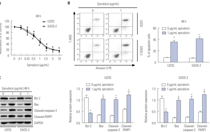
First, to determine the effect of epirubicin on osteosarcoma cells, U2OS and SAOS-2 cells were treated with epirubicin at various concentrations. The results of CCK-8 assays showed that epirubicin inhibited osteosarcoma cell proliferation in a dose-dependent manner (Fig. 1A). Furthermore, 1 μg/mL of epirubicin was regarded as a standard concentration, since it provided effective inhibition of cell viability and a good amount of surviving cells. Then, the effect of epirubicin on cell apop- tosis was further evaluated. We demonstrated that cell apop- totic rate was higher in cells with epirubicin exposure than that of cells treated with 0 μg/mL of epirubicin (Fig. 1B). More-
Fig. 1. Effects of epirubicin on proliferation and apoptosis in osteosarcoma cells. (A) Osteosarcoma cell lines U2OS and SAOS-2 were treated with differ- ent concentrations of epirubicin for 48 h, and Cell Counting Kit-8 assay was performed to evaluate cell proliferation. (B) U2OS and SAOS-2 cells were ex- posed to 0 μg/mL or 1 μg/mL of epirubicin for 48 h, respectively. Flow cytometry was introduced to determine cell apoptotic rate. (C) Western blot was conducted to examine the expressions of Bcl-2, Bax, cleaved-caspase-3, and cleaved-poly (ADP-ribose) polymerase-1 (cleaved-PARP1) in U2OS and SAOS-2 cells with 48 h of 0 μg/mL or 1 μg/mL epirubicin exposure. *p<0.05. Bcl-2, B-cell lymphoma-2; Bax, Bcl-2 assaciated X protein; GAPDH, glyceral- dehyde-3-phosphate dehydrogenase.
120 100 80 60 40 20 0
60
40
20
0
1.5
1.0
0.5
0.0
1.5
1.0
0.5
0.0
U2OS
Bcl-2 Bax Cleaved- Bcl-2 Bax
caspase-3 Cleaved-
caspase-3 Cleaved-
PARP1 Cleaved-
PARP1 U2OS
0 µg/mL epirubicin 1 µg/mL epirubicin
0 µg/mL epirubicin 1 µg/mL epirubicin
0 µg/mL epirubicin 1 µg/mL epirubicin 0 0.1 0.25 0.5 1 2.5 5 10
U2OS SAOS-2 48 h
48 h
U2OS SAOS-2
*
* *
* * * *
* *
* Epirubicin (µg/mL)
Epirubicin (µg/mL) 48 h
0 1
0 1 0 1
Annexin V-PE Epirubicin (µg/mL)
Bcl-2 Bax
Cleaved-caspase-3 Cleaved-PARP1 GAPDH
Absorbance (% of control) % of apoptotic cells
Relative protein expression Relative protein expression
7-ADD U2OSSAOS-2
SAOS-2
SAOS-2 A
C
B
over, the detailed molecular mechanisms of epirubicin affect- ing apoptosis were subsequently explored. The level of anti- apoptosis protein Bcl-2 was decreased, while the expressions of Bax, cleaved-caspase-3 and cleaved-PARP1 were increased in U2OS and SAOS-2 cells treated with 1 μg/mL of epirubicin, compared with cells without epirubicin treatment (Fig. 1C), indicating that epirubicin participates in pro-apoptosis and anti-proliferation in osteosarcoma in vitro.
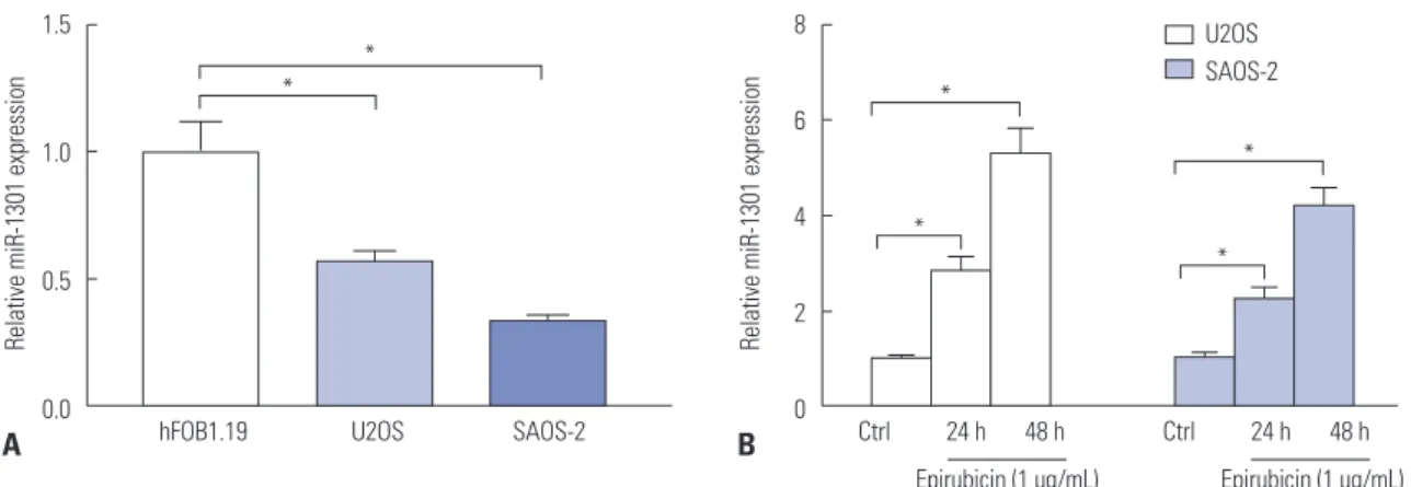
Epirubicin stimulates the expression of miR-1301 in U2OS and SAOS-2 cells
Then, we continued to examine the levels of miR-1301 in osteo- sarcoma cells. We observed that miR-1301 was downregulat- ed in U2OS and SAOS-2 cells, compared with that of hFOB1.19 human osteoblast cells (Fig. 2A), suggesting that miR-1301 may act as a tumor suppressor in osteosarcoma. Moreover, whether epirubicin regulates the expression of miR-1301 was further addressed. As expected, epirubicin increased the abundance of miR-1301 in U2OS and SAOS-2 cells relative to that of the Ctrl group (Fig. 2B), demonstrating that miR-1301 may play a vital role in epirubicin-mediated effects in osteo- sarcoma cells.
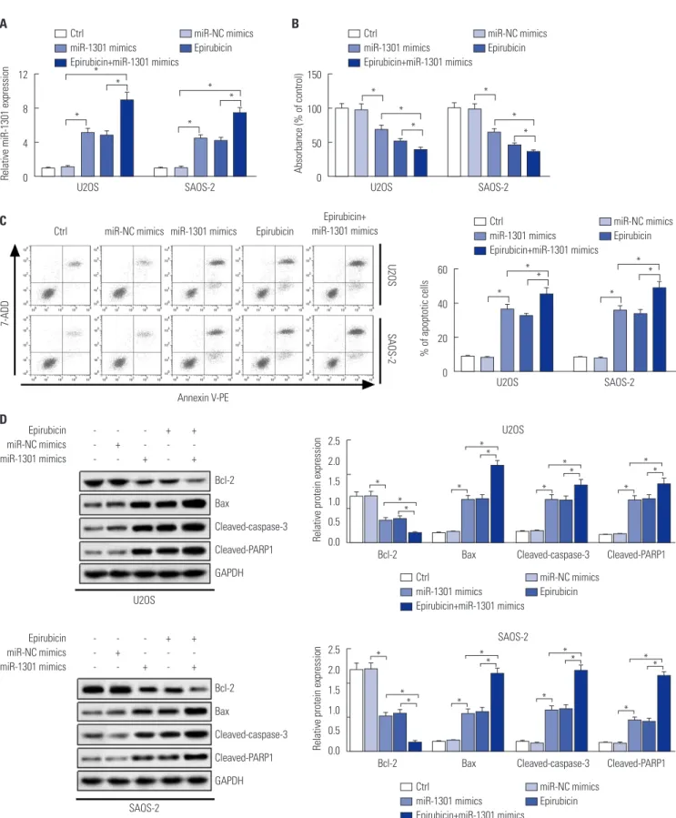
Upregulation of miR-1301 contributes to the anti-cancer effect of epirubicin in osteosarcoma Considering that the level of miR-1301 was increased in os- teosarcoma cells exposed to epirubicin, the role of miR-1301 in epirubicin-mediated anti-osteosarcoma was subsequently investigated. The results of transfection efficiency revealed that the introduction of miR-1301 mimics and epirubicin alone induced the enforced expression of miR-1301. Moreover, the promotion effect was enhanced by combination of epirubicin and miR-1301 mimics (Fig. 3A). As shown in Fig. 3B, the data further demonstrated that cell proliferation was impeded in the miR-1301 mimics group. More importantly, the inhibition effect was strengthened by a combination of them. Further- more, the results of flow cytometry presented that cell apoptotic
rate was elevated in cells transfected with miR-1301 mimics or treated with epirubicin. Intriguingly, a combination of miR- 1301 overexpression and epirubicin reinforced the boosted effect on cell apoptosis (Fig. 3C). In addition, the expressions of apoptosis-related proteins were also analyzed in U2OS and SAOS-2 cells using Western blot. Our data showed that the combination of epirubicin and miR-1301 mimics increased the effects observed with separate treatment of them on the ex- pressions of apoptosis-related protein (Fig. 3D), which was consistent with the data of flow cytometry. Consequently, our findings demonstrated that miR-1301 contributes to the epi- rubicin-mediated anti-osteosarcoma effect via a regulatory ef- fect on cell proliferation and apoptosis.
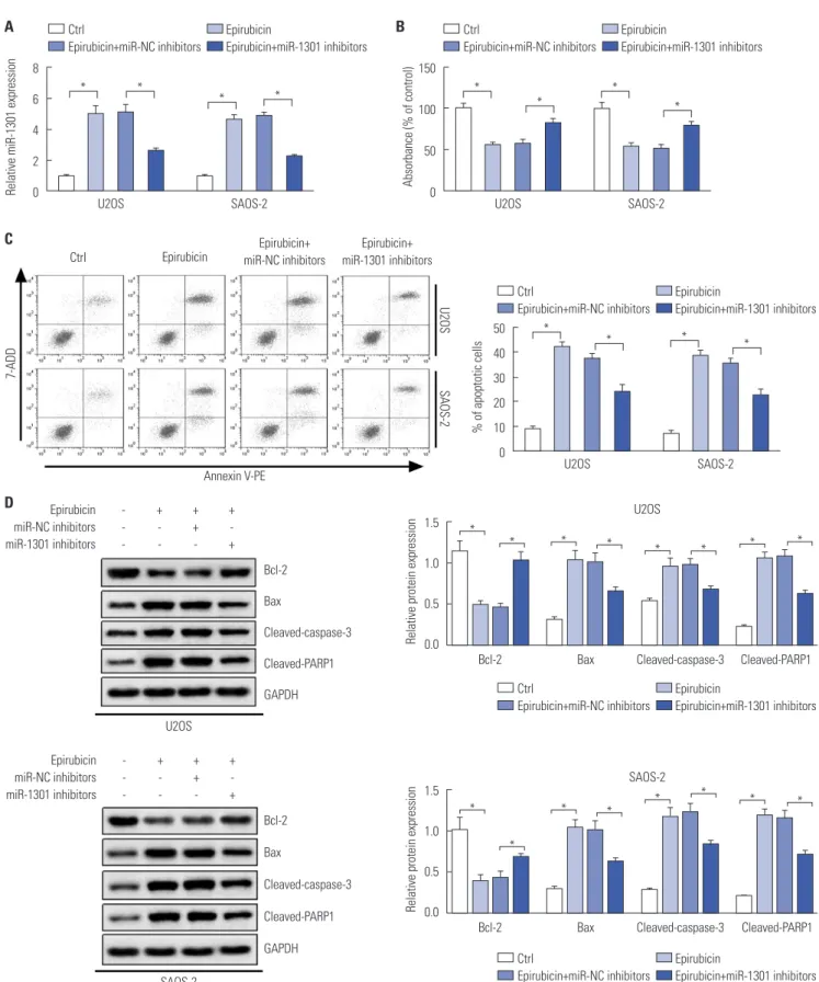
MiR-1301 depletion weakens the anti-cancer effect of epirubicin in osteosarcoma
Based on the above results, we further conducted cell trans- fection to examine whether miR-1301 depletion weakens the anti-osteosarcoma effect of epirubicin. As displayed in Fig.
4A, the transfection of miR-1301 inhibitors abolished epirubi- cin-induced miR-1301 promotion. Simultaneously, cell prolif- eration and apoptosis continued to be evaluated. Downregu- lation of miR-1301 abated the epirubicin-mediated inhibition of cell proliferation in U2OS and SAOS-2 cells (Fig. 4B). More- over, epirubicin promoted cell apoptosis, which was under- mined by downregulation of miR-1301 (Fig. 4C). To expound on this observation, Western blot analysis of apoptosis-related proteins was also performed (Fig. 4D). The data further veri- fied that miR-1301 knockdown overturned the pro-apoptosis effect of epirubicin in osteosarcoma cells.
TRIAP1 is a target of miR-1301
In order to explore the molecular mechanisms of miR-1301 involvement in epirubicin-mediated anti-osteosarcoma ef- fects, we used the TargetScan online database to search for potential candidate targets of miR-1301 and found that TRI- AP1 contained binding sites with miR-1301 (Fig. 5A). More-
Fig. 2. Epirubicin stimulates the expression of microRNA-1301 (miR-1301) in U2OS and SAOS-2 cells. (A) qRT-PCR was employed to measure the expres- sion of miR-1301 in human osteoblast cells hFOB1.19, U2OS cells, and SAOS-2 cells. (B) The expression of miR-1301 in U2OS or SAOS-2 cells treated with 1 μg/mL of epirubicin for 24 h or 48 h. *p<0.05. qRT-PCR, quantitative real-time polymerase chain reaction.
1.5
1.0
0.5
0.0
8
6
4
2
U2OS 0
hFOB1.19 Ctrl 24 h 48 h
Epirubicin (1 µg/mL) Epirubicin (1 µg/mL) Ctrl 24 h 48 h
*
* *
*
*
* U2OS SAOS-2
Relative miR-1301 expression Relative miR-1301 expression
SAOS-2
A B
Fig. 3. Upregulation of microRNA-1301 (miR-1301) contributes to the anti-cancer effect of epirubicin in osteosarcoma. U2OS and SAOS-2 cells were trans- fected with miR-NC mimics or miR-1301 mimics, and after 24-h transfection, cells were treated with epirubicin (1 μg/mL) for 48 h. Cells were divided into five groups: Ctrl, miR-NC mimics, miR-1301 mimics, Epirubicin, or Epirubicin+miR-1301 mimics. (A) qRT-PCR was introduced to detect the levels of miR- 1301. (B) Cell Counting Kit-8 was applied to analyze cell proliferation in U2OS and SAOS-2 cells. (C) Flow cytometry was utilized to evaluate cell apoptotic rate. (D) The expressions of Bcl-2, Bax, cleaved-caspase-3, and cleaved-poly (ADP-ribose) polymerase-1 (cleaved-PARP1) in U2OS and SAOS-2 cells were measured using Western blot. The + respensents Epirubicin or mimic was used to treat cells, while the meaning of - was opposite with +. *p<0.05.
Bcl-2, B-cell lymphoma-2; Bax, Bcl-2 assaciated X protein; GAPDH, glyceraldehyde-3-phosphate dehydrogenase.
U2OS
Ctrl miR-NC mimics miR-1301 mimics Epirubicin
Epirubicin+
miR-1301 mimics U2OS
U2OS
U2OS
SAOS-2 Bcl-2
Bcl-2
Bax
Bax
Cleaved-caspase-3
Cleaved-caspase-3
Cleaved-PARP1
Cleaved-PARP1 U2OS
SAOS-2
Annexin V-PE
Epirubicin - - - + + miR-NC mimics - + - - - miR-1301 mimics - - + - +
Epirubicin - - - + + miR-NC mimics - + - - - miR-1301 mimics - - + - +
Bcl-2 Bax
Cleaved-caspase-3 Cleaved-PARP1 GAPDH
Bcl-2 Bax
Cleaved-caspase-3 Cleaved-PARP1 GAPDH
U2OSSAOS-2
SAOS-2 SAOS-2
SAOS-2
A B
C
D 12
8
4
0
150
100
50
0
60
40
20
0
2.5 2.0 1.5 1.0 0.5 0.0
2.5 2.0 1.5 1.0 0.5 0.0
Relative miR-1301 expression Absorbance (% of control) % of apoptotic cells
Relative protein expressionRelative protein expression
Ctrl miR-NC mimics
miR-1301 mimics Epirubicin Epirubicin+miR-1301 mimics
Ctrl miR-NC mimics
miR-1301 mimics Epirubicin Epirubicin+miR-1301 mimics
Ctrl miR-NC mimics
miR-1301 mimics Epirubicin Epirubicin+miR-1301 mimics
Ctrl miR-NC mimics
miR-1301 mimics Epirubicin Epirubicin+miR-1301 mimics
Ctrl miR-NC mimics
miR-1301 mimics Epirubicin Epirubicin+miR-1301 mimics
* *
*
*
*
*
*
*
*
*
*
*
*
*
*
*
*
*
*
*
*
*
*
*
*
*
*
*
*
*
*
*
*
*
*
* *
* *
* *
*
7-ADD
Fig. 4. MicroRNA-1301 (MiR-1301) depletion weakens the anti-cancer effect of epirubicin in osteosarcoma. U2OS and SAOS-2 cells were transfected with miR-NC inhibitors or miR-1301 inhibitors, and after 24-h transfection, cells were treated with epirubicin (1 μg/mL) for 48 h. Cells were divided into four groups: Ctrl, Epirubicin, Epirubicin+miR-NC inhibitors, or Epirubicin+miR-1301 inhibitors. (A) The expression levels of miR-1301 in cells were determined using qRT-PCR. (B) Cell proliferation was measured in U2OS and SAOS-2 cells. (C) Flow cytometry was utilized to analyze the cell apoptotic rate. (D) Western blot was employed to examine the expressions of Bcl-2, Bax, cleaved-caspase-3, and polymerases (PARP1) in U2OS and SAOS-2 cells. The + respensents Epirubicin or mimic was used to treat cells, while the meaning of - was opposite with +. *p<0.05. Bcl-2, B-cell lymphoma-2; Bax, Bcl-2 assa- ciated X protein; GAPDH, glyceraldehyde-3-phosphate dehydrogenase.
U2OS U2OS
U2OS Ctrl Epirubicin Epirubicin+
miR-NC inhibitors
Epirubicin+
miR-1301 inhibitors
U2OS
SAOS-2 Bcl-2
Bcl-2
Bax
Bax
Cleaved-caspase-3
Cleaved-caspase-3
Cleaved-PARP1
Cleaved-PARP1 U2OS
SAOS-2
Annexin V-PE Epirubicin - + + + miR-NC inhibitors - - + - miR-1301 inhibitors - - - +
Epirubicin - + + + miR-NC inhibitors - - + - miR-1301 inhibitors - - - +
Bcl-2 Bax
Cleaved-caspase-3 Cleaved-PARP1 GAPDH
Bcl-2 Bax
Cleaved-caspase-3 Cleaved-PARP1 GAPDH
U2OSSAOS-2
SAOS-2 SAOS-2
SAOS-2
A B
C
D 8 6 4 2 0
150
100
50
0
50 40 30 20 10 0
1.5
1.0
0.5
0.0
1.5
1.0
0.5
0.0
Relative miR-1301 expression Absorbance (% of control) % of apoptotic cells
Relative protein expressionRelative protein expression
Ctrl Epirubicin
Epirubicin+miR-NC inhibitors Epirubicin+miR-1301 inhibitors
Ctrl Epirubicin
Epirubicin+miR-NC inhibitors Epirubicin+miR-1301 inhibitors
Ctrl Epirubicin
Epirubicin+miR-NC inhibitors Epirubicin+miR-1301 inhibitors
Ctrl Epirubicin
Epirubicin+miR-NC inhibitors Epirubicin+miR-1301 inhibitors
Ctrl Epirubicin
Epirubicin+miR-NC inhibitors Epirubicin+miR-1301 inhibitors
*
*
* *
*
*
*
*
*
*
*
*
*
*
*
*
*
*
*
*
*
*
*
*
*
*
*
*
7-ADD
over, luciferase activity was dramatically reduced in the cells overexpressing miR-1301 in the TRIAP1 WT group, while it showed no significant change in the TRIAP1-MUT group (Fig.
5B and C), suggesting that TRIAP1 is a target of miR-1301.
Furthermore, we also determined the effect of miR-1301 on expression of TRIAP1. The results showed a distinct suppres- sion of TRIAP1 in miR-1301-upregulated cells, whereas inhi-
bition of miR-1301 boosted it in U2OS and SAOS-2 cells (Fig.
5D and E), indicating that miR-1301 negatively regulates the expression of TRIAP1.
Epirubicin regulates TRIAP1 expression by modulating miR-1301
Finally, we also studied whether miR-1301/TRIAP1 axis is as- Fig. 5. TP53-regulated inhibitor of apoptosis 1 (TRIAP1) is a target of microRNA-1301 (miR-1301). (A) The binding sites between miR-1301 and TRIAP1 were predicted by the TargetScan online database, and luciferase reporter plasmids containing the wild-type (WT) or mutated (MUT) TRIAPT1 binding sites of miR-1301 were established. (B) The luciferase activity was measured in U2OS cells co-transfected with TRIAP1-WT or TRIAP1-MUT luciferase reporter plasmids and miR-1301 mimics or miR-NC mimics. (C) The luciferase activity was examined in SAOS-2 cells co-transfected with TRIAP1-WT or TRIAP1- MUT luciferase reporter plasmids and miR-1301 mimics or miR-NC mimics. (D) The expression of TRIAPT in U2OS and SAOS-2 cells transfected with miR- 1301 mimics or miR-NC mimics. (E) The expression of TRIAP1 in U2OS and SAOS-2 cells transfected with miR-1301 inhibitors or miR-NC inhibitors. (D and E) The + respensents Epirubicin or mimic was used to treat cells, while the meaning of - was opposite with +. *p<0.05. GAPDH, glyceraldehyde-3-phos- phate dehydrogenase.
U2OS
U2OS SAOS-2 U2OS SAOS-2 SAOS-2
miR-NC mimics + - + -
miR-1301 mimics - + - +
miR-NC inhibitors + - + - miR-1301 inhibitors - + - + TRIAP1-WT
U2OS U2OS
TRIAP1 TRIAP1
TRIAP1-WT TRIAP1-MUT
SAOS-2 SAOS-2
GAPDH GAPDH
TRIAP1-MUT A
B C
D E
1.5
1.0
0.5
0.0
1.5
1.0
0.5
0.0
2.0
1.5
1.0
0.5
0.0 1.5
1.0
0.5
0.0
Relative luciferase activityTRIAP1/GAPDH ratio TRIAP1/GAPDH ratioRelative luciferase activity
miR-NC mimics miR-1301 mimics
miR-NC mimics miR-1301 mimics
miR-NC inhibitors miR-1301 inhibitors
miR-NC mimics miR-1301 mimics
*
* *
* *
*
sociated with epirubicin-mediated anti-osteosarcoma effects.
qRT-PCR was subsequently employed to determine the mRNA levels of TRIAP1. We found that the mRNA levels of TRIAP1 were remarkably lower in epirubicin-treated osteo- sarcoma cells, which was attenuated by transfection of miR- 1301 inhibitors (Fig. 6A). Similarly, Western blot also demon- strated that epirubicin inhibited protein levels of TRIAP1 in
U2OS and SAOS-2 cells, which was rescued by miR-1301 knockdown (Fig. 6B and C). Subsequently, rescue-of-function was also performed to determine the effect of TRIAP1 upregu- lation on epirubicin-mediated TRIAP1 inhibition. Our data further indicated that overexpression of TRIAP1 abated lower expression of TRIAP1 induced by epirubicin at mRNA (Fig.
6D) and protein (Fig. 6E and F) levels. These data revealed Fig. 6. Epirubicin regulates TP53-regulated inhibitor of apoptosis 1 (TRIAP1) expression by modulating microRNA-1301 (miR-1301). (A-C) U2OS and SAOS- 2 cells were transfected with miR-NC inhibitors or miR-1301 inhibitors, and after 24-h transfection, cells were treated with epirubicin (1 μg/mL) for 48 h.
Cells were divided into four groups: Ctrl, Epirubicin, Epirubicin+miR-NC inhibitors, or Epirubicin+miR-1301 inhibitors. (D-G) U2OS and SAOS-2 cells were transfected with vector or TRIAP1 overexpression plasmid (TRIAP1), and after 24-h transfection, cells were treated with epirubicin (1 μg/mL) for 48 h. Cells were divided into four groups: Ctrl, Epirubicin, Epirubicin+vector, or Epirubicin+TRIAP1. The mRNA levels of TRIAP1 in U2OS and SAOS-2 cells were eval- uated (A and D). Western blot was applied to examine the levels of TRIAP1 in U2OS (B and E) and SAOS-2 cells (C and F). (G) Cell viability was determined by Cell Counting Kit-8 assay. (B, C, E, and F) The + respensents Epirubicin or mimic was used to treat cells, while the meaning of - was opposite with +.
*p<0.05. GAPDH, glyceraldehyde-3-phosphate dehydrogenase.
U2OS
U2OS
U2OS
SAOS-2
SAOS-2
SAOS-2 A
D
G 1.5
1.0
0.5
0.0
1.5
1.0
0.5
0.0
150
100
50
0
1.5 1.0 0.5 0.0
1.5 1.0 0.5 0.0
1.5 1.0 0.5 0.0
1.5 1.0 0.5 0.0
Relative mRNA expression TRIAP1Relative mRNA expression TRIAP1Absorbance (% of control) TRIAP1/GAPDH ratioTRIAP1/GAPDH ratio TRIAP1/GAPDH ratioTRIAP1/GAPDH ratio
*
*
*
*
*
*
*
*
*
*
*
*
*
*
*
*
*
*
*
* Ctrl
Epirubicin
Epirubicin+miR-NC inhibitors Epirubicin+miR-1301 inhibitors
Ctrl Epirubicin Epirubicin+vector Epirubicin+TRIAP1
Ctrl Epirubicin Epirubicin+vector Epirubicin+TRIAP1
U2OS
U2OS
SAOS-2
SAOS-2 Epirubicin - + + +
miR-NC inhibitors - - + - miR-1301 inhibitors - - - +
Epirubicin - + + + Vector - - + - TRIAP1 - - - +
Epirubicin - + + + miR-NC inhibitors - - + - miR-1301 inhibitors - - - +
Epirubicin - + + + Vector - - + - TRIAP1 - - - + TRIAP1
TRIAP1
TRIAP1
TRIAP1 GAPDH
GAPDH
GAPDH
GAPDH B
E
C
F
that epirubicin repressed TRIAP1 expression by upregulating miR-1301. In addition, ectopic expression of TRIAP1 rescued the epirubicin-mediated decrease of cell viability (Fig. 6G).
These results supported the hypothesis that miR-1301/ TRI- AP1 may be implicated in epirubicin-mediated pro-apoptosis and anti-proliferation in osteosarcoma.
DISCUSSION
Epirubicin, an anti-tumor drug, has been used in the treat- ment of osteosarcoma.20 In the current study, we found that epirubicin curbed cell proliferation and induced apoptosis in osteosarcoma cells, which was in line with a previous study.20 Emerging evidence suggests that miR-1301 could serve as a tumor-suppressor in a variety of cancers. For example, Zhi, et al.23 disclosed that miR-1301 is downregulated in glioma tis- sues and cell lines and inhibits cell proliferation by targeting neuroblastoma ras viral oncogene homolog. miR-1301-3p is decreased in breast cancer tissues and cells, induces cell cycle G0/G1 phase arrest and apoptosis, and inhibits cell prolifera- tion by targeting immature colon carcinoma transcript-1.24 Here, we demonstrated that miR-1301 is expressed at low lev- els in osteosarcoma cells, suggesting that miR-1301 might contribute to the anti-osteosarcoma effect. Interestingly, the expression of miR-1301 showed an evident increase in cells after epirubicin exposure. These data indicated that miR-1301 may play an important role in osteosarcoma therapy in com- bination with epirubicin. Therefore, we subsequently investi- gated its role and the underlying molecular mechanisms in respect to epirubicin-mediated anti-osteosarcoma. Our study indicated that overexpression of miR-1301 hinders cell prolif- eration and promotes apoptosis in osteosarcoma cells, which is similar to the functions of epirubicin. To gain a deeper in- sight into the molecular mechanisms of epirubicin and miR- 1301 affecting apoptosis, the main regulatory molecules that govern the main basic mechanisms, such as the expression of apoptosis-related protein Bcl-2, Bax, cleaved-caspase-3, and poly (ADP-ribose) polymerase-1 (PARP1), were further exam- ined. Bcl-2 is an anti-apoptosis protein, and dysregulation of Bcl-2-mediated apoptosis underlies a plethora of diseases.25 Bax is a pro-apoptotic member of the BCL-2 family.26 Also, de- tection of cleaved-caspase-3 in cells is an important method for apoptosis induced by a wide variety of apoptotic signals.27,28 PARP1 is also a pro-apoptosis gene and could restore miR- 520-induced cell apoptosis.29 Our findings demonstrated that epirubicin and upregulation of miR-1301 both reduces the ex- pression of Bcl-2 and increases the expressions of Bax, cleaved- caspase-3, and PARP1, further reflecting the miR-1301-medi- ated pro-apoptosis effect. Importantly, we also revealed that miR-1301 enhances the function of epirubicin-induced anti- proliferation and pro-apoptosis in U2OS and SAOS-2 cells, in- dicating that miR-1301 increases the epirubicin effect. In con-
trast, miR-1301 knockdown attenuated the effect of epirubicin on osteosarcoma cells. Therefore, we inferred that miR-1301 fa- cilitates epirubicin-mediated chemosensitivity in osteosarcoma.
miRNAs can regulate post-transcriptional gene expression and silence a broad set of targets genes. TRIAP1, a novel apoptosis inhibitor, has been shown to be upregulated in many types of cancers.30 Stable silencing of TRIAP1 induced late apoptosis in RPMI8226 cells.31 In addition, miR-18a inhibited ovarian cancer proliferation and induced apoptosis by target- ing TRIAP1.32 miR-320b suppressed nasopharyngeal carcino- ma cell proliferation and enhanced mitochondrial fragmenta- tion and apoptosis by regulating TRIAP1. These data implied that TRIAP1 could be modulated by miRNAs. Here, we showed that miR-1301 repressed TRIAP1 expression by directly bind- ing to the 3’UTR of TRIAP1. Moreover, the level of TRIAP1 was repressed by epirubicin in osteosarcoma cells, which was also rescued by introduction of miR-1301 inhibitors or TRIAP1 overexpression plasmid. Interestingly, restoration of TRIAP1 abolished the epirubicin-mediated cell proliferation inhibi- tion. It is tempting to speculate that miR-1301 may participate in epirubicin-mediated chemosensitivity by targeting TRIAP1.
Nevertheless, our study only investigated the role of miR- 1301/TRIAP1 axis in vitro, and it needs to be further confirmed in xenograft models. Moreover, the role of TRIAP1 alone in osteosarcoma should be addressed in the future.
Taken together, our data highlighted the pivotal role of miR- 1301 in epirubicin-mediated chemosensitivity in osteosarcoma.
A novel miR-1301/TRIAP1 axis was first observed in osteosar- coma. Importantly, we found that an epirubicin-mediated miR-1301/TRIAP1 axis is involved in cell proliferation and apoptosis in osteosarcoma. Our findings provide powerful ev- idence of the molecular mechanism of chemosensitivity to epirubicin in osteosarcoma, which will help support clinical applications of a combination of epirubicin and miR-1301 in osteosarcoma.
AUTHOR CONTRIBUTIONS
Conceptualization: Lijun Yu and Min Meng. Data curation: Min Meng and Yun Bao. Formal analysis: Chao Zhang. Funding acquisi- tion: Bei Gao and Rina Sa. Investigation: Wenyuan Luo and Lijun Yu.
Methodology: Lijun Yu and Bei Gao. Project administration: Rina Sa and Lijun Yu. Resources: Wenyuan Luo. Software: Wenyuan Luo. Su- pervision: Bei Gao. Validation: Yun Bao. Visualization: Min Meng.
Writing—original draft: Bei Gao and Lijun Yu. Writing—review & ed- iting: Lijun Yu and Yun Bao.
ORCID iDs
Lijun Yu https://orcid.org/0000-0001-8366-5179 Min Meng https://orcid.org/0000-0002-8133-3127 Yun Bao https://orcid.org/0000-0002-4019-6445 Chao Zhang https://orcid.org/0000-0003-2752-3373 Bei Gao https://orcid.org/0000-0001-5426-0252 Rina Sa https://orcid.org/0000-0003-1881-2077
Wenyuan Luo https://orcid.org/0000-0003-0482-2231
REFERENCES
1. Moore DD, Luu HH. Osteosarcoma. Cancer Treat Res 2014;162:
65-92.
2. Biazzo A, De Paolis M. Multidisciplinary approach to osteosarco- ma. Acta Orthop Belg 2016;82:690-8.
3. Harrison DJ, Geller DS, Gill JD, Lewis VO, Gorlick R. Current and future therapeutic approaches for osteosarcoma. Expert Rev An- ticancer Ther 2018;18:39-50.
4. Fan XL, Cai GP, Zhu LL, Ding GM. Efficacy and safety of ifos- famide-based chemotherapy for osteosarcoma: a meta-analysis.
Drug Des Devel Ther 2015;9:5925-32.
5. Luetke A, Meyers PA, Lewis I, Juergens H. Osteosarcoma treat- ment - where do we stand? A state of the art review. Cancer Treat Rev 2014;40:523-32.
6. Friebele JC, Peck J, Pan X, Abdel-Rasoul M, Mayerson JL. Osteo- sarcoma: a meta-analysis and review of the literature. Am J Or- thop (Belle Mead NJ) 2015;44:547-53.
7. Petrioli R, Roviello G, Zanotti L, Roviello F, Polom K, Bottini A, et al. Epirubicin-based compared with docetaxel-based chemo- therapy for advanced gastric carcinoma: a systematic review and meta-analysis. Crit Rev Oncol Hematol 2016;102:82-8.
8. Wu J, Xue X, Zhang B, Cao H, Kong F, Jiang W, et al. Enhanced an- titumor activity and attenuated cardiotoxicity of Epirubicin com- bined with Paeonol against breast cancer. Tumour Biol 2016;37:
12301-13.
9. Liu ZL, Wang G, Shu Y, Zou PA, Zhou Y, Yin QS. Enhanced antitu- mor activity of epirubicin combined with cerulenin in osteosarco- ma. Mol Med Rep 2012;5:326-30.
10. Basaran M, Bavbek ES, Saglam S, Eralp L, Sakar B, Atalar AC, et al.
A phase II study of cisplatin, ifosfamide and epirubicin combina- tion chemotherapy in adults with nonmetastatic and extremity os- teosarcomas. Oncology 2007;72:255-60.
11. Acunzo M, Romano G, Wernicke D, Croce CM. MicroRNA and cancer--a brief overview. Adv Biol Regul 2015;57:1-9.
12. Waresijiang N, Sun J, Abuduaini R, Jiang T, Zhou W, Yuan H. The downregulation of miR-125a-5p functions as a tumor suppressor by directly targeting MMP-11 in osteosarcoma. Mol Med Rep 2016;
13:4859-64.
13. Bach DH, Hong JY, Park HJ, Lee SK. The role of exosomes and miRNAs in drug-resistance of cancer cells. Int J Cancer 2017;141:
220-30.
14. Wang J, Yang M, Li Y, Han B. The role of MicroRNAs in the che- moresistance of breast cancer. Drug Dev Res 2015;76:368-74.
15. Li Q, Liu M, Ma F, Luo Y, Cai R, Wang L, et al. Circulating miR-19a and miR-205 in serum may predict the sensitivity of luminal A subtype of breast cancer patients to neoadjuvant chemotherapy with epirubicin plus paclitaxel. PLoS One 2014;9:e104870.
16. Tan X, Fu Y, Chen L, Lee W, Lai Y, Rezaei K, et al. miR-671-5p in- hibits epithelial-to-mesenchymal transition by downregulating
FOXM1 expression in breast cancer. Oncotarget 2016;7:293-307.
17. Zhang W, Qiao B, Fan J. Overexpression of miR-4443 promotes the resistance of non-small cell lung cancer cells to epirubicin by targeting INPP4A and regulating the activation of JAK2/STAT3 pathway. Pharmazie 2018;73:386-92.
18. Chen D, Liu D, Chen Z. Potential therapeutic implications of miRNAs in osteosarcoma chemotherapy. Tumour Biol 2017;39:
1010428317705762.
19. Wang F, Yu D, Liu Z, Wang R, Xu Y, Cui H, et al. MiR-125b func- tions as a tumor suppressor and enhances chemosensitivity to cisplatin in osteosarcoma. Technol Cancer Res Treat 2016;15:
NP105-NP12.
20. Zhang Y, Hu H, Song L, Cai L, Wei R, Jin W. Epirubicin-mediated expression of miR-302b is involved in osteosarcoma apoptosis and cell cycle regulation. Toxicol Lett 2013;222:1-9.
21. Wang L, Hu K, Chao Y. MicroRNA-1301 inhibits migration and in- vasion of osteosarcoma cells by targeting BCL9. Gene 2018;679:
100-7.
22. Li Y, Tang X, He Q, Yang X, Ren X, Wen X, et al. Overexpression of mitochondria mediator gene TRIAP1 by miR-320b loss is associ- ated with progression in nasopharyngeal carcinoma. PLoS Genet 2016;12:e1006183.
23. Zhi T, Jiang K, Zhang C, Xu X, Wu W, Nie E, et al. MicroRNA-1301 inhibits proliferation of human glioma cells by directly targeting N-Ras. Am J Cancer Res 2017;7:982-98.
24. Peng X, Yan B, Shen Y. MiR-1301-3p inhibits human breast cancer cell proliferation by regulating cell cycle progression and apopto- sis through directly targeting ICT1. Breast Cancer 2018;25:742-52.
25. Kvansakul M, Caria S, Hinds MG. The Bcl-2 family in host-virus interactions. Viruses 2017;9. pii: E290.
26. Raemy E, Martinou JC. Involvement of cardiolipin in tBID-in- duced activation of BAX during apoptosis. Chem Phys Lipids 2014;179:70-4.
27. Choudhary GS, Al-Harbi S, Almasan A. Caspase-3 activation is a critical determinant of genotoxic stress-induced apoptosis. Meth- ods Mol Biol 2015;1219:1-9.
28. Elmore S. Apoptosis: a review of programmed cell death. Toxicol Pathol 2007;35:495-516.
29. Dong X, Yang L, Wang H. miR-520 promotes DNA-damage-in- duced trophoblast cell apoptosis by targeting PARP1 in recurrent spontaneous abortion (RSA). Gynecol Endocrinol 2017;33:274-8.
30. Adams C, Cazzanelli G, Rasul S, Hitchinson B, Hu Y, Coombes RC, et al. Apoptosis inhibitor TRIAP1 is a novel effector of drug resis- tance. Oncol Rep 2015;34:415-22.
31. Fook-Alves VL, de Oliveira MB, Zanatta DB, Strauss BE, Colleoni GW. TP53 Regulated Inhibitor of Apoptosis 1 (TRIAP1) stable si- lencing increases late apoptosis by upregulation of caspase 9 and APAF1 in RPMI8226 multiple myeloma cell line. Biochim Biophys Acta 2016;1862:1105-10.
32. Liu P, Qi X, Bian C, Yang F, Lin X, Zhou S, et al. MicroRNA-18a in- hibits ovarian cancer growth via directly targeting TRIAP1 and IPMK. Oncol Lett 2017;13:4039-46.