Korean Journal of Neuromuscular Disorders 2018 37 Geum-Bong Lee, et al. | SOD with Family History
Received: August 24, 2017 / Revised: October 11, 2018 / Accepted: October 29, 2018 Address for correspondence: Byeol A Yoon, MD
Department of Neurology, Dong-A University College of Medicine, 26 Daesingongwon-ro, Seo-gu, Busan 49201, Korea Tel: +82-51-240-5266, Fax: +82-51-244-8338, E-mail: [email protected]
ISSN 2093-3312
Korean Journal of Neuromuscular Disorders Vol. 10 No. 2, December 2018
Case Report
가족력이 있는 중격 시신경 형성 장애 증례
동아대학교 의과대학 신경과학교실1, 안과학교실2, 고신대학교 의과대학 신경과학교실3
이금봉1, 김지선1, 류원열2, 이새미2, 이원구3, 김종국1, 윤별아1
A Case of Septo-Optic Dysplasia with Family History
Geum-Bong Lee, MD1, Ji Sun Kim, MD1, Won Yeol Ryu, MD, PhD2, Sae Mi Lee, MD2, Won-Gu Lee, MD3, Jong Kuk Kim, MD, PhD1, Byeol A Yoon, MD1
Departments of 1Neurology, 2Ophthalmology, Dong-A University College of Medicine, Busan; 3Department of Neurology, Kosin University College of Medicine, Busan, Korea
KEYWORDS Septo-optic dysplasia, Optic nerve hypoplasia, familial bilateral
Septo-optic dysplasia (SOD), also known as de-Morsier's syndrome, is a rare disorder with more than two of optic nerve hypoplasia, pituitary gland hypoplasia, or midline brain abnormalities including absence of septum pellucidum and/or corpus callosal agenesis. Most cases of SOD are sporadic, but there are cases of patients who have familial history of SOD.
중격 시신경 형성 장애(septo-optic dysplasia, SOD)는 선 천성 시신경병의 드문 원인 중 하나로 시신경형성 저하, 뇌 하수체호르몬기능 이상, 투명사이막공간(septum pellucidum) 이나 뇌들보무 발생(corpus callosal agenesis)과 같은 중간선 뇌이상 중 두 가지 이상을 만족할 때 진단할 수 있으며 대 부분의 경우 산발적으로 발생하므로 가족력이 있는 경우는 거의 알려진 바가 없다.1 이에 저자들은 아급성으로 진행하 는 시력 저하를 주소로 내원한 50세 남자 환자에서 SOD를 진단하였고 동일한 증상이 있는 남동생을 통하여 가족력이 있음을 확인하였기에 이를 보고하고자 한다.
증 례
50세 남자가 2개월 전부터 진행하는 시력 저하로 내원하 였다. 주야간 교대로 금속 단조작업을 하는 환자로 2개월
전부터 야간근무를 시작하면서 양안이 흐리게 보이는 증상 이 발생하였다. 증상은 근무시간에 따라 변화하는 양상으로 주간근무를 할 때는 증상이 호전되고 야간근무를 할 때 악 화되어 한 달 전부터 주간에만 근무를 하고 있었다. 환자는 20년 전부터 전신경련으로 뇌전증을 진단받고 zonisamide 를 복용하였으나 6년 전부터 증상이 완전히 소실되어 복용 을 중단하였다. 가족력은 3남 1녀 중 셋째로 막내인 남동생 이 6세부터 뇌전증을 앓았으나 증상 조절이 잘 되지 않아 심한 인지기능 저하가 있었고 시력 저하 증상으로 시각장 애 2급을 진단받았다. 부모를 포함한 다른 가족은 시력 저 하 및 뇌전증의 병력은 없었다. 혈압, 당뇨 등의 과거력은 없었다.
신경학적 검사에서 인지기능과 근력 및 감각신경검사는 정상이었다. 뇌신경검사에서 시력이 우안 0.6, 좌안 0.2로 감소되었고 한식 색각검사표를 이용한 적녹색각검사에서 양안 모두 감소된 반응을 보였다(우안 10/12, 좌안 9/12). 안
대 한 신 경 근 육 질 환 학 회 지 2 0 18 38
Korean Journal of Neuromuscular Disorders Vol. 10 No. 2, 2018
A B
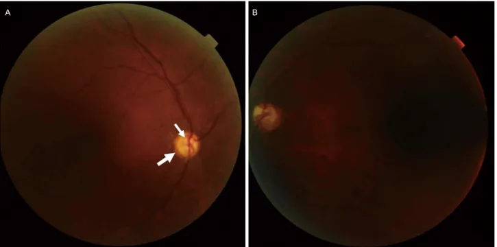
Figure 1. Fundus photographs of the patient. (A) Right eye of the patient. Optic nerve was very small with double ring sign consistent with optic nerve hypoplasia. The outer arrow demarcates the abnormal yellow ring that encircles the optic disc (demarcated by the small ar- row). (B) Left eye of the patient showed very small optic nerve with diffuse optic disc pallor.
저검사에서 양안의 시신경 창백과 작은 시신경유두 소견이 보였고(Fig. 1) 자동시야검사(Humphrey visual field test, cen- tral 30-2 threshold) 상 우안 관자위(superotemporal) 1/4분맹 (quadrantanopsia)이 확인되었다. 지속적인 양안의 자발안진 이 관찰되었는데 어릴 때부터 증상이 있어 정확한 발생 시 점을 기억하지 못하였다. 다른 뇌신경기능의 이상은 없었고 심부건반사도 정상 범위에 해당되었다.
콩팥 및 간기능검사, 갑상샘기능검사, 전해질검사는 정상 범위였고 류마티스인자, 항핵항체검사 및 혈액의 항-아쿠아 포린 4 (anti-aquaporin 4) 항체를 포함한 자가항체검사도 음 성이었다. 뇌 자기공명영상에서 투명사이막공간이 소실 및 양측 시신경과 시신경교차(optic chiasm)의 위축이 확인되 었고 대뇌피질의 뭇미세이랑증(polymicrogyria)이 보였는데 타 대학병원에서 촬영한 동생의 뇌 자기공명영상에서도 투 명사이막공간의 소실과 함께 뇌갈림증(schizencephaly)이 관 찰되었다(Fig. 2). 뇌하수체호르몬검사에서는 갑상샘자극호 르몬 2.79 uIU/mL, 유리 T4 1.399 ng/dL, 프로락틴 11.082 ng/mL, 인슐린유사성장인자(insulin-like growth factor)-1 236.8 ng/mL, 황체형성호르몬 2.55 mIU/mL, 난포자극호르몬 4.07 mIU/mL, 테스토스테론 4.34 ng/mL로 정상 범위에 해당되었고 부신 피질자극호르몬이 126.545 pg/mL로 증가되어 있었으나 한 달 뒤 재검한 결과에서 정상 범위로 회복되어 스트레스유
발고코르티졸증(stress induced hypercortisolism)으로 판단되 었다. 투명사이막공간의 소실과 시신경 및 시신경교차의 위 축을 동반한 SOD로 진단하였고 동생의 가족력을 고려하였 을 때 상염색체열성유전의 가능성을 고려할 수 있었다. Whole exome sequencing을 통하여 대표적인 돌연변이 유전 자로 알려져 있는 HESX1의 검사를 고려하였으나 검사를 원하지 않았고 주기적인 안과검진을 포함하여 외래추적 중 이다.
고 찰
SOD는 1941년 Reeves2가 투명사이막공간의 소실과 시신 경의 이상이 동반된 7개월 된 아기를 처음 보고하였고 1954 년 De Morsier3가 후각망울형성이상, 뇌들보무발생 및 앞 맞교차(anterior commissure)의 이상을 syndrome of dysplasie olfacto-génitale로 이름하면서 de Morsier syndrome으로 불 리기도 하였다. 대부분 산발적으로 발생하며 그 원인에 대 해서는 거의 알려진 바가 없으나 Riedl 등4이 보고한 코호 트 연구에 따르면 산모의 나이가 어리거나 임신 기간에 약 물이나 알코올을 남용하는 경우 SOD의 발생이 증가하였다.
임신 4-6주가 앞뇌(forebrain)를 형성하는 앞신경판(anterior
Korean Journal of Neuromuscular Disorders 2018 39 Geum-Bong Lee, et al. | SOD with Family History
A B C
D E F
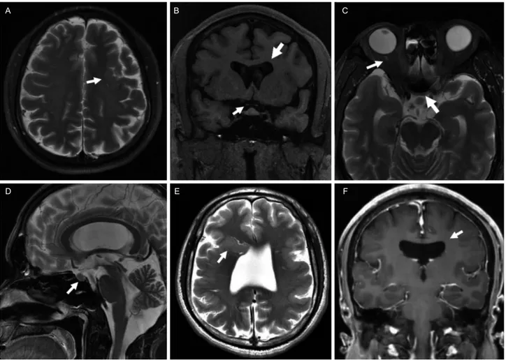
Figure 2. Brain magnetic resonance imaging of the patient (A-D) and his younger brother (E, F). (A) Axial T2 weighted image showing polymicrogyria (arrow). (B) Coronal T1 weighted image showing the absence of septum pellucidum (large arrow) and optic chiasmal hy- poplasia (small arrow). (C) Axial T2 weighted image showing optic nerve hypoplasia (small arrow) and optic chiasmal hypoplasia (large arrow). (D) Sagittal T2 weighted image showing small pituitary (arrow). Axial T2 weighted image and post-enhanced coronal T1 image showing closed type schizencephaly (E, arrow) and absence of septum pellucidum (F, arrow) respectively.
neural plate)이 발달하는데 가장 중요한 시기이므로 이 때 발생하는 뇌손상이 SOD를 유발할 것으로 추측된다.4 가족 력이 있는 경우는 거의 알려져 있지 않으며 Benner 등5이 남매 간에 발생한 SOD를 보고하여 상염색체 열성 유전의 가능성을 시사하였다. SOD와 관계된 유전자는 HESX1과 SOX2가 알려져 있다. HESX1은 뇌하수체의 발달과정에서 가장 초기에 발현되는 전사인자(transcription factor) 중 하 나로 동종접합 돌연변이(homozygous mutation)를 가진 경 우 다음 세대에 모두 발현(penetrance)되었고 이형접합 돌연 변이(heterozygous mutation)의 경우는 다양하게 발현되며 경미한 증상을 보였다.6,7 SOX2는 뇌하수체의 줄기세포에 발현되어 있는 전사인자로 돌연변이가 있는 환자에서 무안 구증(anophthalmia)이나 작은안구증(microphthalmia)과 같은 심각한 눈증상, 뇌들보무발생과 뇌하수체형성 저하가 발생
하였고, 작은 키와 발달 장애 그리고 감각신경난청 등이 동 반되었다.1,8 본 증례의 경우 가족력이 있어 상염색체 열성 유전이 의심되었으나 환자가 검사를 원하지 않아 확인할 수 없었다. 다만, SOD의 경우 가족력이 있는 경우가 1% 미 만이므로 형제가 우연히 산발적으로 증상이 생겼을 가능성 이 있다.
위 환자는 아급성으로 진행하는 시신경이상과 함께 뇌 자기공명영상에서 중간선뇌이상이 발견되어 SOD로 진단 되었으며 가족력이 있는 SOD는 현재까지 국내에서 보고된 바가 없다. SOD는 내분비기능 장애를 동반하는 경우 발달 지연, 저혈당 및 부신위기(adrenal crisis)에 의한 사망에도 이를 수 있어 조기 진단과 치료가 중요하며 최소 6개월 간 격으로 안과 진료를 포함한 주기적인 추적관찰이 필요하다.
또한 가족력이 없는 경우에는 1% 미만에서 다음 세대에 나
대 한 신 경 근 육 질 환 학 회 지 2 0 18 40
Korean Journal of Neuromuscular Disorders Vol. 10 No. 2, 2018
타나지만 가족력이 있는 경우에는 유전형에 따라 발현의 차이가 있어 유전상담이 반드시 필요하다. 따라서, 임상의 는 아급성으로 진행하는 시신경병이 있는 환자에서 SOD을 감별진단에 포함하고 뇌 자기공명영상의 촬영을 고려해야 한다.
REFERENCES
1. Webb EA, Dattani MT. Septo-optic dysplasia. Eur J Hum Genet 2010;18:393-397.
2. Reeves DL. Congenital absence of the septum pellucidum. Bull Johns Hopkins Hosp 1941;69:67-71.
3. De Morsier G. Studies on malformation of cranio-encephalic sutures. III. Agenesis of the septum lucidum with malformation of the optic tract. Schweiz Arch Neurol Psychiatr 1956;77:
267-292.
4. Riedl S, Vosahlo J, Battelino T, Stirn-Kranjc B, Brugger PC, Prayer D, et al. Refining clinical phenotypes in septo-optic dysplasia based on MRI findings. Eur J Pediatr 2008;167:
1269-1276.
5. Benner JD, Preslan MW, Gratz E, Joslyn J, Schwartz M, Kelman S. Septo-optic dysplasia in two siblings. Am J Ophthalmol 1990;
109:632-637.
6. Thomas PQ, Dattani MT, Brickman JM, McNay D, Warne G, Zacharin M, et al. Heterozygous HESX1 mutations associated with isolated congenital pituitary hypoplasia and septo-optic dysplasia. Hum Mol Genet 2001;10:39-45.
7. Cohen RN, Cohen LE, Botero D, Yu C, Sagar A, Jurkiewicz M, et al. Enhanced repression by HESX1 as a cause of hypopituitarism and septooptic dysplasia. J Clin Endocrinol Metab 2003;88:
4832-4839.
8. Alatzoglou KS, Kelberman D, Dattani MT. The role of SOX proteins in normal pituitary development. J Endocrinol 2009;
200:245-258.