Korean J Gastroenterol Vol. 71 No. 1, 54-57 https://doi.org/10.4166/kjg.2018.71.1.54 pISSN 1598-9992 eISSN 2233-6869
IMAGE OF THE MONTH
Korean J Gastroenterol, Vol. 71 No. 1, January 2018 www.kjg.or.kr
간 피막을 통과한 담도 배액관의 근위부 이동
최영훈, 백우현
서울대학교 의과대학 서울대학교병원 내과 및 간연구소
Proximal Migration of a Biliary Stent through the Liver Capsule
Young Hoon Choi and Woo Hyun Paik
Department of Internal Medicine and Liver Research Institute, Seoul National University Hospital, Seoul National University College of Medicine, Seoul, Korea
CC This is an open access article distributed under the terms of the Creative Commons Attribution Non-Commercial License (http://creativecommons.org/licenses/
by-nc/4.0) which permits unrestricted non-commercial use, distribution, and reproduction in any medium, provided the original work is properly cited.
Copyright © 2018. Korean Society of Gastroenterology.
교신저자: 백우현, 03080, 서울시 종로구 대학로 101, 서울대학교 의과대학 서울대학교병원 내과학교실 및 간연구소
Correspondence to: Woo Hyun Paik, Department of Internal Medicine and Liver Research Institute, Seoul National University Hospital, Seoul National University College of Medicine, 101 Daehak-ro, Jongno-gu, Seoul 03080, Korea. Tel: +82-2-2072-3536, Fax: +82-2-762-9662, E-mail: [email protected]
Financial support: None. Conflict of interest: None.
A B
Fig. 1. Initial CT scan. (A, B) CT scan shows 2.8×2.6 cm ill-defined low attenuating lesion with peripheral enhancement at the liver segment 8 with dilation of the right intrahepatic duct. CT, computed tomography.
증례: 95세 여자 환자가 3일 전부터 발생한 복통과 발열을 주 소로 내원하였다. 환자는 기저 질환으로 대동맥판협착증, 울혈 성 심부전, 고혈압이 있었다. 1년 3개월 전 구불결장암으로 전방 절제술을 시행받았고, 당시 병기는 T4aN0M0였으나 고령으로 보조 화학 요법은 시행 받지 않았다. 내원 당시 혈압 220/86 mmHg, 맥박 88회/분, 호흡수 18회/분, 체온 37.9°C였다. 신체검진에서 우상복부에서 경한 압통이 관찰되었다. 혈액 검사에서 백혈구
14,000/mm3, 혈색소 10.7 g/dL, 간기능 검사에서 아스파르테이 트아미노전달효소 171 U/L, 알라닌아미노전달효소 198 U/L, 알칼 리인산분해효소 639 U/L, 감마글루타밀트랜스펩티다제 348 U/L, 총빌리루빈 3.12 mg/dL였고, C-반응 단백 4.3 mg/dL (정상 0-0.5 mg/dL)였다. 복부 조영증강 컴퓨터단층촬영(computed tomography, CT)에서 간 제8분절에 약 2.8×2.6 cm 크기의 경계가 불분명하면서 음영이 저하되고, 변연의 조영증강을 동반
Choi YH and Paik WH. Proximal Migration of a Biliary Stent through the Liver Capsule
55
Vol. 71 No. 1, January 2018
A B
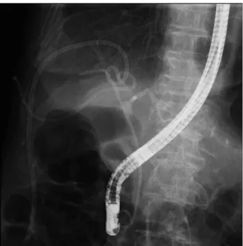
Fig. 2. Endoscopic retrograde cholangiopancreatography. (A) Cholangiogram shows focal stricture at the origin of the right intrahepatic duct.
(B) A 7 F 15 cm straight plastic stent was inserted into the right intrahepatic duct.
A B C
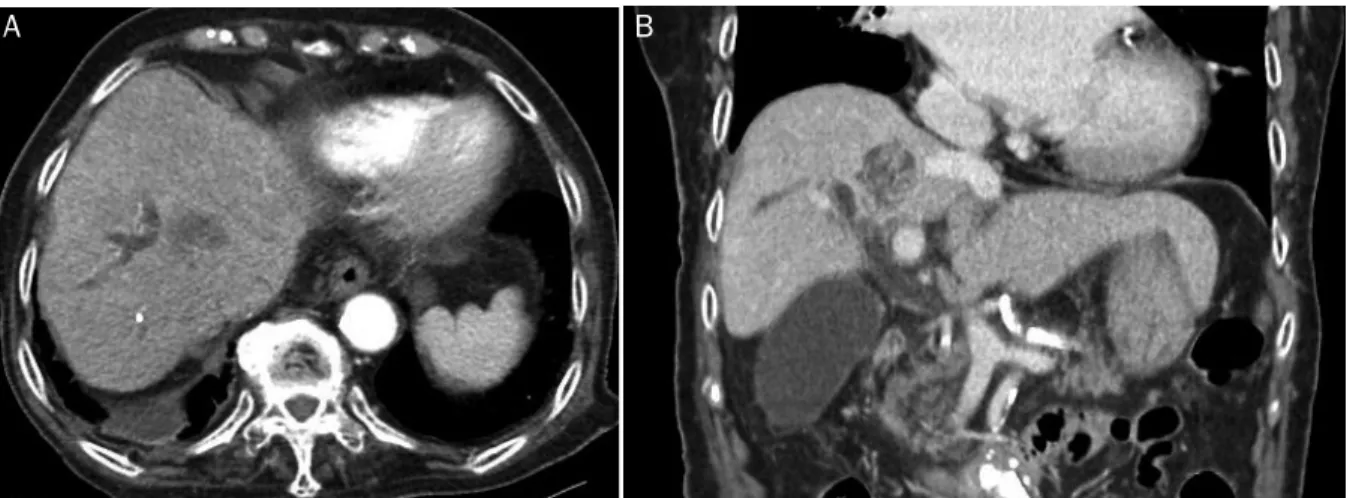
Fig. 3. (A) CT scan shows proximal migration of a previously inserted plastic stent that passes through the liver capsule. (B) CT scan shows 5.3×2.9 cm central low attenuating lesion with rim-like enhancement at the liver segment 8. (C) CT scan shows 5.3×2.7 cm central low attenu- ating lesion with rim-like enhancement at liver segment 7 and right pleural effusion. CT, computed tomography.
한 병변이 관찰되며, 우측 간내담도의 확장이 관찰되었다(Fig.
1). 대장암의 간 전이에 의한 담도 폐쇄와 동반된 급성 담관염이 의심되는 소견으로 정맥내 3세대 Cephalosporin 투여를 시작 하였고, 입원 다음 날 내시경적 역행성 담췌관조영술(endosco- pic retrograde cholangiopancreatography, ERCP)을 시행하였 다. ERCP를 통해 조임근절개술 후 총담관내 결석 및 괴사성 물 질을 제거하고, 우측 간내담도 기시부에 국소적인 협착 소견을 보여 담도내 조직 검사를 시행하였으며, 담도 우전측분지로 7 F 15 cm의 직선형 플라스틱 배액관을 삽입하였다(Fig. 2). 담도 협 착 부위의 조직 검사 결과는 만성 염증이었다. 시술 후 환자는 복 통 및 전신상태가 호전되어 퇴원하였다. 퇴원 3개월 뒤 발열 및 복통을 주소로 재내원하였고, 당시 혈압 126/60 mmHg, 맥박 67회/분, 호흡수 20회/분, 체온 38.5°C였다. 신체검진에서 복부
는 경직되어 있었고, 복부 전반에 압통 및 반발압통을 보였다.
혈액 검사에서 백혈구 23,520/mm3, 혈색소 10.5 g/dL, 간기능 검사에서 아스파르테이트아미노전달효소 107 U/L, 알라닌아미 노전달효소 69 U/L, 알칼리인산분해효소 244 U/L, 감마글루타 밀트랜스펩티다제 85 U/L, 총빌리루빈 0.52 mg/dL였다. 복부 CT에서 이전에 삽입한 담도 플라스틱 배액관이 근위부로 이동 하여 간 피막을 뚫고 나간 소견이 관찰되며, 간 제8분절에 5.3×2.9 cm, 간 제7분절에 5.3×3.7 cm 크기의 경계가 비교적 분 명하고 테두리에 조영증강을 보이는 저음영 결절과 우측 흉수가 관찰되었다(Fig. 3). 담도 배액관의 근위부 이동 및 그와 연관된 우측 흉수 및 간농양 소견으로 3세대 Cephalosporin 및 Metro- nidazole 투여를 시작하였고, 간농양과 우측 흉수에 대해 각각 경피적 배액관을 삽입하였다. 이후 환자 상태는 안정화되어 입원
56
최영훈, 백우현. 간 피막을 통과한 담도 배액관의 근위부 이동The Korean Journal of Gastroenterology
A B
Fig. 5. Computed tomography scan shows decreased size of central low attenuating lesion with rim-like enhancement at (A) liver segment 8 (4.4×3.5 cm) and (B) segment 7 (2.2×1.8 cm).
Fig. 4. A 7 F 10 cm double pigtail stent was inserted into the right posterior sectoral duct. Percutaneous catheter drainage tubes in- serted for liver abscess and right pleural effusion were also ob- served in this image.
6일 후 ERCP를 시행하였고, 이전에 삽입한 담도 배액관을 rat tooth forceps로 제거한 뒤 확인한 담도조영술에서 간 실질 외부 로 조영제의 누출 소견이 관찰되어 우측 담도로 7 F 10 cm의 이 중 돼지꼬리형 플라스틱 배액관을 삽입하였다(Fig. 4). 환자는 입 원 치료를 지속하였으며, 입원 2주째에 우측 흉수가 호전되어 우 측 흉수 배액관을 제거하였고, 입원 27일째 촬영한 복부 CT에서 간 제8분절과 제7분절의 농양은 크기가 줄어드는 양상을 보였다 (Fig. 5). 그러나 입원 29일째 기저 질환(대동맥판협착증, 울혈성
심부전)의 진행과 연관된 호흡곤란이 갑작스럽게 발생하였고, 고령과 동반된 기저 질환의 악화로 내원 30일째 사망하였다.
진단: 담도 배액관의 근위부 이동
담도 배액관을 이용한 내시경적 담도 배액술은 담도 협착의 치료에 있어 널리 사용되고 있는 방법이다. 담도 배액관과 관 련된 합병증으로는 담관염, 담낭염, 췌장염, 십이지장 천공, 배액관 막힘, 배액관 이동 등이 대표적이다.1,2 이 중 배액관 이동은 약 6% 정도의 비율로 발생하는 것으로 알려져 있다.2,3 배액관 이동은 원위부 이동(distal migration)과 근위부 이동 (proximal migration)으로 구분되며, 배액관의 원위부 이동이 발생한 경우 대부분은 배액관이 문제 없이 장을 통과하여 배 출되지만 장에 걸리게 되는 경우 문제를 일으킬 수 있는데, 십이지장 천공, 대장 게실에 매복되는 합병증 등이 보고된 바 있다.4,5 배액관의 근위부 이동의 경우 기술적으로 제거가 어 려울 수 있으며 원위부 이동에 비하여 좀 더 심각한 합병증을 발생시킬 가능성이 있다.6,7 근위부로 이동한 배액관의 원위부 말단에 의한 총담관 천공 증례 외에도, 췌장-위 누공, 흉막-담 도 누공, 간-위 누공이 보고되었다.8-11 본 증례의 경우 근위부 로 이동한 배액관이 간 피막을 뚫고 우측 횡격막 인접 부위까 지 올라가서 간농양 및 우측 흉수가 발생한 경우이다.
현재까지 담도 배액관의 근위부 이동의 위험인자에 대해서 Johanson 등이 악성 담도 협착인 경우, 배액관의 직경이 클 수록, 배액관의 길이가 짧을수록 근위부 이동의 위험성이 증 가한다고 보고하였다.3 그러나 Kawaguchi 등의 보고에서는 오히려 양성 담도 협착에서 배액관의 근위부 이동 위험도가 올라간다고 보고하였으며, 그 외에 원위부 담도 협착, 10 mm 를 초과하는 담도 직경, 1개월을 초과하는 배액관의 거치 기
Choi YH and Paik WH. Proximal Migration of a Biliary Stent through the Liver Capsule
57
Vol. 71 No. 1, January 2018
간, 돼지꼬리형이 아닌 직선형 배액관의 사용, 직경이 7 F보다 10 F인 배액관에서 근위부 이동의 위험도가 증가한다고 보고 하였다.12 두 연구의 공통적인 연구 결과는 직경이 넓은 배액 관에서 근위부 이동의 위험도가 증가하는 것으로, 이는 직경 이 클수록 축 방향 힘(axial force)이 크기 때문인 것으로 추측 해 볼 수 있다.
본 증례의 경우는 근위부 악성 담도 협착이면서 7 F 배액관 을 사용하였기 때문에 담도 배액관의 근위부 이동이 높지 않 을 것으로 기대할 수 있었으나, 고령 및 동반 질환으로 인하여 반복적인 ERCP 시술이 어려웠기 때문에 장기간 담도 배액관 을 거치하였고, 직선형 배액관을 사용하였기 때문에 이들이 배액관 근위부 이동의 원인이 될 수 있겠다.
근위부로 이동된 담도 배액관은 내시경적으로 풍선카테터 를 이용한 간접 견인 혹은 바스켓, 올가미, 겸자를 이용한 직 접 견인을 통해 80-90%의 경우 내시경적으로 담도 배액관을 제거할 수 있으며, 내시경적으로 배액관 제거가 어려운 경우 수술적 치료를 통한 제거를 고려해야 한다.6,7,13
요약하자면, 본 증례는 악성 근위부 담도 협착 환자에서 발 생한 담도 배액관의 근위부 이동 및 이로 인한 배액관의 간피 막 및 흉막 관통으로, 장기간 담도 배액관을 거치할 경우에는 이와 같은 드문 합병증의 발생도 염두에 두어야 하겠다.
REFERENCES
1. Somogyi L, Chuttani R, Croffie J, et al. Biliary and pancreatic stents. Gastrointest Endosc 2006;63:910-919.
2. Mueller PR, Ferrucci JT Jr, Teplick SK, et al. Biliary stent endopros- thesis: analysis of complications in 113 patients. Radiology 1985;156:637-639.
3. Johanson JF, Schmalz MJ, Geenen JE. Incidence and risk factors for biliary and pancreatic stent migration. Gastrointest Endosc 1992;38:341-346.
4. Paikos D, Gatopoulou A, Moschos J, Soufleris K, Tarpagos A, Katsos I. Migrated biliary stent predisposing to fatal ERCP-re- lated perforation of the duodenum. J Gastrointestin Liver Dis 2006;15:387-388.
5. Ruffolo TA, Lehman GA, Sherman S, Aycock R, Hayes A. Biliary stent migration with colonic diverticular impaction. Gastrointest Endosc 1992;38:81-83.
6. Lahoti S, Catalano MF, Geenen JE, Schmalz MJ. Endoscopic re- trieval of proximally migrated biliary and pancreatic stents: expe- rience of a large referral center. Gastrointest Endosc 1998;47:
486-491.
7. Chaurasia OP, Rauws EA, Fockens P, Huibregtse K. Endoscopic techniques for retrieval of proximally migrated biliary stents: the Amsterdam experience. Gastrointest Endosc 1999;50:780-785.
8. Elewaut A, De Vos M, Huble F, De Cock J. Unusual migration of a straight Amsterdam-type endoprosthesis for bile duct stones. Am J Gastroenterol 1989;84:674-676.
9. Heyries L, Desjeux A, Sahel J. Bile duct-duodenum and pancre- atic-gastric fistulas: two exceptional complications of biliary and pancreatic stenting. Gastrointest Endosc 1999;50:571-574.
10. Richenberg JI, Amin Z, Hatfield AR, Lees WR. Pleurobiliary fistula following Tannenbaum biliary stent insertion. Eur Radiol 1996;6:697-699.
11. Mahadeva S, Ranjeev P, Goh KL. Hepaticogastric fistulation from a proximally migrated biliary stent. Gastrointest Endosc 2003;
58:295-297.
12. Kawaguchi Y, Ogawa M, Kawashima Y, et al. Risk factors for prox- imal migration of biliary tube stents. World J Gastroenterol 2014;20:1318-1324.
13. Tarnasky PR, Cotton PB, Baillie J, et al. Proximal migration of bili- ary stents: attempted endoscopic retrieval in forty-one patients.
Gastrointest Endosc 1995;42:513-520.