Biomedical Science Letters 2019, 25(3): 237~246 https://doi.org/10.15616/BSL.2019.25.3.237 eISSN : 2288-7415
PDGFC, MARK3 and BCL2 Polymorphisms are Associated with Left Ventricular Hypertrophy in Korean Population
Tae-Eun Jeon
*and Hyun-Soek Jin
†,* *Department of Biomedical Laboratory Science, College of Life and Health Sciences, Hoseo University, Asan, Chungnam 31499, Korea
Left ventricular hypertrophy (LVH) refers to the expansion and the enlarged myocardium due to the increased resistance to ejection from the left ventricle to the aorta and/or the periphery, or the long-term burden imposed by the blood increase.
Hypertension is a major risk factor that accounts for more than 50% of the causes of cardiovascular disease. If hypertension endure in the long term, the myocardium responds to abnormal heartbeat in the heart. Therefore, the prevalence of left ventricular hypertrophy also increases. As a result of genome-wide association study (GWAS) analysis for European people, PDGFC, MARK3, and BCL2 were related to blood pressures. In this study, the genetic polymorphisms of PDGFC, MARK3, and BCL2 were extracted and selected based on Korean genomic and epidemiologic data, and then logistic regression analysis was performed on LVH. As a result, one SNP (rs9307953) in PDGFC gene, four SNPs (rs6575983, rs17679475, rs2273703 and rs10141388) in MARK3 gene and two SNPs (rs17756073 and rs17070739) in BCL2 gene were statistically significant. The rs6575983 of the MARK3 gene showed the highest significance level (P=7.2 × 10
-3) among the SNPs and the relative risk of 1.08 (95% confidence interval: 1.06 to 1.45). These results suggest that the polymorphisms of PDGFC, MARK3, and BCL2 not only affect European blood pressures but also correlate with LVH in Korean. These results suggest that increased understanding of the genetic correlations of the pathogenesis of LVH.
Key Words: BCL2, Candidate gene association study, Hypertension, Left ventricular hypertrophy, MARK3, PDGFC
서 론
좌심실 비대(Left Ventricular Hypertrophy)는 비정상적인 심부하에 대한 심근의 반응 형태이다. 좌심실에서 대동맥 혹은 말초로의 박출저항이 높아지거나 혈액량 증가로 인 해 장기적인 부담이 가해지면 심근이 늘어나고 확장이 일어나게 된다(Im, 1995; Chung and Song, 2007). 또한 관상 동맥질환, 급사, 심부전 등의 심혈관계 질환에서 독립적 인 위험 요인으로 작용한다. 좌심실 비대를 진단하는 검
사로는 심전도 검사, 심장초음파검사, 단순 흉부 방사선 검사 등이 시행되며, 이 중 심전도 검사가 좌심실 비대 의 선별검사에서 가장 많이 사용되고 있다(Reichek and
Devereux, 1981).
고혈압은 우리나라 인구 중 30세 이상에서 약 30%의 유병률을 나타내는 대표적인 만성질환으로 수축기 혈압
(Systolic Blood Pressure, SBP) 또는 이완기 혈압(Diastolic Blood Pressure, DBP)이 지속적으로 높게 유지되는 상태를
말한다(Ahn, 2007). 최근에는 서구화된 식습관과 스트레스 등으로 인하여 비만인 인구가 증가하고 있다. 이에 따라Original Article
Received: July 24, 2019 / Revised: September 11, 2019 / Accepted: September 16, 2019
*Undergraduate student, **Professor.
†Corresponding author: Hyun-Seok Jin. Department of Biomedical Laboratory Science, College of Life and Health Sciences, Hoseo University, Asan, Chungnam 31499, Korea.
Tel: +82-41-540-9968, Fax: +82-41-540-9997, e-mail: [email protected]
○CThe Korean Society for Biomedical Laboratory Sciences. All rights reserved.
○CCThis is an Open Access article distributed under the terms of the Creative Commons Attribution Non-Commercial License (http://creativecommons.org/licenses/by-nc/3.0/) which permits unrestricted non-commercial use, distribution, and reproduction in any medium, provided the original work is properly cited.
혈관에 과부하가 지속되면서 고혈압이 유발되고 있다. 고 혈압이 장기적으로 지속될 경우 심장에 비정상적인 심부 하로 인해 심근이 반응하여 좌심실 비대의 유병률 역시 증가된다(Levy et al., 1990; Sullivan et al., 1993; Oh, 1996;
Chung and Song, 2007). 고혈압은 심혈관계 질환의 발생 원
인 중 주요 위험요소로, 원인의 약 50% 이상을 차지한다(WHO, 2002). 고혈압의 발생 원인은 혈압에 따라 신장에
서 Na+과 수분의 재흡수를 조절하고 심혈관 기능의 조절 에 관여하는 레닌-안지오텐진-알도스테론 시스템(Renin-Angioten-Aldosterone System, RAAS) 및 교감 신경계(Sym- pathetic Nervous System, SNS)의 장애에 의한 것들이 대표
적으로 알려져 있다(Drummond et al., 2019). 따라서 이러한 고혈압은 유전적 요인과 식습관 및 생활환경 등의 환경적 요인이 복합적으로 작용되어 나타난다.앞서 보고된 논문(Evangelou et al., 2018)에서는 유럽계 사람들(UK Biobank data: 502,620명, ICBP data: 299,024명) 을 대상으로 혈압과 관련된 특성[SBP, DBP, Pulse Pressure
(PP)]에 대해 genome-wide association study (GWAS) 분석
을 하였다. 그 결과 535개의 새로운 유전좌위(gene loci)가 발견되었다. 본 연구에서는 발표된 유전좌위 중 SBP, DBP,PP의 세 가지 항목에서 공통적으로 유의성을 보인 유전
좌위(PDGFC, MARK3, BCL2)를 한국인 유전체 역학 데이 터를 기반으로 각 유전자의 다형성과 좌심실 비대의 상 관관계를 분석하였다.재료 및 방법
연구 대상자
이번 연구를 위한 한국인 연구 대상자는 한국인 유전체 역학 조사 사업(Korean genome and epidemiology; KoGES) 의 일환인 Korean Association Resource (KARE)를 기반으로 하였다(Cho et al., 2009). 연구에 사용된 자료는 질병관리본 부 인체자원은행에서 분양을 받아 사용하였다(KBN-2017-
046). 이는 질병관리본부에서 한국인 역학 및 유전체 연
구를 위해 경기도 안성 지역과 안산 지역 거주자들을 대 상으로 한 코호트 연구이다. 대상자는 40세에서 69세 사 이의 10,038명을 모집하였고, 이 중 QC (Quality Control) 과정을 통해 분석 기준에 적합하지 않은 1,198명이 제외 되어 8,840명(남성: 4,182명, 여성: 4,658명)을 분석에 가능 한 연구 대상자로 선정하였다. 이번 연구는 좌심실 비대 에 대한 유전 변이와의 상관성 연구가 목적이므로 8,840명 의 대상자 중에서 심전도 검사를 하지 않은 대상자를 제외하여 최종적으로 8,828명을 연구 대상으로 선정한 후, 좌심실 비대 환자와 건강 대조군을 선별하였다. 좌심실 비대 환자군으로는 심전도 검사에서 좌심실 비대의 진단 기준에 해당하는 1,274명을 선정하였고, 건강 대조군으로 는 7,581명 중 특별한 질환이 없는 2,772명을 선정하였다.
좌심실 비대 환자군과 건강 대조군의 평균 나이는 각각
54.41 ± 9.08세와 50.66 ± 8.70세로 두 그룹 사이에 유
의한 차이가 있었다. 이번 연구에 활용한 유전 정보는 질 병관리본부(KNIH, National Institutes of Health)와 호서대 학교에서 연구 윤리 승인을 받은 후 분석을 진행하였다(1041231-170822-BR-062-01).
좌심실 비대 진단 기준
좌심실 비대는 Minnesota Code Classification 시스템에 기초한 심전도 결과를 기반으로 진단되었으며, 진단 기준 은 R 진폭이 V5, V6에서 26.0 mm를 초과하거나 I, II, III,
aVL 유도에서 20.0 mm를 초과하거나 혹은 aVL 유도에
서 12.0 mm를 초과했을 경우에 좌심실 비대로 판단하였 다(Tuinstra et al., 1982). 또한 만약 이가 나타나지 않은 경 우에는 I 유도에서 R 진폭이 전체적으로 15.0 mm를 초과 하지만 20.0 mm 이하이거나 혹은 V5 또는 V6에서의 R 진폭과 V1에서의 S 진폭이 35.0 mm를 초과하는 대상자 를 좌심실 비대로 진단하였다.유전형 분석과 Single Nucleotide Polymorphism (SNP) 선별
이번 연구에서는 KARE 유전형 자료를 기반으로 SNP 을 선별하였다. DNA 시료는 연구 참여자의 말초 혈액에 서 분리 추출하였고, 유전형 판독을 위해서는 Affymetrix
Genome-Wide human SNP array 5.0 (Affymetrix, Inc., Santa Clara CA, USA)을 사용하였다. 유전형 판독 정확도가 98%
이하이거나, 4% 이상의 높은 missing genotype call rate을 보이거나, 30% 초과의 heterozygosity를 가지거나, 성별 불 일치가 존재하는 대상자들은 제외되었고 또한 암을 갖 고 있던 대상자들도 제외되었다. 이번 연구에서 분석한
PDGFC, MARK3, BCL2 유전자들의 영역은 각각의 전사
체(209,783 bp, 118,466 bp, 196,035 bp) 양 말단에서 5 kb씩 확장하여 이 범위에 존재하는 각각 15개, 18개, 35개의SNP들을 대상으로 하였다. 이 SNP들의 염색체 상의 위
치는 UCSC Genome Browser on Human Mar. 2006 (NCBIhuman genome build 36)을 기준으로 하였다.
상관성 분석과 통계 분석
대부분의 통계 분석에는 PLINK version 1.07 (http://pngu.-
mgh.harvard.edu/~purcell/plink)과 PASW Statistics version 18.0 (SPSS Inc. Chicago, IL, USA)을 사용하였다. 좌심실 비대 환
자군과 건강 대조군에 대한 유전적 변이의 상관성 분석은 로지스틱 회귀 분석을 사용하였으며 additive genetic model 을 기반으로 하였다. 회귀 분석 시행 시 공변수에 나이, 지역, 성별을 처리하여 분석하였고, 분석값에 대한 유의 수준은 0.05 이하를 기준으로 하였다. Regional associationplots을 확인하기 위하여 웹 기반 프로그램인 Locuszoom version 1.1 (http://csg.sph.umich.edu/locuszoom)을 사용하였다.
결 과
PDGFC
,MARK3
,BCL2
유전자와 좌심실 비대와의 로 지스틱 회귀 분석연구 대상자들에 대한 임상 표현형 특징은 Table 1에 정리하였다. 좌심실 비대 환자(n=1,247)의 평균 나이, 성 별 비율은 각각 50.66 ± 8.70, 남성 63%와 여성 37%로
나타났다. 성별 비율은 좌심실 비대 환자군에서 남성의 비율은 63%로 건강 대조군의 남성이 비율인 48%에 비해 서 더 많은 비중을 차지하였다.
이번 연구 대상인 PDGFC, MARK3, BCL2 유전자들은
UCSC Genome Browser on Human Mar. 2006 (NCBI human genome build 36)을 기준으로 각 유전자의 전사체 양 말단
에서 5 kb씩 확장하여 유전자 영역을 설정한 후, KARE 유전형 정보에서 SNP들을 선별하였다. 그 결과 PDGFC 유전자의 분석 대상 SNP들은 4번 염색체에서 15개의SNP이 확인되었으며, MARK3 유전자는 14번 염색체에서 18개의 SNP이 확인되었고, BCL2 유전자는 18번 염색체
에서 35개의 SNP이 확인되었다(Supplementary Table 1). 선 별된 SNP을 대상으로 좌심실 비대 환자군과 건강 대조군 에 대해서 로지스틱 회귀 분석을 시행한 결과 PDGFC 유 전자에서는 1개의 SNP (rs9307953), MARK3 유전자에서는4개의 SNP (rs6575983, rs17679475, rs2273703, rs10141388), BCL2 유전자에서는 2개의 SNP (rs17756073, rs17070739)에
서 통계적으로 유의한 상관관계(P<0.05)를 확인할 수 있 었다. 이 중 MARK3 유전자의 rs6575983에서 가장 높은 유의 수준(P=7.2×10⁻³)이 나타났으며, 상대적 위험도는
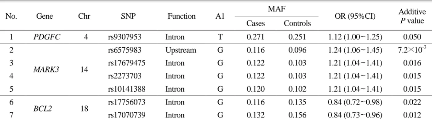
Table 2. Results of the case-control association analysis between SNPs in the PDGFC, MARK3, BCL2 genes and left ventricular hypertrophy in the KARE (Additive P value<0.05)
No. Gene Chr SNP Function A1 MAF
OR (95%CI) Additive P value Cases Controls
1 PDGFC 4 rs9307953 Intron T 0.271 0.251 1.12 (1.00~1.25) 0.050
2
MARK3 14
rs6575983 Upstream G 0.116 0.096 1.24 (1.06~1.45) 7.2×10⁻³
3 rs17679475 Intron G 0.122 0.103 1.21 (1.04~1.41) 0.016
4 rs2273703 Intron G 0.122 0.103 1.21 (1.04~1.41) 0.015
5 rs10141388 Intron G 0.120 0.102 1.21 (1.04~1.41) 0.015
6 BCL2 18 rs17756073 Intron G 0.116 0.135 0.84 (0.72~0.98) 0.022
7 rs17070739 Intron G 0.132 0.156 0.84 (0.73~0.96) 0.012
Abbreviations: SNP, single nucleotide polymorphism; A1, minor allele; MAF, minor allele frequency; OR, odds ration; CI, confidence interval; KARE, Korean association resource
Table 1. Basic characteristics of the subjects in the KARE
Characteristics Total LVH
P value
Cases Controls
Number of subjects 8,840 1,247 2,772
Age (M years ± SD) 52.22 ± 8.91 54.41 ± 9.08 50.66 ± 8.70 0.002
Gender [men (%) / women (%)] 4,182 (47) / 4,658 (53) 790 (63) / 457 (37) 1,332 (48) / 1,440 (52) <0.0001 Abbreviations: KARE, Korean association resource; M, mean value; SD, standard deviation
1.24(95% 신뢰구간: 1.06~1.45)로 나타났다. 그러나 BCL2
유전자 SNP 중 유의한 2개의 SNP (rs17756073, rs17070739) 는 상대적 위험도가 두 SNP 모두 0.84로 확인되었으며, 이는 Minor allele (G)를 가질수록 좌심실 비대 발생의 상 대적 위험도를 감소시키는 방향으로 상관성이 있었다(Table 2).
PDGFC
,MARK3
,BCL2
유전자의 유의한 SNP에 대한in silico
기능 분석PDGFC, MARK3, BCL2 유전자에서 통계적으로 유의성
을 가진 총 7개의 SNP들이 유전자 혹은 단백질 발현에 어떻게 영향을 미치는지 확인하기 위해서 Regulome DB(http://www.reguloumdb.org/index)와 HaploReg (http://archive.- broadinstitute.org/mammals/haploreg/haploreg_v3.php)를 이용
하여 in silico 기능 분석을 하였다. 그 결과 MARK3 유전 자의 SNP 중 2개의 SNP (rs17679475, rs2273703)에서 1fscore의 의미 있는 score (score<3a)가 나타났다(Table 3). 1f score는 eQTL뿐 만 아니라 해당 SNP이 전사 인자 결합
반응에 영향을 미칠 수 있거나 DNase peak에서 차이가 있다는 것을 의미한다. 또한 rs2273703은 HaploReg에서motif의 변화를 예측하고 있으므로 유전형에 따라 MARK3
유전자 발현에 영향을 미칠 가능성이 있다는 것을 보여 준다.PDGFC
,MARK3
,BCL2
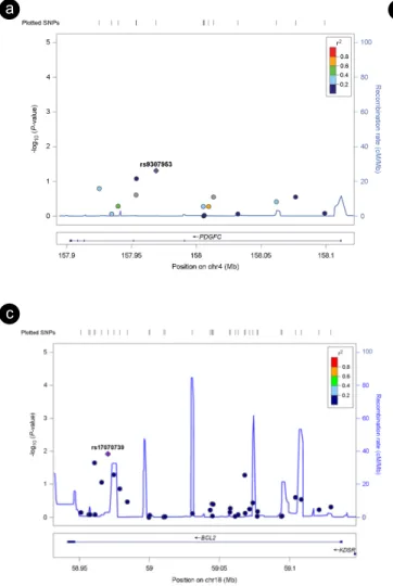
유전자의 Regional plot 확인PDGFC, MARK3, BCL2 유전자 SNP과 좌심실 비대 환
자군 사이의 상관관계를 기반으로 LocusZoom Version 1.1(http://csg.sph.umich.edu/locuszoom), (Pruim et al., 2010) 프로
그램을 사용하여 regional plot을 확인하였다. 분석 시에는 기준을 hg 19 version ASN (Asian population)으로 하였다.상관 분석 결과 가장 높은 유의 수준을 나타낸 SNP은 자 주색의 다이아몬드로 표시되고, 이는 regional plot의 기준
SNP이 된다(Fig. 1). MARK3 유전자의 분석 결과 rs6575983
을 기준으로 하였을 때 3개의 SNP에서 r2> 0.8 이상으로
나타났다. 따라서 rs6575983과 주위에 있는 유의 수준을 갖는 MARK3 유전자의 SNP들은 서로 연관되어 있다는 것을 확인할 수 있었다.고 찰
선행된 논문(Evangelou et al., 2018)에서는 UKB (UK
Biobank)와 ICBP (International Consortium of Blood Pressure Genome-wide Association Studies)로부터 얻은 유럽계 사람
들의 혈압과 관련된 특성(SBP, DBP, PP)에 대해 GWAS 분석을 하였다. 그 결과 535개의 새로운 유전좌위를 발견 하였으며, 이 중 SBP, DBP, PP의 세 가지 항목과 모두 관 Table 3. Results of the Regulome DB and HaploReg of SNPs in the PDGFC, MARK3, BCL2 geneGene Chr SNP A1 A2
Regulome DB HaploReg
Score TFBS DNase Proteins
bound Motifs
Promoter histone
marks
Enhancer histone
marks
Motifs
PDGFC 4 rs9307953 T A 6 - - - Myogenin/
NF-1 - 5 tissues 4 altered motifs
MARK3 14
rs6575983 G C 6 - - - 17 tissues Pax-5
rs17679475 G A 1f - + - - FAT 8 tissues -
rs2273703 G C 1f - + - - - 4 tissues 7 altered
motifs
rs10141388 G A 5 + - NFIC,
FOXM1 HOXA13 - - AIRE,
CTCF
BCL2 18
rs17756073 G A 4 + + GATA2,
GATA3 - BLD, GI 12 tissues -
rs17070739 G T 3a + + RUNX3 Sox12,
BCL6B BLD 7 tissues ATF3
Abbreviations: SNP, single nucleotide polymorphism; A1, minor allele; A2, major allele; TFBS, transcription binding factor site; +, affect;
Regulome DB is a database that annotates SNPs with known and predicted regulatory elements in the intergenic regions of the H. sapiens genome (http://www.regulomedb.org/index); HaploReg is a tool for exploring annotations of candidate regulatory SNPs (http://archive.- broadinstitute.org/mammals/haploreg/haploreg_v3.php).
련된 유전좌위는 총 12개(BCL2, KLF14, L2HGDH, LRP4,
MARK3, PDGFC, RXFP2, TERT, ARMC4, FGFR2, SNRNP70, YTHDF3)였다.
고혈압이 오래 지속되는 경우 심장에 비정상적인 심부 하로 인해 심근이 반응하여 좌심실 비대의 유병률 역시 증가되므로 고혈압과 좌심실 비대는 서로 연관성이 있다 고 볼 수 있다(WHO, 2002; Chung and Song, 2007). 그러므 로 이번 연구는 유럽인에서 혈압과 연관성 있다고 새롭 게 발견된 12개의 유전좌위를 가지고 한국인을 대상으로 유전자의 변이와 좌심실 비대에 대한 상관 분석을 시행 하여 각 유전자의 유전적 다형성이 좌심실 비대에 영향 을 미칠 가능성이 있는지 확인하고자 하였다. 따라서 각 각의 유전자에서 로지스틱 회귀 분석을 통해 좌심실 비대 환자군과 건강 대조군 간의 SNP에 따른 통계적 유의성 을 분석하여 유전적 변이에 따른 좌심실 비대 발생과의 상관성에 대하여 확인해 보았다. 그 결과 PDGFC 유전자
에서 1개의 SNP (rs9307953), MARK3 유전자에서 4개의
SNP (rs6575983, rs17679475, rs2273703, rs10141388), BCL2
유전자에서 2개의 SNP (rs17756073, rs17070739)들이 좌심 실 비대와 유의한 상관관계(P<0.05)를 나타냈다.Regulome DB와 HaploReg를 이용하여 SNP이 유전자
조절이나 단백질 발현에 어떻게 영향을 미치는지 조사 하였다. 그 결과 MARK3 유전자의 SNP 중 rs17679475와rs2273703은 Regulome DB score 1f를 나타냈다. 이는 minor allele를 보유함에 따라 MARK3 유전자의 발현에 차이를
나타내는 expression quantitative trait loci (eQTL) 영역임을 의미한다. rs17679475에서는 피하지방, 췌장, 뇌하수체, 전 립선, 혈관 등의 17개 조직에서 유전자 발현에 차이가 있음을 확인하였다. rs2273703에서는 피하지방, 정강신경, 췌장, 뇌하수체, 혈액 등 16개 조직에서 유전자의 발현 차이가 나타나는 것을 확인하였다. 또한 rs17679475와rs2273703은 DNase peak에서 차이가 있음을 확인하였다.
Fig. 1. The results of associations between PDGFC, MARK3, BCL2 SNPs and Left ventricular hypertrophy in KARE. The Figure 1-a shows the association results of the genetic variation on PDGFC, 1-b shows those of the MARK3 and 1-c shows those of the BCL2. The statistical significances (-log10 P-value) of association with the SNPs are plotted. At the bottom of the figure, the nucleotide positions within the chromosomes where each gene locus matches appears. The recombination rates estimated using HapMap Asian population data is shown by a blue line. The purple diamond with a SNP number represents the SNP most strongly associated with left ventricular hypertrophy, and its correlations with other SNPs are shown by colors indicating the levels of linkage disequilibrium (r2).
a
c
b
DNase는 염색질의 특정 영역을 활성화시켜 DNA를 노출
시킨다. 전사 단계에 필요한 여러 인자들은 노출된 DNA에 결합할 수 있게 되고 전사가 이루어진다. 결국 rs17679475,rs2273703은 DNase의 sensitivity를 변화시켜 MARK3 유전
자 전사 활성에 영향을 미칠 것으로 예상한다. PDGFC 유전자의 rs9307953은 myogenin/NF-1 motif를 형성하고MARK3 유전자의 rs10141388은 HOXA13 motif를, BCL2
유전자의 rs17070739는 Sox12와 BCL6B motif를 형성한다.따라서 각각의 SNP들은 유전자 발현에 영향을 미칠 가능 성을 보여주고 있다.
PDGF는 혈관의 발달과 동맥 경화증의 발병에 관여한다 (Ross, 1993; Betsholtz and Raines, 1997). PDGFC와 PDGFD
는 PDGF 유전자 군에 속하며, PDGF-C는 민무늬근 세포 에서 발현되고, PDGF-D는 섬유모세포의 외막세포에서 대 부분 발현되어 진다. 두 유전자는 배양된 내피세포 및 다 양한 종양 세포주에서 활성을 보이는 것으로 보고되었다.PDGF-C와 PDGF-D는 사람의 관상동맥 민무늬근 세포를
자극하며(Uutela et al., 2001), PDGFC와 PDGFD가 심장에서 과발현 될 때, PDGF-C는 심근의 섬유아세포 증식을 증가 시키는 것으로 나타났다(Li et al., 2000).MARK3의 기능은 아직 밝혀지지 않았으나, MARKs와
상동관계가 있는 것으로 밝혀졌다(Ono et al., 1997). MARKs 는 미세소관과 관련된 tau 단백질, MAP2 및 MAP4의 특 이적 인산화에 관여하는 새로운 키나아제이며, 결과적으 로 in vitro 및 배양된 세포에서 미세소관으로부터의 분리 를 촉매한다(Drewes et al., 1997).BCL2의 발현은 미토콘드리아에서 나오는 사이토크롬 C의 방출을 억제시키고 Apaf-1 complex의 생성을 방해하
므로 세포의 죽음을 예방할 수 있다(Yang et al., 1997). 따 라서 고혈압과 같은 병태생리학적 상태에서 세포자멸사 가 증가하는 현상은 BCL2 발현이 억제되는 것과 관계가 있다고 한다(Yang et al., 1997; Lee et al., 2006). 따라서 세 포자멸사를 억제시키는 작용을 가진 단백질의 증가는 심 근을 손상으로부터 보호하는 효과를 가지고 있다고 한다(Fehrenbach and Northoff, 2001).
이번 연구는 대규모 유전체 역학 코호트를 이용한 연 구로서 PDGFC, MARK3, BCL2 유전자의 특정 SNP들이 좌심실 비대에 영향을 미칠 수 있을 것이라는 가능성을 제시해주고 있으나 in silico를 통해 진행한 분석이므로 결 과를 간접적으로 확인할 수 있는 한계점이 있다. 현재까 지 여러 질병들의 발생과 환경적 요인 및 유전적 요인과 의 상관관계에 대한 다양한 연구들이 진행되었다(Jin et
al., 2018; Ko and Jin, 2019). 이번 연구는 좌심실 비대에 대
해서 한국인의 유전체 역학 자료를 기반으로 분석하였을 때 PDGFC, MARK3, BCL2 유전자 변이들이 좌심실 비대 와의 상관관계가 성립되는지 확인하고자 하였다. 연구 결 과를 통해 PDGFC, MARK3, BCL2 유전자는 유럽인의 혈 압에 영향을 미칠 뿐 만 아니라 한국인에서 좌심실 비대 에도 영향을 미치는 유전자임을 유추할 수 있다.ACKNOWLEDGEMENT
This research was supported by Basic Science Research Program through the National Research Foundation of Korea (NRF) grant (NRF-2017R1D1A3B03034752) funded by the Ministry of Education. This study was conducted with bio- resources from National Biobank of Korea, the Centers for Disease Control and Prevention, Republic of Korea (KBN- 2017-046).
CONFLICT OF INTEREST
The authors declare that they have no competing interests.
REFERENCES
Ahn YH. Characteristics of subgroups on patients with hypertension for hypertension management -based on knowledge, attitudes, and behavior related to medication and health lifesytle. Korean Academy of Community Health Nursing. 2007. 18: 112-122.
Betsholtz C, Raines EW. Platelet-derived growth factor: a key regulator of connective tissue cells in embryogenesis and pathogenesis. Kidney International. 1997. 51: 1361-1369.
Cho YS, Go MJ, Kim YJ, Heo JY, Oh JH, Ban HJ, Yoon D, Lee MH, Kim DJ, Park M, Cha SH, Kim JW, Han BG, Min H, Ahn Y, Park MS, Han HR, Jang HY, Cho EY, Lee JE, Cho NH, Shin C, Park T, Park JW, Lee JK, Cardon L, Clarke G, McCarthy MI, Lee JY, Lee JK, Oh B, Kim HL. A large-scale genome-wide association study of asian populations uncovers genetic factors influencing eight quantitative traits. Nature Genetics. 2009. 41: 527-534.
Chung JH, Song CH. Factors associated with left ventricular hypertrophy on ECG in middle-aged normotensive healthy men. Korean Journal of Family Medicine. 2007. 28: 92-99.
Drewes G, Ebneth A, Preuss U, Mandelkow EM, Mandelkow E.
MARK, a novel family of protein kinases that phosphorylate microtubule-associated proteins and trigger microtubule dis-
ruption. Cell. 1997. 89: 297-308.
Drummond GR, Vinh A, Guzik TJ, Sobey CG. Immune mechanisms of hypertension. Nat Rev Immunol. 2019. 19: 517-532.
Evangelou E, Warren HR, Mosen-Ansorena D, Mifsud B, Pazoki R, Gao H, Ntritsos G, Dimou N, Cabrera CP, Karaman I, Ng FL, Evangelou M, Witkowska K, Tzanis E, Hellwege JN, Giri A, Velez Edwards DR, Sun YV, Cho K, Gaziano JM, Wilson PWF, Tsao PS, Kovesdy CP, Esko T, Mägi R, Milani L, Almgren P, Boutin T, Debette S, Ding J, Giulianini F, Holliday EG, Jackson AU, Li-Gao R, Lin WY, Luan J, Mangino M, Oldmeadow C, Prins BP, Qian Y, Sargurupremraj M, Shah N, Surendran P, Thériault S, Verweij N, Willems SM, Zhao JH, Amouyel P, Connell J, de Mutsert R, Doney ASF, Farrall M, Menni C, Morris AD, Noordam R, Paré G, Poulter NR, Shields DC, Stanton A, Thom S, Abecasis G, Amin N, Arking DE, Ayers KL, Barbieri CM, Batini C, Bis JC, Blake T, Bochud M, Boehnke M, Boerwinkle E, Boomsma DI, Bottinger EP, Braund PS, Brumat M, Campbell A, Campbell H, Chakravarti A, Chambers JC, Chauhan G, Ciullo M, Cocca M, Collins F, Cordell HJ, Davies G, de Borst MH, de Geus EJ, Deary IJ, Deelen J, Del Greco MF, Demirkale CY, Dörr M, Ehret GB, Elosua R, Enroth S, Erzurumluoglu AM, Ferreira T, Frånberg M, Franco OH, Gandin I, Gasparini P, Giedraitis V, Gieger C, Girotto G, Goel A, Gow AJ, Gudnason V, Guo X, Gyllensten U, Hamsten A, Harris TB, Harris SE, Hartman CA, Havulinna AS, Hicks AA, Hofer E, Hofman A, Hottenga JJ, Huffman JE, Hwang SJ, Ingelsson E, James A, Jansen R, Jarvelin MR, Joehanes R, Johansson Å, Johnson AD, Joshi PK, Jousilahti P, Jukema JW, Jula A, Kähönen M, Kathiresan S, Keavney BD, Khaw KT, Knekt P, Knight J, Kolcic I, Kooner JS, Koskinen S, Kristiansson K, Kutalik Z, Laan M, Larson M, Launer LJ, Lehne B, Lehtimäki T, Liewald DCM, Lin L, Lind L, Lindgren CM, Liu Y, Loos RJF, Lopez LM, Lu Y, Lyytikäinen LP, Mahajan A, Mamasoula C, Marrugat J, Marten J, Milaneschi Y, Morgan A, Morris AP, Morrison AC, Munson PJ, Nalls MA, Nandakumar P, Nelson CP, Niiranen T, Nolte IM, Nutile T, Oldehinkel AJ, Oostra BA, O'Reilly PF, Org E, Padmanabhan S, Palmas W, Palotie A, Pattie A, Penninx BWJH, Perola M, Peters A, Polasek O, Pramstaller PP, Nguyen QT, Raitakari OT, Ren M, Rettig R, Rice K, Ridker PM, Ried JS, Riese H, Ripatti S, Robino A, Rose LM, Rotter JI, Rudan I, Ruggiero D, Saba Y, Sala CF, Salomaa V, Samani NJ, Sarin AP, Schmidt R, Schmidt H, Shrine N, Siscovick D, Smith AV, Snieder H, Sõber S, Sorice R, Starr JM, Stott DJ, Strachan DP, Strawbridge RJ, Sundström J, Swertz MA, Taylor KD, Teumer A, Tobin
MD, Tomaszewski M, Toniolo D, Traglia M, Trompet S, Tuomilehto J, Tzourio C, Uitterlinden AG, Vaez A, van der Most PJ, van Duijn CM, Vergnaud AC, Verwoert GC, Vitart V, Völker U, Vollenweider P, Vuckovic D, Watkins H, Wild SH, Willemsen G, Wilson JF, Wright AF, Yao J, Zemunik T, Zhang W, Attia JR, Butterworth AS, Chasman DI, Conen D, Cucca F, Danesh J, Hayward C, Howson JMM, Laakso M, Lakatta EG, Langenberg C, Melander O, Mook-Kanamori DO, Palmer CNA, Risch L, Scott RA, Scott RJ, Sever P, Spector TD, van der Harst P, Wareham NJ, Zeggini E, Levy D, Munroe PB, Newton-Cheh C, Brown MJ, Metspalu A, Hung AM, O'Donnell CJ, Edwards TL; Million Veteran Program, Psaty BM, Tzoulaki I, Barnes MR, Wain LV, Elliott P, Caulfield MJ. Publisher correction: genetic analysis of over 1 million people identifies 535 new loci associated with blood pressure traits. Nature Genetics. 2018. 50: 1755-018-0297-3.
Fehrenbach E, Northoff H. Free radicals, exercise, apoptosis, and heat shock proteins. Exercise Immunology Review. 2001. 7:
66-89.
Im SA. Patterns of left ventricular hypertrophy and gemotric re- modeling in essential hypertension. Korean Circulation Journal.
1995. 25: 423-433.
Jin HS, Lee SI, Park S. Association between ITGB2 genetic poly- morphisms and tuberculosis. Korean J Clin Lab Sci. 2018. 50:
118-125.
Ko B, Jin HS. MACROD2 polymorphisms are associated with hypertension in korean population. Korean J Clin Lab Sci.
2019. 51: 57-63.
Lee J, Cho HS, Kim WK. The effects of regular exercise on the expression of Bcl-2 and apoptosis in myocardium of L-NAME induced hypertensive rat. Korean Journal of Sport Science.
2006. 17: 45-54.
Levy D, Garrison RJ, Savage DD, Kannel WB, Castelli WP. Pro- gnostic implications of echocardiographically determined left ventricular mass in the framingham heart study. The New England Journal of Medicine. 1990. 322: 1561-1566.
Li X, Pontén A, Aase K, Karlsson L, Abramsson A, Uutela M, Bäckström G, Hellström M, Boström H, Li H, Soriano P, Betsholtz C, Heldin CH, Alitalo K, Ostman A, Eriksson U.
PDGF-C is a new protease-activated ligand for the PDGF alpha-receptor. Nature Cell Biology. 2000. 2: 302-309.
Oh JY. Patterns of left ventricular hypertrophy by echocardiography in coronary artery diseases. Korean Circulation Journal. 1996.
26: 473-482.
Ono T, Kawabe T, Sonta S, Okamoto T. Assignment of MARK3
alias KP78 to human chromosome band 14q32.3 by in situ hybridization. Cytogenetics and Cell Genetics. 1997. 79: 101 -102.
Pruim RJ, Welch RP, Sanna S, Teslovich TM, Chines PS, Gliedt TP, Boehnke M, Abecasis GR, Willer CJ. LocusZoom: regional visualization of genome-wide association scan results. Bio- informatics. 2010. 26:2336-2337.
Reichek N, Devereux RB. Left ventricular hypertrophy: relation- ship of anatomic, echocardiographic and electrocardiographic findings. Circulation. 1981. 63: 1391-1398.
Ross R. The pathogenesis of atherosclerosis: a perspective for the 1990s. Nature. 1993. 362: 801-809.
Sullivan JM, Vander Zwaag RV, el-Zeky F, Ramanathan KB, Mirvis DM. Left ventricular hypertrophy: effect on survival.
Journal of the American College of Cardiology. 1993. 22: 508 -513.
Tuinstra CL, Rautaharju PM, Prineas RJ, Duisterhout JS. The per- formance of three visual coding procedures and three computer programs in classification of electrocardiograms according to the Minnesota Code. Journal of Electrocardiology. 1982. 15:
345-350.
Uutela M, Laurén J, Bergsten E, Li X, Horelli-Kuitunen N, Eriksson U, Alitalo K. Chromosomal location, exon structure, and vascular expression patterns of the human PDGFC and PDGFD genes. Circulation. 2001. 103: 2242-2247.
World Health Organization. Cardiovascular death and disability can be reduced more than 50 percent. Indian J Med Sci. 2002.
57: 117-121.
Yang J, Liu X, Bhalla K, Kim CN, Ibrado AM, Cai J, Peng TI, Jones DP, Wang X. Prevention of apoptosis by Bcl-2: release of cytochrome c from mitochondria blocked. Science. 1997.
275: 1129-1132.
https://doi.org/10.15616/BSL.2019.25.3.237
Cite this article as: Jeon TE, Jin HS. PDGFC, MARK3
and BCL2 Polymorphisms are Associated with Left
Ventricular Hypertrophy in Korean Population. Biomedical
Science Letters. 2019. 25: 237-246.
Supplementary Table 1. Results of the case-control association analysis between SNPs in the PDGFC, MARK3, BCL2 genes and left ventricular hypertrophy in the KARE
Gene Chr No. SNP BP Function A1 MAF
OR (95%CI) Additive P value Cases Controls
PDGFC 4
1 rs7672622 157925001 Intron C 0.257 0.243 1.08 (0.97~1.21) 0.161 2 rs17035257 157934512 Intron C 0.165 0.170 0.99 (0.87~1.13) 0.862 3 rs4691380 157939574 Intron A 0.288 0.285 1.04 (0.93~1.16) 0.520 4 rs10517653 157953440 Intron A 0.056 0.064 0.89 (0.72~1.09) 0.247 5 rs10033375 157953762 Intron C 0.077 0.065 1.18 (0.98~1.42) 0.083 6 rs9307953 157968941 Intron T 0.271 0.251 1.12 (1.00~1.25) 0.050 7 rs10517657 158005478 Intron A 0.152 0.160 0.96 (0.84~1.10) 0.528 8 rs17035367 158005675 Intron G 0.063 0.065 1.00 (0.82~1.22) 0.981 9 rs7662187 158006222 Intron C 0.064 0.065 1.01 (0.82~1.23) 0.939 10 rs342317 158009437 Intron G 0.249 0.243 1.04 (0.93~1.16) 0.529 11 rs17035387 158013333 Intron A 0.009 0.012 0.76 (0.46~1.25) 0.279 12 rs2343116 158032128 Intron G 0.110 0.111 1.01 (0.87~1.18) 0.864 13 rs10517660 158061846 Intron A 0.382 0.392 0.96 (0.86~1.06) 0.380 14 rs6851803 158076643 Intron C 0.010 0.012 0.77 (0.48~1.24) 0.282 15 rs17035464 158099171 intron T 0.119 0.118 1.02 (0.87~1.18) 0.832
MARK3 14
1 rs6575983 102939602 Upstream G 0.116 0.096 1.24 (1.06~1.45) 0.007 2 rs4906321 102948635 Upstream T 0.137 0.121 1.15 (0.99~1.33) 0.063 3 rs2065018 102948940 Upstream C 0.307 0.318 0.93 (0.84~1.04) 0.193 4 rs975892 102953102 Intron T 0.454 0.442 1.03 (0.93~1.14) 0.565 5 rs17679127 102959844 Intron G 0.045 0.034 1.26 (0.99~1.61) 0.064 6 rs8015723 102965662 Intron A 0.367 0.359 1.04 (0.94~1.15) 0.486 7 rs17841064 102970362 Intron G 0.456 0.446 1.02 (0.93~1.13) 0.633 8 rs6575988 102970580 Intron G 0.430 0.424 1.01 (0.91~1.11) 0.884 9 rs12894275 102971177 Intron A 0.314 0.325 0.93 (0.83~1.03) 0.160 10 rs2065015 102973814 Intron G 0.311 0.322 0.93 (0.84~1.04) 0.188 11 rs17095251 102983535 Intron A 0.115 0.125 0.87 (0.75~1.01) 0.069 12 rs17679475 102986711 Intron G 0.122 0.103 1.21 (1.04~1.41) 0.016 13 rs2273703 102987782 Intron G 0.122 0.103 1.21 (1.04~1.41) 0.015 14 rs2273702 102987935 Intron C 0.439 0.429 1.02 (0.92~1.12) 0.708 15 rs10141388 103009432 Intron G 0.120 0.102 1.21 (1.04~1.41) 0.015 16 rs3783397 103017042 Intron G 0.304 0.317 0.92 (0.83~1.03) 0.137 17 rs3783398 103019131 Intron C 0.305 0.316 0.93 (0.84~1.04) 0.196 18 rs9671414 103037052 Intron C 0.431 0.422 1.02 (0.92~1.13) 0.706
BCL 18
1 rs10503078 58951168 Intron A 0.399 0.396 1.02 (0.92~1.12) 0.715 2 rs4987839 58956729 Intron C 0.408 0.410 0.99 (0.90~1.09) 0.831 3 rs1531697 58957586 Intron A 0.426 0.424 1.01 (0.92~1.12) 0.820 4 rs17756073 58960763 Intron G 0.116 0.135 0.84 (0.72~0.98) 0.022 5 rs12457831 58961220 Intron C 0.392 0.389 1.01 (0.92~1.12) 0.836 6 rs1542578 58965895 Intron A 0.458 0.435 1.09 (0.99~1.20) 0.088 7 rs17070739 58970363 Intron G 0.132 0.156 0.84 (0.73~0.96) 0.012 8 rs4941185 58974534 Intron T 0.479 0.456 1.10 (1.00~1.21) 0.051
Supplementary Table 1. Results of the case-control association analysis between SNPs in the PDGFC, MARK3, BCL2 genes and left ventricular hypertrophy in the KARE (Continued)
Gene Chr No. SNP BP Function A1 MAF
OR (95%CI) Additive P value Cases Controls
BCL 18
9 rs2199937 58978769 Intron C 0.476 0.459 1.08 (0.98~1.19) 0.136 10 rs7228914 58984206 Intron A 0.498 0.491 1.05 (0.95~1.16) 0.338 11 rs4940576 58999619 Intron A 0.407 0.409 1.00 (0.90~1.10) 0.989 12 rs4941187 59000196 Intron C 0.409 0.408 1.01 (0.91~1.12) 0.868 13 rs899966 59010221 Intron T 0.401 0.403 1.00 (0.90~1.10) 0.976 14 rs12457700 59011226 Intron T 0.399 0.402 1.00 (0.90~1.10) 0.955 15 rs17757541 59030666 Intron G 0.176 0.178 0.98 (0.86~1.11) 0.761 16 rs4987774 59043342 Intron G 0.291 0.301 0.97 (0.87~1.08) 0.602 17 rs1893506 59044660 Intron G 0.138 0.128 1.06 (0.92~1.23) 0.390 18 rs1775798 59044959 Intron G 0.049 0.049 1.01 (0.81~1.27) 0.915 19 rs9959874 59045526 Intron A 0.138 0.128 1.06 (0.92~1.23) 0.398 20 rs17070861 59057460 Intron C 0.051 0.050 1.05 (0.83~1.31) 0.703 21 rs11152375 59057664 Intron C 0.484 0.481 1.03 (0.93~1.14) 0.534 22 rs17685559 59061198 Intron A 0.048 0.048 1.01 (0.81~1.27) 0.920 23 rs10503079 59063346 Intron T 0.046 0.049 0.97 (0.77~1.23) 0.830 24 rs17070904 59068036 Intron T 0.061 0.074 0.82 (0.68~1.00) 0.052 25 rs7240326 59068331 Intron G 0.237 0.229 1.02 (0.91~1.14) 0.755 26 rs8096380 59071461 Intron G 0.332 0.346 0.97 (0.87~1.08) 0.564 27 rs7234941 59073831 Intron T 0.182 0.173 1.06 (0.93~1.21) 0.364 28 rs3744951 59076960 Intron G 0.047 0.047 1.01 (0.80~1.27) 0.931 29 rs12457893 59077141 Intron T 0.382 0.386 0.98 (0.89~1.08) 0.671 30 rs3810031 59094075 Intron G 0.238 0.239 1.01 (0.90~1.13) 0.933 31 rs8089331 59094686 Intron G 0.199 0.200 0.99 (0.88~1.12) 0.909 32 rs4987724 59104259 Intron T 0.020 0.023 0.82 (0.58~1.15) 0.251 33 rs1381548 59108376 Intron A 0.338 0.348 0.95 (0.85~1.05) 0.290
34 rs4941195 59121016
Intron variant, synonymous codon
G 0.237 0.232 1.03 (0.92~1.16) 0.597
35 rs12458289 59129566 Intron A 0.266 0.260 1.04 (0.93~1.16) 0.492 P-value<0.05 are indicated in bold. Abbreviaitons: SNP, single nucleotide polymorphism; BP, base pair; A1, minor allele; MAF, minor allele frequency; OR, odds ration; CI, confidence interval; KARE, Korean association resource