The Indoor Level of House Dust Mite Allergen Is Associated with Severity of Atopic Dermatitis in Children
We attempted to investigate the correlation between the severity of atopic dermatitis (AD) in children and the indoor level of house dust mite (HDM) allergens. Ninety-five patients (31.1 ± 19.5 months of age) with AD were enrolled in this study, and serum specific IgE against Dermatophagoides pteronyssinus and D. farinae was measured. The severity of AD was assessed using the visual analogue scale on the same day of house dust collection.
Living rooms and mattresses where the child usually slept were vacuumed for 2 minutes and concentrations of Der f 1 were measured by enzyme-linked immunosorbent assay. The skin symptoms were more severe in patients with Der f 1 concentrations in living room
> 2 μg/g dust than ≤ 2 μg/g dust (P = 0.018). This difference was noted in AD patients without sensitization to HDM (P = 0.004), but not in patients with sensitization. There was no difference in symptom severity according to Der f 1 concentrations in mattresses (P = 0.062). The severity of skin symptoms is associated with indoor concentrations of HDM in children with AD, and it is likely to act as nonspecific irritants as well as allergens in AD skin lesions.
Key Words: Atopic Dermatitis; Allergens; House Dust Mite Jihyun Kim,1,2 Sangwoon Lee,2
Sook-young Woo,3 Youngshin Han,2 Jung Hyun Lee,4 In-Yong Lee,5 In-Seok Lim,6 Eung-Sang Choi,6 Byoung-Whi Choi,7 Hae-Kwan Cheong,8 Sang-Il Lee,1,2 and Kangmo Ahn1,2
1Department of Pediatrics, Samsung Medical Center, Sungkyunkwan University School of Medicine, Seoul; 2Environmental Health Center for Atopic Diseases, Samsung Medical Center, Seoul;
3Biostatistics Team, Samsung Biomedical Research Institute, Seoul; 4Department of Pediatrics, Gwangmyeong Sungae Hospital, Gwangmyeong;
5Department of Environmental Medical Biology, Yonsei University College of Medicine, Seoul;
Departments of 6Pediatrics and 7Internal Medicine, Chung-Ang University College of Medicine, Seoul;
8Department of Social and Preventive Medicine, Sungkyunkwan University School of Medicine, Suwon, Korea
Received: 23 July 2012 Accepted: 11 October 2012 Address for Correspondence:
Kangmo Ahn, MD
Department of Pediatrics, Samsung Medical Center, 81 Irwon-ro, Gangnam-gu, Seoul 135-710, Korea Tel: +82.2-3410-3530, Fax: +82.2-3410-0043 E-mail: [email protected]
This study was funded by Environmental Health Research Center Project by the Ministry of Environment, Republic of Korea.
http://dx.doi.org/10.3346/jkms.2013.28.1.74 • J Korean Med Sci 2013; 28: 74-79
INTRODUCTION
Dermatophagoides pteronyssinus and D. farinae are common inhabitants in homes in temperate climates and are major con- tributors to the allergen concentrations of house dust (1). Previ- ous reports have demonstrated that about 35% of patients with allergic diseases are sensitized to house dust mites (HDM) (2).
It is well established that exposure to HDM is associated with development of allergic rhinitis or asthma in children (3, 4), and removal of HDM has been suggested to improve bronchial hy- perresponsiveness in asthmatic patients (5).
Atopic dermatitis (AD) is a chronic and highly pruritic inflam- matory skin disease with a prevalence of 12.8%-26.5% in chil- dren (6, 7). Previous studies have attempted to document the relationship between indoor HDM levels and the development
of AD (8, 9), but there has been relatively little information in the literature regarding the effect of HDM concentrations on skin symptoms in patients with AD. Moreover, there are contro- versies about the relationship between HDM and AD, whereas asthma or allergic rhinitis shows a strong relationship with ex- posure to HDM (4, 5). For example, it has been demonstrated that the skin and homes of patients with eczema have higher concentrations of mites than those of healthy people, and con- sequently, reduction of exposure to HDM may result in clinical improvement of eczema (9, 10). On the other hand, it has been reported that domestic HDM exposure was not correlated with SCORing of AD (SCORAD), and no improvement of disease ac- tivity was observed in adult patients with AD undertaking 1 yr of HDM avoidance measures (11, 12).
A better understanding of the relationship between AD and
HDM exposure in areas where exposure to HDM is ubiquitous may help us to prevent aggravation of skin symptoms in patients with eczema. This is especially relevant for children with AD, since AD requires a comprehensive long-term strategy in the set- ting of limited therapeutic options (13). Therefore, we attempt- ed to investigate the relationship between the severity of AD in children and the indoor level of HDM allergens in this study.
MATERIALS AND METHODS Study population
Ninety-five patients (median age: 23.0 months; range: 2-168 months) with AD as defined by the criteria of Hanifin and Rajka (14) were included in this study. None of the patients had re- ceived systemic corticosteroids during the 2 months prior to the clinical evaluation. During the study period, all of the patients were asked to take a bath once daily with warm water for 5 to 10 min and apply moisturizers frequently. Intermittent use of low potency topical corticosteroids (TCS) was allowed in patients who present with erythema and itching. For the patients requiring TCS as rescue medicine, we offered prednisolone valeroacetate or 1% hydrocortisone, and educated the patients to cover the body area equivalent to 2 hands using one fingertip unit of TCS.
The severity of atopic dermatitis
The severity of AD was evaluated by the use of the visual ana- logue scale (VAS) (15). Parents were asked to quantify the over- all AD symptoms on a VAS ranging from 0 (no symptoms at all) to 10 (very severe symptoms) on the day of house dust collec- tion. The answer was recorded to E-VAS in response to the ques- tion, “How was the eczema in the last month?”; I-VAS to “How were itching symptoms in the last month?”; and S-VAS to “How were sleep-disturbing symptoms in the last month?” E-VAS, I- VAS, and S-VAS were added up to produce T-VAS (VAS of 0-30).
The use of medications was recorded as rescue medicine con- sumption index (RMCI) to compare their treatment during the last 1 month (15). Allowed medications for AD were for short courses (3 days) of TCS and/or oral hydroxyzine on demand in the case of worsening pruritus, itching, edema, or oozing. When the bacterial infection was suspected, the patients were pre- scribed a 7-day course of 1st generation cephalosporin. The use of medications was scored 1 point for each dose of oral hydroxi- zine or topical prednosolone valeroacetate ointment and 2 points for each dose of oral antibiotics over the 7-day course.
Total IgE and allergen specific IgE
Blood samples were collected for measurement of the total IgE level and specific IgE (sIgE) level at the initial visit. The sIgE an- tibodies to D. pteronyssinus, D. farinae, and common food al- lergens including egg white, cow’s milk, wheat, soy, peanut, and buckwheat were measured by an ImmunoCAP system (Thermo
Fisher Scientific Inc., Waltham, MA, USA), with concentrations
≥ 0.35 kU/L being regarded as sensitization.
Dust sampling and allergen determination
Dust samples were taken from living rooms and mattresses. A standard household vacuum cleaner (Majestic 360, 1050 W;
HMI Industries, Strongsville, OH, USA) equipped with 10 μm filter paper was used. Living room and mattress samples were standardized by vacuuming an area of 1 × 1 m for 2 min. Ex- posed filter papers were stored at -20°C until allergen analysis.
Each sample was sieved through a 355 nm mesh sieve, record- ing weights before and after sieving, and 2.0 mL PBS-T (0.05%
Tween 20 in phosphate buffered saline, pH 7.4) was added to 100 mg of sieved dust. A proportionate amount of buffer was added if the weight of the dust was between 30 and 100 mg. The dust samples were constantly rotated at room temperature for 2 hr and the aqueous layer was removed after centrifugation at 2,500 rpm at 4°C for 20 min and stored at -20°C. Der f 1 concen- trations were determined using monoclonal antibody (mAb) enzyme-linked immunosorbent assay (Indoor Biotechnologies, Cardiff, UK) as described previously (16). The lower limit of de- tection for the allergen assays was 0.1 μg/g dust, with no upper limit.
Statistical analysis
The data were analyzed using SAS version 9.1.3 (SAS, Cary, NC, USA). The concentration of Der f 1 was divided into two groups with a reference concentration of 2 μg/g dust, which is a known cutoff value for HDM sensitization (17). Differences in VAS ac- cording to Der f 1 concentrations in each location among the AD patients were analyzed using the Wilcoxon two-sample test. AD patients were classified as being in the sensitized group or not according to the result of sIgE antibodies to D. pteronyssinus or D. farinae. Differences in VAS according to Der f 1 concentrations in each group were also analyzed. In addition, patients were di- vided into 2 age groups; less than 24 months and 24 months or older. Because the use of separate univariate t tests leads to an inflated type 1 error, the Bonferroni’s correction was applied to the subgroup analysis by adjusting the P value. A P value < 0.05 was considered to be significant.
Ethics statement
This study was approved by the institutional review board at Samsung Medical Center in Seoul (IRB approval number: 2009- 02-034). Written informed consent was obtained from parents prior to participation in the study.
RESULTS
Patients’ characteristics
AD patients comprised 62 boys and 33 girls. Their clinical and
demographic characteristics were shown in Table 1. Sensitiza- tion to HDM was found in 44 out of 95 patients with AD (Table 1). The mean VAS was 5.4 in eczema, 5.3 in itching symptom, 4.1 in sleep-disturbing symptom, and 20.1 in total.
RMCIs were 17.0 ± 16.6 in patients with high (> 2 μg/g dust) Der f 1 levels from living rooms and 13.3 ± 14.4 in those with low (≤ 2 μg/g dust) Der f 1 levels. In addition, RMCIs were 14.7 ± 16.2 in patients with high Der f 1 levels from mattresses and 13.6 ± 14.1 in those with low Der f 1 levels. There were no differences of RMCI between 2 groups with high and low Der f 1 levels from living rooms and mattresses (P = 0.418 and 0.788).
Skin symptoms according to domestic Der f 1 concentrations Der f 1 allergen levels were detectable in 24 (25.3%) of 95 sam- ples from living rooms and 79 (83.2%) of 95 samples from mat- tresses. The highest level was found from mattresses, and Der f
1 levels from mattresses were higher than those from living rooms without statistical significance (P = 0.082). Mean values for Der f 1 (± SD) were 1.4 ± 5.0 (range, 0-33.7) μg/g dust from living rooms and 5.1 ± 19.7 (range, 0-187.0) μg/g dust from mattress- es, respectively.
VAS was higher in patients with Der f 1 concentration from living rooms > 2 μg/g dust than in patients with Der f 1 concen- tration ≤ 2 μg/g dust (P = 0.018) (Fig. 1). In particular, the dif- ference of VAS according to Der f 1 from living rooms was also found in AD patients without sensitization to HDM (P = 0.004).
However, the difference of VAS according to Der f 1 from living rooms was not found in AD patients who were sensitized to HDM (P = 1.000). In addition, there were no differences in VAS
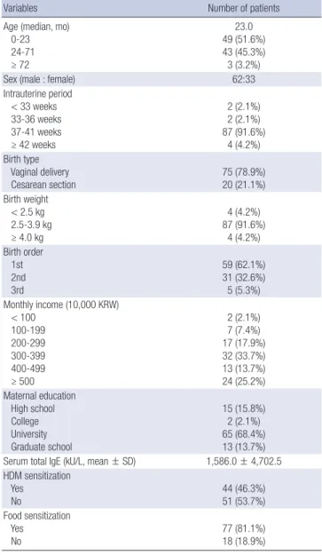
Table 1. General characteristics of the subjects (n = 95)
Variables Number of patients
Age (median, mo) 0-23 24-71 ≥ 72
23.0 49 (51.6%) 43 (45.3%) 3 (3.2%)
Sex (male : female) 62:33
Intrauterine period < 33 weeks 33-36 weeks 37-41 weeks ≥ 42 weeks
2 (2.1%) 2 (2.1%) 87 (91.6%)
4 (4.2%) Birth type
Vaginal delivery
Cesarean section 75 (78.9%)
20 (21.1%) Birth weight
< 2.5 kg 2.5-3.9 kg ≥ 4.0 kg
4 (4.2%) 87 (91.6%)
4 (4.2%) Birth order
1st 2nd 3rd
59 (62.1%) 31 (32.6%) 5 (5.3%) Monthly income (10,000 KRW)
< 100 100-199 200-299 300-399 400-499 ≥ 500
2 (2.1%) 7 (7.4%) 17 (17.9%) 32 (33.7%) 13 (13.7%) 24 (25.2%) Maternal education
High school College University Graduate school
15 (15.8%) 2 (2.1%) 65 (68.4%) 13 (13.7%) Serum total IgE (kU/L, mean ± SD) 1,586.0 ± 4,702.5 HDM sensitization
Yes No
44 (46.3%) 51 (53.7%) Food sensitization
Yes No
77 (81.1%) 18 (18.9%) HDM, house dust mite.
Fig. 1. Visual analogue scale in patients with atopic dermatitis according to the indoor level of Der f 1. *P < 0.05. VAS, visual analogue scale; AD, atopic dermatitis.
VAS
Living room Mattress
30
20
10
0
VAS
Living room Mattress
30
20
10
0
Total patients with AD Der f 1 ≤ 2 µg/g dust Der f 1 > 2 µg/g dust
AD with sensitization to HDM Der f 1 ≤ 2 µg/g dust Der f 1 > 2 µg/g dust
AD without sensitization to HDM Der f 1 ≤ 2 µg/g dust Der f 1 > 2 µg/g dust
*
VAS
Living room Mattress
30
20
10
0
*
according to Der f 1 levels from living rooms in younger (< 24 months old) age group and older (≥ 24 months old) age group (P = 0.069 and 0.306).
We did not find differences in VAS according to concentra- tions of Der f 1 (P = 0.062) from mattresses (Fig. 1). No signifi- cant differences in the VAS were apparent between high and low concentrations of Der f 1 from mattresses in AD patients not sensitized to HDM (P = 0.126). There was also no significant dif- ference in the VAS according to concentrations of Der f 1 from mattresses in AD patients sensitized to HDM (P = 1.000). In younger age group, VAS was higher in patients with Der f 1 level from mattresses > 2 μg/g dust than in those with Der f 1 level
≤ 2 μg/g dust (P = 0.007). However, older age group did not show any significant difference in VAS between high and low Der f 1 concentrations from mattresses (P = 1.000)
DISCUSSION
Our results in this study indicate that indoor HDM levels are as- sociated with the severity of skin symptoms, especially in AD patients without sensitization to HDM. This evidence supports the assertion that we need to reduce the level of HDM to pre- vent exacerbations of AD. In addition, our findings suggest the possibility that Der f 1 could act as a nonspecific irritant as well as an allergen. The effect of HDM on skin symptoms of AD was not dependent on HDM sensitization, showing their obvious relationship in the non-sensitized group in the present study.
These results support the finding that repeated application of D.
farinae extract to the skin causes clinical and histological symp- toms similar to those in human AD (18). Similarly, in a subgroup of patients with eczema, epicutaneous challenge with HDM ex- tracts resulted in a delayed-type response that resembled natu- rally-occurring eczema (19). This atopy patch test reaction is re- garded as a proteolytic irritancy as well as an IgE-mediated re- sponse (9, 19).
Dominant allergens of HDM such as group 1 cysteine prote- ases, trypsin, chymotrypsin, and a serine protease, possess pep- tidase activity and have direct effects on mucosal permeability and epithelial function (9, 20). A recent study reported that HDM and cockroach allergens with protease activity can influence homeostasis of the epidermal permeability barrier through pro- tease-activated receptor 2 activation and consequent modula- tion of the calcium ions in the skin (21). A disruption of the per- meability barrier in AD lesions can lead to an increased pene- tration of environmental allergens into the skin, which initiates immunologic reactions (21). In particular, the disruption of the skin barrier by other stimulants such as volatile organic com- pounds, can enhance the adverse effects of HDM on provoca- tion testing (22), suggesting that barrier dysfunction of AD skin lesion might lead to a direct pathophysiological role of HDM in AD patients. HDM allergens can be danger signals in the skin
through nucleotide-binding oligomerization domain, leucine- rich repeat and pyrin-domain containing 3 (NLRP 3), which plays a central role in both innate immunity and inflammation (23). The results obtained in our study along with previous re- ports indicate that the proteolytic activities of HDM allergens might play a role as a nonspecific irritant in AD patients.
Although there was no association between indoor HDM level and AD severity in those who were sensitized to HDM, it does not mean that HDM is not an aggravating factor in these patients. IgE-mediated response can be shown even at low al- lergen levels in selected patients with sensitization. For exam- ple, an environmental challenge study demonstrated that simi- lar pulmonary responses between low- and high- allergen con- ditions in subjects with documented IgE-mediated allergic re- actions to rats (24). A previous study showing clinical effect of HDM immunotherapy indicates that HDM can act as an aggra- vating factor of AD through IgE-mediated mechanism (25).
Therefore, in AD patients, regardless of sensitization, HDM is an important aggravating factor which should be reduced in in- door environment.
D. farinae often predominates in Korean homes, although slight variation of mite species has been documented depend- ing on each region (26). Previous studies conducted in the Unit- ed States also revealed a variable prevalence of mite species be- tween different locations in the same country (27). Few homes showed detectable levels of Der p 1 with a Der f 1/Der p 1 ratio of 11.8 in our preliminary data, which is in agreement with those found in Seoul (Der f 1/Der p 1 ratios of 3.7-27) (26, 28). Prelimi- nary data also showed no significant differences of VAS between high and low Der p 1 levels from living rooms and mattresses (data not shown). For this reason, we used the indoor concen- tration of Der f 1 not Der p 1 for analyses in this study.
Our study has limitations, mostly stemming from the mea- surement of HDM. We collected house dust for HDM allergen analysis at only one point of time in various seasons. However, previous studies have reported that Der p 1 and Der f 1 concen- trations in children’s mattress dust are highly stable, indicating that a single measurement of HDM levels in house dust could be a good indication of exposure to HDM during a 1-yr period (29, 30). We also tried to overcome this limitation by evaluating the severity on the same day of the dust collection, not on the day of subject enrollment. In addition, we evaluated AD severi- ty using VAS, not objective scales such as SCORAD or EASI (ec- zema area and severity index), because clinicians were not ac- companied on the day of measurement. Another potential prob- lem in our study was the use of anti-inflammatory treatment and antihistamine which affect skin lesions and pruritus. How- ever, we compared their medication use through RMCI and found no differences between the two groups.
In conclusion, the severity of skin symptoms is associated with indoor HDM levels in children with AD. These findings are
independent of sensitization to HDM, suggesting that, along with the allergenic effects, HDM can act as nonspecific irritants in skin lesions of AD patients. Avoidance of exposure to HDM would be important to prevent exacerbations of skin symptoms in AD, regardless of sensitization to HDM.
ACKNOWLEDGMENTS
We thank to Jong Mok Ha for laboratory assistance. The authors have no conflicts of interest to disclose.
REFERENCES
1. Voorhorst R, Spieksma-Boezeman MI, Spieksma FT. Is a mite (Derma- tophagoides sp.) the producer of the house-dust allergen? Allerg Asthma (Leipz) 1964; 10: 329-34.
2. Kim KH, Kim KT, Lee SK, Park HS, Lee YM, Nahm DH, Son CH, Yang DK, Roh MS, Choi PJ, et al. Sensitization rates for inhalant allergens in patients with respiratory allergy in Busan. Korean J Asthma Allergy Clin Immunol 2005; 25: 59-63.
3. Kuehr J, Frischer T, Meinert R, Barth R, Schraub S, Urbanek R, Karmaus W, Forster J. Sensitization to mite allergens is a risk factor for early and late onset of asthma and for persistence of asthmatic signs in children. J Allergy Clin Immunol 1995; 95: 655-62.
4. Arshad SH, Bateman B, Sadeghnejad A, Gant C, Matthews SM. Preven- tion of allergic disease during childhood by allergen avoidance: the Isle of Wight prevention study. J Allergy Clin Immunol 2007; 119: 307-13.
5. Htut T, Higenbottam TW, Gill GW, Darwin R, Anderson PB, Syed N.
Eradication of house dust mite from homes of atopic asthmatic subjects:
a double-blind trial. J Allergy Clin Immunol 2001; 107: 55-60.
6. Yu JS, Lee CJ, Lee HS, Kim J, Han Y, Ahn K, Lee SI. Prevalence of atopic dermatitis in Korea: analysis by using national statistics. J Korean Med Sci 2012; 27: 681-5.
7. Oh JW, Pyun BY, Choung JT, Ahn KM, Kim CH, Song SW, Son JA, Lee SY, Lee SI. Epidemiological change of atopic dermatitis and food allergy in school-aged children in Korea between 1995 and 2000. J Korean Med Sci 2004; 19: 716-23.
8. Colloff MJ. Exposure to house dust mites in homes of people with atopic dermatitis. Br J Dermatol 1992; 127: 322-7.
9. Kramer U, Lemmen C, Bartusel E, Link E, Ring J, Behrendt H. Current eczema in children is related to Der f 1 exposure but not to Der p 1 expo- sure. Br J Dermatol 2006; 154: 99-105.
10. Teplitsky V, Mumcuoglu KY, Babai I, Dalal I, Cohen R, Tanay A. House dust mites on skin, clothes, and bedding of atopic dermatitis patients. Int J Dermatol 2008; 47: 790-5.
11. Gutgesell C, Heise S, Seubert S, Seubert A, Domhof S, Brunner E, Neu- mann C. Double-blind placebo-controlled house dust mite control mea- sures in adult patients with atopic dermatitis. Br J Dermatol 2001; 145:
70-4.
12. Gutgesell C, Seubert A, Junghans V, Neumann C. Inverse correlation of domestic exposure to Dermatophagoides pteronyssinus antigen patch test reactivity in patients with atopic dermatitis. Clin Exp Allergy 1999;
29: 920-5.
13. Krakowski AC, Eichenfield LF, Dohil MA. Management of atopic der-
matitis in the pediatric population. Pediatrics 2008; 122: 812-24.
14. Hanifin J, Rajka G. Diagnostic features of atopic dermatitis. Acta Derm Venereol Suppl (Stockh) 1980; 92: 44-7.
15. Pajno GB, Caminiti L, Vita D, Barberio G, Salzano G, Lombardo F, Ca- nonica GW, Passalacqua G. Sublingual immunotherapy in mite-sensi- tized children with atopic dermatitis: a randomized, double-blind, pla- cebo-controlled study. J Allergy Clin Immunol 2007; 120: 164-70.
16. Zock JP, Heinrich J, Jarvis D, Verlato G, Norback D, Plana E, Sunyer J, Chinn S, Olivieri M, Soon A, et al. Distribution and determinants of house dust mite allergens in Europe: the European Community Respira- tory Health Survey II. J Allergy Clin Immunol 2006; 118: 682-90.
17. Kuehr J, Frischer T, Meinert R, Barth R, Forster J, Schraub S, Urbanek R, Karmaus W. Mite allergen exposure is a risk for the incidence of specific sensitization. J Allergy Clin Immunol 1994; 94: 44-52.
18. Yamamoto M, Haruna T, Yasui K, Takahashi H, Iduhara M, Takaki S, Deguchi M, Arimura A. A novel atopic dermatitis model induced by topical application with dermatophagoides farinae extract in NC/Nga mice. Allergol Int 2007; 56: 139-48.
19. Darsow U, Laifaoui J, Kerschenlohr K, Wollenberg A, Przybilla B, Wuth- rich B, Borelli S Jr, Giusti F, Seidenari S, Drzimalla K, et al. The preva- lence of positive reactions in the atopy patch test with aeroallergens and food allergens in subjects with atopic eczema: a European multicenter study. Allergy 2004; 59: 1318-25.
20. Wan H, Winton HL, Soeller C, Tovey ER, Gruenert DC, Thompson PJ, Stewart GA, Taylor GW, Garrod DR, Cannell MB, et al. Der p 1 facilitates transepithelial allergen delivery by disruption of tight junctions. J Clin Invest 1999; 104: 123-33.
21. Jeong SK, Kim HJ, Youm JK, Ahn SK, Choi EH, Sohn MH, Kim KE, Hong JH, Shin DM, Lee SH. Mite and cockroach allergens activate protease- activated receptor 2 and delay epidermal permeability barrier recovery.
J Invest Dermatol 2008; 128: 1930-9.
22. Huss-Marp J, Eberlein-Konig B, Breuer K, Mair S, Ansel A, Darsow U, Kramer U, Mayer E, Ring J, Behrendt H. Influence of short-term expo- sure to airborne Der p 1 and volatile organic compounds on skin barrier function and dermal blood flow in patients with atopic eczema and healthy individuals. Clin Exp Allergy 2006; 36: 338-45.
23. Dai X, Sayama K, Tohyama M, Shirakata Y, Hanakawa Y, Tokumaru S, Yang L, Hirakawa S, Hashimoto K. Mite allergen is a danger signal for the skin via activation of inflammasome in keratinocytes. J Allergy Clin Immunol 2011; 127: 806-14.e1-4.
24. Eggleston PA, Ansari AA, Adkinson NF Jr, Wood RA. Environmental challenge studies in laboratory animal allergy. Effect of different airborne allergen concentrations. Am J Respir Crit Care Med 1995; 151: 640-6.
25. Bussmann C, Maintz L, Hart J, Allam JP, Vrtala S, Chen KW, Bieber T, Thomas WR, Valenta R, Zuberbier T, et al. Clinical improvement and immunological changes in atopic dermatitis patients undergoing subcu- taneous immunotherapy with a house dust mite allergoid: a pilot study.
Clin Exp Allergy 2007; 37: 1277-85.
26. Ree HI, Jeon SH, Lee IY, Hong CS, Lee DK. Fauna and geographical dis- tribution of house dust mites in Korea. Korean J Parasitol 1997; 35: 9-17.
27. Arlian LG, Bernstein D, Bernstein IL, Friedman S, Grant A, Lieberman P, Lopez M, Metzger J, Platts-Mills T, Schatz M, et al. Prevalence of dust mites in the homes of people with asthma living in eight different geo- graphic areas of the United States. J Allergy Clin Immunol 1992; 90:
292-300.
28. Choi JY, Sohn MH, Jang GC, Lee KE, Kim KE. Concentrations of dust mite in the dust of childhood bedclothing, cloth wrappers, and sewing dolls. Pediatr Allergy Respir Dis 2002; 12: 185-91.
29. Heinrich J, Hölscher B, Douwes J, Richter K, Koch A, Bischof W, Fahl- busch B, Kinne RW, Wichmann HE. Reproducibility of allergen, endo-
toxin and fungi measurements in the indoor environment. J Expo Anal Environ Epidemiol 2003; 13: 152-60.
30. Gent JF, Belanger K, Triche EW, Bracken MB, Beckett WS, Leaderer BP.
Association of pediatric asthma severity with exposure to common house- hold dust allergens. Environ Res 2009; 109: 768-74.