Copyright © 2011 by Th e Korean Orthopaedic Association
Th is is an Open Access article distributed under the terms of the Creative Commons Attribution Non-Commercial License (http://creativecommons.org/licenses/by-nc/3.0) which permits unrestricted non-commercial use, distribution, and reproduction in any medium, provided the original work is properly cited.
Clinics in Orthopedic Surgery • pISSN 2005-291X eISSN 2005-4408
Medial Unicompartmental Knee
Arthroplasty in Patients with Spontaneous Osteonecrosis of the Knee
Won-Sik Choy, MD, Kap Jung Kim, MD, Sang Ki Lee, MD, Dae Suk Yang, MD, Choon Myeon Kim, MD, Ju Sang Park, MD
Department of Orthopaedic Surgery, Eulji University School of Medicine, Daejeon, Korea
Received April 13, 2011; Accepted August 24, 2011 Correspondence to: Kap Jung Kim, MD
Department of Orthopaedic Surgery, Eulji University School of Medicine, 1306 Dunsan-dong, Seo-gu, Daejeon 322-799, Korea
Tel: +82-42-611-3278, Fax: +82-42-259-1289 E-mail: [email protected]
Spontaneous osteonecrosis of the knee (SPONK) was fi rst reported by Ahlback et al.1) in 1968. Since then, the terms SPONK and Ahlback’s disease have been used synony- mously.1) The typical SPONK patient is fifty-five years of age or older, has no risk factors, has unilateral monoar- ticular pain, has limited involvement of the periarticular
Background: We analyzed the clinical and radiologic results of patients with spontaneous osteonecrosis of the knee treated by minimally invasive medial unicompartmental arthroplasty using Oxford Uni.
Methods: We reviewed 22 knees in 21 patients which were treated for spontaneous osteonecrosis between 2002 and 2006. Pa- tients included one male and 20 females. The mean age was 70.8 years (range, 53 to 82 years). The mean follow-up period was 70.3 months (range, 48 to 93 months). The clinical results were evaluated using the Hospital for Special Surgery (HSS) knee score and the range of motion of the knee preoperatively and at the fi nal follow-up. Preoperative plain radiographs and magnetic reso- nance images were analyzed to determine the size and stage of osteonecrotic lesions.
Results: The mean HSS knee score was 64.3 (range, 54 to 75) preoperatively and 92.0 (range, 71 to 100) at the fi nal follow-up.
The mean preoperative fl exion contracture was 8.9o (range, 0 to 15o) and 0.2o (range, 0 to 5o) at the fi nal follow-up. The mean fur- ther fl exion increased from 138.6o (range, 100 to 145o) preoperatively to 145.6o (range, 140 to 150o) at the fi nal follow-up. Active full fl exion was possible within 2 months of the operation. The squatting position was possible in 16 patients (84.2%) out of 19, except one case of bronchiectasis and one case of spine fracture. The cross-leg posture was possible in 19 patients (90.5%) out of 21. The mean tibiofemoral angle was improved from varus 0.98o to valgus 3.22o. Meniscal bearing dislocation occurred in 2 cases and femoral component loosening occurred in 1 case.
Conclusions: Unicompartmental knee arthroplasty using Oxford Uni could be an alternative treatment option in spontaneous os- teonecrosis of the knee.
Keywords: Knee, Spontaneous osteonecrosis, Unicompartmental knee arthroplasty
bone, and is involved mainly in medial femoral condyle.2) The clinical presentation of SPONK commonly includes sudden onset of acute pain which is frequently worse at night.2) The sudden acute onset of pain correlates with reports in the literature stating that insuffi ciency fractures with natural and expected surrounding necrosis of bone may be the underlying clinical pathology of this disorder.
However, the exact pathogenesis is still unknown. The treatment options of SPONK are microfracture technique, high tibial osteotomy, unicompartmental knee arthroplas- ty, or total knee arthroplasty depending on the location, size, and progression of lesion, and on the patient's age and activity. In stages described as advanced by Mont clas-
sifi cation which show a crescent sign in plain radiographs or magnetic resonance imaging (MRI), arthroplasty could be one of the best therapeutic options.3-8) In stage 3 or 4 of SPONK in medial femoral condyle, unicompartmental knee arthroplasty is a treatment option and there are some authors who reported good clinical results using this mini- mally invasive technique.9,10) However, a range of opera- tive indications and clinical outcomes are controversial in various reports and the outcome of total knee arthroplasty is better than that of unicompartmental knee arthroplas- ty.11,12)
We retrospectively analyzed the clinical and radio- logical results of unicompartmental knee arthroplasty in
SPONK localized in medial femoral condyle.
METHODS
Approval for the present study was obtained from the in- stitutional review board. From June 2002 to March 2006, 21 patients of 22 knees were included without loss of fol- low-up. One patient was male and 20 patients were female.
Th e mean age was 70.3 years (range, 53 to 82 years). Th e mean follow-up period was 70.3 months (range, 48 to 93 months). All unicompartmental knee arthroplasty using Oxford Uni (Biomet Ltd., Bridgend, UK) was performed by the senior author.
We measured the location, size, stage, condylar ra- tio, and the volume of the necrotic lesion via plain radio- graphs and MRI. Th e patients were over 50 years old, had non-specifi c past medical histories, had tenderness or pain confi ned to the medial side of the knee, and were stage 3 or 4 by Mont classifi cation.2) Other indications were intact anterior cruciate ligament (ACL) confi rmed by MRI, varus deformity less than 15o, fl exion contracture less than 15o, intact lateral compartment, and no translation via varus- valgus stress view. We excluded a patient who had both medial and lateral side lesions, painful arthritis in patello- femoral joint, and secondary osteonecrosis.
The stage of SPONK was evaluated by Mont clas- sification.2) In plain radiographs, stage 1 was normal appearance, stage 2 was existence of cystic and osteoscle- rotic lesions in distal femur or proximal tibia, stage 3 was existence of crescent sign and subchondral collapse, and stage 4 was narrowed joint space. Th e size of lesions was measured by condylar ratio, as described by Lotke et al.13) Condylar ratio is the ratio of the width of lesion to the me- dial femoral condyle in anteroposterior radiograph (Fig.
Fig. 1. The condylar ratio is the ratio of the width of lesion (b) to the width of the medial femoral condyle (a) in anteroposterior radiographs.
The condylar ratio = b/a × 100 (%).
Fig. 2. The volume of the lesion is measured with the magnetic resonance imaging as the product of the width and height in T1-weighted coronal images (A) and the depth in T1-weighted sagittal images (B).
1). Th e volume of lesions was product of the width (a) and height (b) in the T1-weighted coronal images and depth (c) in T1-weighted sagittal images from MRI (Fig. 2). Th e vol- ume was then classifi ed as small (0-10 cm3), medium (10- 20 cm3), or large (20-30 cm3).2)
All patients were placed supine position after spi- nal anesthesia on a routine operating table with the lower leg rest bent downward. Th e thigh was fi xed with a thigh holder with the hip flexed about 30o and the thigh tour- niquet infl ated. Th is placement of the leg permits passive knee fl exion at least 120o during the procedure. Frequent fl exion-extension manipulations are necessary during the procedure because some medial structures are preferen- tially visualized at either low or high degree of fl exion.14) Medial parapatellar incision was used and the patella was not everted. The average length was 6.3 cm (range, 6 to 6.5 cm). The average thickness of bearings was 3.7 mm (range, 3 to 5 mm). All bearings used in this study were non-anatomic bearings, which were non-specifi c and us- able on either side. From the fi rst day aft er surgery passive range of motion was started. Th e maximum passive fl ex- ion was performed from about postoperative 7 days until discharge.
Th e clinical results were evaluated using the range of knee motion and Hospital for Special Surgery (HSS) knee score, preoperatively and at the final follow-up. We also checked squatting and cross leg sitting postures which are common in Korean daily life. In radiologic assessments, weight-bearing anteroposterior and lateral radiographs of the knee and hip to ankle fi lms for measuring tibiofemoral angle were taken at each visit. Mechanical failures, such as component loosening or rotation, and component migration or subsidence were also checked. Either tibial or femoral components were considered loosening when radiolucency was greater than 2 mm around the compo- nents. Component rotation was considered when greater than 10o.15,16) The end point for survival was defined as revision for any reason. Th e 95% confi dence intervals were calculated using the method of Kaplan-Meier. All collected data were analyzed statistically using SPSS ver.18.0 (SPSS Inc., Chicago, IL, USA). For all tests, a p-value less than 0.05 was considered signifi cant. To improve the inter-observer reliability of each measurement, the measurements of two major orthopedic specialists and one resident were aver- aged (intra-class coeffi cient, 0.961).
RESULTS
The SPONK lesion was limited to the medial condyle of the distal femur in all cases. Th e lesions were all stage 4 in
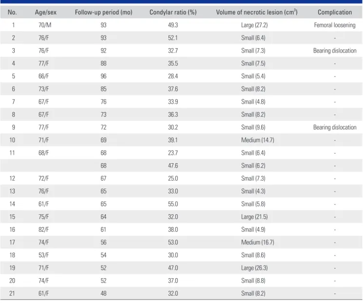
Mont classifi cation. Sixteen knees (72.7%) were lower that 40% of condylar ratio and 6 knees (27.3%) were higher than 40% of condylar ratio. Th e volume of lesion was small in 17 cases (77.3%), medium in 2 cases (9.1%), and large in 3 cases (13.6%) (Table 1).
In all patients, passive full flexion of the knee was possible within 7 days of the operation and painless active full fl exion was possible 2 months aft er the operation. Th e mean HSS score increased from 64.3 (range, 54 to 75) to 92.0 (range, 71 to 100) at the time of the final follow-up (p < 0.05). Th e mean preoperative fl exion contracture was 8.9o (range, 0 to 15), which improved to mean 0.2o (range, 0 to 5) at the fi nal follow-up. Th e mean preoperative full flexion was 138.6o (range, 100 to 145o), which improved to mean 145.6o (range, 140 to 150o) at the fi nal follow-up (p < 0.05). Th e mean preoperative tibiofemoral angle was varus 0.98o and was corrected to mean valgus 3.22o. Th us, the mean correction degree was 4.2o. Th e squatting posi- tion was possible in 16 patients (84.2%) out of 19, except one case of bronchiectasis and one case of spine fracture.
The one case of bronchiectasis could not be checked for squatting due to the intolerable medical condition at the fi nal follow-up. Th e other case of spine fracture could not be checked for squatting because of severe back pain. Th e cross-leg posture was possible in 19 patients (90.5%) out of 21.
Complications occurred in 3 knees of 3 patients (13.6%). Bearing dislocations occurred in 2 cases (9.1%) (Table 1). They returned to the pre-dislocation level of activity with the insertion of thicker bearing. Beneath the tibial components, either complete or partial radiolucent lines were seen in 15 knees out of 22. However, the forma- tion of radiolucent lines was not related with tibial compo- nent loosening. Femoral component loosening occurred in one case (4.5%). This case had a preoperative lesion of 49.3% in condylar ratio and was large in volume (22.8 cm3). It was converted to revision total knee arthroplasty.
Th e survival rate at 6-years was 78%, with revision for any reason as the end point (95% confi dence interval) (Fig. 3).
DISCUSSION
Numerous reports have been published on clinical and radiological outcomes of unicompartmental knee arthro- plasty for treatment of osteoarthritis, while only limited data are available for SPONK. Fewer studies on SPONK indicate that the incidence is less frequent. This study is confi ned to patients who had SPONK limited to the me- dial condyle of the distal femur.
There are several treatment options for managing
SPONK, including high tibial osteotomy, debridement, and autologous bone graft. If other treatments failed or if the stage of SPONK was 3 or higher, the treatment of choice is arthroplasty.4) Arthoplasties of the knee can be either total or unicompartmental. Reports have shown variable results of each method.11,12,17) Marmor6) obtained good clinical results from 30 cases in 34 patients who had unicompartmental knee arthroplasty. However, two pa- tients got revision arthroplasty due to depression around where they had surgery and two patients got revision ar- throplasty due to newly forming lesions in the lateral con- dyle of the femur. Radke et al.12) reviewed 39 cases. Among them, 23 were unicompartmental knee arthroplasty and 16 were total knee arthroplasty. With and average follow- up of more than 5 years, they reported the patients who underwent total knee arthroplasty showed clinically bet- Table 1. Details of Patient Data
No. Age/sex Follow-up period (mo) Condylar ratio (%) Volume of necrotic lesion (cm3) Complication
1 70/M 93 49.3 Large (27.2) Femoral loosening
2 76/F 93 52.1 Small (6.4) -
3 76/F 92 32.7 Small (7.3) Bearing dislocation
4 77/F 88 35.5 Small (7.5) -
5 66/F 96 28.4 Small (5.4) -
6 73/F 85 37.6 Small (8.2) -
7 67/F 76 33.9 Small (4.8) -
8 67/F 73 36.3 Small (8.2) -
9 77/F 72 30.2 Small (9.6) Bearing dislocation
10 71/F 69 39.1 Medium (14.7) -
11 68/F 68 23.7 Small (6.4) -
68 47.6 Small (6.2) -
12 72/F 67 25.0 Small (7.3) -
13 76/F 65 33.0 Small (4.3) -
14 61/F 65 55.0 Small (5.8) -
15 75/F 64 32.0 Large (21.5) -
16 82/F 61 38.0 Small (4.9) -
17 74/F 56 53.0 Medium (16.7) -
18 53/F 54 30.0 Small (8.6) -
19 71/F 52 47.0 Large (26.3) -
20 74/F 52 37.0 Small (8.8) -
21 61/F 48 32.0 Small (8.2) -
Fig. 3. Cumulative rate of survival of the prosthesis was 78% 6 years after operation (95% confi dence interval).
ter results. In patients who underwent unicompartmental knee arthroplasty, four had a revision arthroplasty. How- ever, they reported that the main causes of the poor results of the unicompartmental knee arthroplasty are inadequate operative technique and patient selection. Recently, good clinical results and high long-term survival rates of uni- compartmental knee arthroplasty have been reported due to improvement of surgical technique, component design, and strict selection criteria.11,18) Th erefore, we applied nar- row and strict indications for the patient selection.
Soucacos et al.19) suggested that patients of stages 1 and 2 be treated by conservative management and of stages 3 and 4 be treated with surgery according to the size of the lesion. Lotke et al.13) suggested that surgical treat- ment should be considered when femoral condyle rate is over 50%. Aglietti et al.20) suggested that prognosis is poor when femoral condyle rate is over 40% and the lesion is over 5 cm2. Consequently, most studies state that progres- sion period and lesion size of SPONK are the most impor- tant factors.5,13,19,20) Th e stage of lesion was classifi ed as 4 or 5 using plain radiographs by many authors5,20-22) and there were several methods to measure the size of lesions. Most authors16,20,23) use anteroposterior plain radiograph for measuring the condylar ratio and lateral plain radiograph for measuring the extent of lesion that can be utilized in therapeutic methods and decisions of prognosis. How- ever, Bjorkengren et al.24) recommend that MRI is needed for early diagnosis of spontaneous osteonecrosis because it can determine more about the range of involved bone marrow and damage of cartilage by collapsed bone com- pared with plain radiographs. Th ey reported the utility of MRI to obtain more information about stage and progno- sis in SPONK. Lotke and Ecker25) reported that the deter- mination of the stage of lesion was more precise in MRI than in plain radiographs. We experienced a few cases that were diffi cult to measure because the lesions were super- imposed on the lateral condyle of the femur in lateral plain radiograph. MRI was taken in all cases before surgery and we classifi ed the lesions into 3 groups according to the vol- ume of lesion.
Complications occurred in 3 cases (13.6%) in our study. We experienced one case of femoral loosening.
The volume of this case was 22.8 cm3 and was classi- fi ed as large. Th e lesion extended to the diaphysis and to the epiphysis. Two cases of bearing dislocation occurred which all were changed to thicker bearing. The exact mechanism of bearing dislocation is still unclear. Lewold et al.26) documented that bearing dislocation attributed to malposition of the components and soft tissue imbalance with subsequent maltracking of the meniscal bearing. An- other probable mechanism is posterior impingement by remaining meniscus or osteophytes, ligament laxity due to release of medial collateral ligament, or physiologic laxity of lateral collateral ligament.27) It was assumed that diff er- ent life styles common in Korea, such as full fl exion, squat- ting, and sitting on the fl oor, might cause strain of anterior cruciate ligament, which might be a causative factor of bearing dislocation. Care must be taken for preventing bearing dislocation.
Even though we applied strict indications for patient selection, the survival rate at 6-years of 78% (95% confi - dence interval) was not very high in this study. We thought that it was due to the fact that we could not establish guidelines for total or partial knee replacement. We were also not able to explain the exact mechanism of bearing dislocation or loosening of femoral component. Th e limi- tations of our study were that we retrospectively analyzed the clinical and radiological results of unicompartmental knee arthroplasty in SPONK without a control group of total knee arthroplasty. In addition, the sample size of our study was small.
In conclusion, minimal invasive unicompartmental knee arthroplasty using Oxford Uni could be an alterna- tive treatment option in spontaneous osteonecrosis of the knee.
CONFLICT OF INTEREST
No potential confl ict of interest relevant to this article was reported.
REFERENCES
Instr Course Lect. 2001;50:495-8.
4. Forst J, Forst R, Heller KD, Adam GB. Core decompression in Ahlback's disease: follow-up and therapy control using MR tomography. Rofo. 1994;161(2):142-6.
5. Koshino T. Th e treatment of spontaneous osteonecrosis of the knee by high tibial osteotomy with and without bone- 1. Ahlback S, Bauer GC, Bohne WH. Spontaneous osteone-
crosis of the knee. Arthritis Rheum. 1968;11(6):705-33.
2. Mont MA, Baumgarten KM, Rifai A, Bluemke DA, Jones LC, Hungerford DS. Atraumatic osteonecrosis of the knee. J Bone Joint Surg Am. 2000;82(9):1279-90.
3. Ecker ML. Spontaneous osteonecrosis of the distal femur.
grafting or drilling of the lesion. J Bone Joint Surg Am.
1982;64(1):47-58.
6. Marmor L. Unicompartmental arthroplasty for osteonecro- sis of the knee joint. Clin Orthop Relat Res. 1993;(294):247- 53.
7. Ritter MA, Eizember LE, Keating EM, Faris PM. Th e surviv- al of total knee arthroplasty in patients with osteonecrosis of the medial condyle. Clin Orthop Relat Res. 1991;(267):108- 14.
8. Rozing PM, Insall J, Bohne WH. Spontaneous osteonecrosis of the knee. J Bone Joint Surg Am. 1980;62(1):2-7.
9. Langdown AJ, Pandit H, Price AJ, et al. Oxford medial uni- compartmental arthroplasty for focal spontaneous osteone- crosis of the knee. Acta Orthop. 2005;76(5):688-92.
10. Pandit H, Jenkins C, Barker K, Dodd CA, Murray DW. Th e Oxford medial unicompartmental knee replacement us- ing a minimally-invasive approach. J Bone Joint Surg Br.
2006;88(1):54-60.
11. Myers TG, Cui Q, Kuskowski M, Mihalko WM, Saleh KJ.
Outcomes of total and unicompartmental knee arthroplasty for secondary and spontaneous osteonecrosis of the knee. J Bone Joint Surg Am. 2006;88 Suppl 3:76-82.
12. Radke S, Wollmerstedt N, Bischoff A, Eulert J. Knee arthro- plasty for spontaneous osteonecrosis of the knee: unicom- partimental vs bicompartimental knee arthroplasty. Knee Surg Sports Traumatol Arthrosc. 2005;13(3):158-62.
13. Lotke PA, Abend JA, Ecker ML. Th e treatment of osteone- crosis of the medial femoral condyle. Clin Orthop Relat Res.
1982;(171):109-16.
14. Christensen NO. Unicompartmental prosthesis for gonar- throsis: a nine-year series of 575 knees from a Swedish hos- pital. Clin Orthop Relat Res. 1991;(273):165-9.
15. Carr A, Keyes G, Miller R, O'Connor J, Goodfellow J. Medial unicompartmental arthroplasty: a survival study of the Ox- ford meniscal knee. Clin Orthop Relat Res. 1993;(295):205- 13.
16. Hodge WA, Chandler HP. Unicompartmental knee replace-
ment: a comparison of constrained and unconstrained de- signs. J Bone Joint Surg Am. 1992;74(6):877-83.
17. Mont MA, Rifai A, Baumgarten KM, Sheldon M, Hunger- ford DS. Total knee arthroplasty for osteonecrosis. J Bone Joint Surg Am. 2002;84(4):599-603.
18. Parratte S, Argenson JN, Dumas J, Aubaniac JM. Unicom- partmental knee arthroplasty for avascular osteonecrosis.
Clin Orthop Relat Res. 2007;464:37-42.
19. Soucacos PN, Xenakis TH, Beris AE, Soucacos PK, Geor- goulis A. Idiopathic osteonecrosis of the medial femoral condyle: classifi cation and treatment. Clin Orthop Relat Res.
1997;(341):82-9.
20. Aglietti P, Insall JN, Buzzi R, Deschamps G. Idiopathic os- teonecrosis of the knee: aetiology, prognosis and treatment.
J Bone Joint Surg Br. 1983;65(5):588-97.
21. Aglietti P, Buzzi R. Idiopathic osteonecrosis of the knee. Ital J Orthop Traumatol. 1984;10(2):217-26.
22. Bayne O, Langer F, Pritzker KP, Houpt J, Gross AE. Osteo- chondral allograft s in the treatment of osteonecrosis of the knee. Orthop Clin North Am. 1985;16(4):727-40.
23. Muheim G, Bohne WH. Prognosis in spontaneous osteone- crosis of the knee: investigation by radionuclide scintimetry and radiography. J Bone Joint Surg Br. 1970;52(4):605-12.
24. Bjorkengren AG, AlRowaih A, Lindstrand A, Wingstrand H, Th orngren KG, Pettersson H. Spontaneous osteonecrosis of the knee: value of MR imaging in determining prognosis.
AJR Am J Roentgenol. 1990;154(2):331-6.
25. Lotke PA, Ecker ML. Osteonecrosis of the knee. J Bone Joint Surg Am. 1988;70(3):470-3.
26. Lewold S, Goodman S, Knutson K, Robertsson O, Lidgren L. Oxford meniscal bearing knee versus the Marmor knee in unicompartmental arthroplasty for arthrosis: a Swedish multicenter survival study. J Arthroplasty. 1995;10(6):722- 31.
27. Emerson RH Jr. Unicompartmental mobile-bearing knee arthroplasty. Instr Course Lect. 2005;54:221-4.