Pre-transplant Predictors for 3-Month Mortality after Living Donor Liver Transplantation
Department of Surgery
1, Division of Gastroenterology, Department of Medicine
2, Department of Anesthesiology and Pain Medicine
3, Samsung Medical Center, Sungkyunkwan University School of Medicine, Seoul, Korea
Nuri Lee, M.D.
1, Jong Man Kim, M.D.
1, Choon Hyuck David Kwon, M.D.
1, Jae-Won Joh, M.D.
1, Dong Hyun Sinn, M.D.
2, Joon Hyeok Lee, M.D.
2, Mi Sook Gwak, M.D.
3,
Seung Woon Paik, M.D.
2and Suk-Koo Lee, M.D.
1Background: High model for end-stage liver disease (MELD) scores (≥35) is closely associated with poor posttransplantation out- comes in patients who undergo living donor liver transplantation (LDLT). There is little information regarding factors that negatively impact the survival of patients with high MELD scores. The aim of this study was to identify factors associated with 3-month mortal- ity of patients after LDLT.
Methods: We retrospectively analyzed 774 patients who underwent adult LDLT with right lobe grafts between 1996 and 2012.
Exclusion criteria were re-transplantation, left graft, auxiliary partial orthotopic liver transplantation, and inadequate medical recording. Preoperative variables were analyzed retrospectively.
Results: The overall 3-month survival rate was 92%. In univariate analysis, acute progression of disease, severity of hepatic ence- phalopathy, Child-Pugh class C, hepatorenal syndrome, use of continuous renal replacement therapy, use of ventilator, intensive care unit (ICU) care before transplantation, and MELD scores ≥35 were identified as potential risk factors. However, only ICU care before transplantation and MELD scores ≥35 were independent risk factors for 3-month mortality after LDLT. Three-month and 1-year patient survival rates for those with no risk factors were 95.5% and 88.6%, respectively. In contrast, patients with at least one risk factor had 3-month and 1-year patient survival rates of 88.4% and 81.1%, respectively, while patients with two risk factors had 3-month and 1-year patient survival rates of 55.6% and 55.6%, respectively.
Conclusions: Patients with both risk factors (ICU care before LDLT and MELD scores ≥35) should be cautiously considered for treatment with LDLT.
Key Words: Living donor liver transplantation, End stage liver disease, Model for end stage liver disease, Pretransplant, Mortality
중심 단어: 생체 부분 간이식, 말기 간질환, MELD 점수, 이식전, 사망률Received October 24, 2014 Revised December 8, 2014 Accepted December 16, 2014 Corresponding author: Jae-Won Joh
Department of Surgery, Samsung Medical Center, Sungkyunkwan University School of Medicine, 81 Irwon-ro, Gangnam-gu, Seoul 135-710, Korea
Tel: 82-2-3410-3466, Fax: 82-2-3410-0040 E-mail: [email protected]
First co-author: Nuri Lee, Jong Man Kim
INTRODUCTION
Liver transplantation is the best treatment option for pa- tients with end-stage liver diseases and early hepatocellular carcinoma. In Eastern countries, in which deceased donors are scarce, living donor liver transplantation (LDLT) has been considered as an active alternative option for deceased donor liver transplantation (DDLT). In 2013, 2,286 liver transplantations were performed in Korea and 69% of the procedures were LDLTs, while at the end of the year 6,334
patient were waiting for liver transplants(1).
Living donor hepatectomy is a highly invasive procedure, and the main cause of short term mortality is early graft loss(2). However, the patient survival rate after LDLT has become comparable to the patient survival rate after DDLT because of improvements in surgical techniques such as re- vascularization, biliary tract reconstruction, and improved postoperative management(3,4).
The model for end-stage liver disease (MELD) score was initially described to predict patient survival rates and com- plications after transjugular intrahepatic portosystemic shunt procedures(5). The MELD score was adopted by the United Network for Organ Sharing as the standard priority rule for determining who should receive liver transplants(6).
However, there is no simple means of predicting patient survival in LDLT, although such prediction is a critical step to achieving the most favorable patient outcomes. A high MELD score should not be considered an absolute contra- indication for LDLT, although it is associated with higher postoperative mortality, higher postoperative complication rates, prolonged intensive care unit (ICU) stays, larger in- traoperative blood transfusions, longer hospital stays, and increases in transplant costs(7). Some studies have reported that high MELD scores in LDLT patients are not associated with graft failure or survival rate(8,9). The meaning of MELD scores in LDLT must be reevaluated.
The aim of this study was to determine factors related to 3-month mortality after LDLT using right lobe grafts, and to identify criteria that might be useful for predicting outcomes of LDLT.
MATERIALS AND METHODS
1. Patients
From June 1996 to May 2012, we reviewed the records of 823 patients who underwent primary adult LDLT at Samsung Medical Center, Korea. Patients with ABO in- compatible LDLT (n=22), and using a left lobe grafts in older than or equal to 18 years age (n=15), and auxiliary partial orthotopic liver transplantation (n=2) were excluded.
Therefore, 774 patients were included in this study.
We analyzed data for the recipients, grafts, donors, and intraoperative variables including gender, age, underlying
diseases (hypertension, diabetes mellitus), presence of hep- atoencephalopathy, hepatorenal syndrome), history of pre- operative ICU management, graft-to-recipient weight ratio (GRWR), and MELD score. The indication of ICU manage- ment in end-stage liver disease was following: (1) abnormal pulse rate (<40 or >150/minutes), (2) mean arterial pres- sure <70 mmHg, (3) septic shock, (4) serum bilirubin >6 mg/dL, (5) severe hepatic encephalopathy, (6) required dial- ysis in hepatic renal syndrome, (7) respiratory rate > 30/minutes, or (8) severe metabolic acidosis (pH <7.2). The primary endpoint of the present study was to identify factors associated with 3-month mortality of patients after LDLT.
2. The evaluation and selection of living liver donors Liver donation should be absolutely voluntary. All poten- tial donors underwent a battery of medical evaluations in- cluding an initial health screening survey, laboratory exami- nations including complete blood count, liver and renal bio- chemistry, coagulation profile, and serologic assays for blood transmittable viruses, electrocardiography, chest ra- diography, and pulmonary function test. Psychiatric assess- ments were performed routinely. Doppler ultrasonography was used to evaluate liver quality. Triple-phase abdominal computed tomography (CT) scans were also obtained to cal- culate liver volume and assess vascular anatomy. The pri- mary selection criteria for a living liver donor were ABO blood group compatibility and adequate size of graft liver and future remnant liver as measured by CT scan. Estimated graft volume (GV) greater than 40% of the recipient's standard liver volume was considered acceptable. Our LDLT program limits donor hepatectomy within 70% of the whole donor liver volume. Absolute exclusion criteria were any underlying medical condition that increases perioperative risk and inoperable hepatic vascular variation. When eligi- bility was confirmed, magnetic resonance cholangiopancrea- tography was acquired to verify biliary anatomy(10).
3. Surgical procedures for living donor liver trans- plantation
Intraoperative ultrasonography for evaluating hepatic ve- nous anatomy was performed to determine adequate re- section plane before donor hepatic resection. Parenchymal resection was carried out with a Cavitron ultrasonic surgical
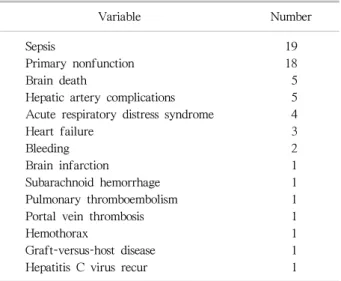
Table 1. Causes of 3-month mortality after living donor liver transplantation
Variable Number
Sepsis 19
Primary nonfunction 18
Brain death 5
Hepatic artery complications 5
Acute respiratory distress syndrome 4
Heart failure 3
Bleeding 2
Brain infarction 1
Subarachnoid hemorrhage 1
Pulmonary thromboembolism 1
Portal vein thrombosis 1
Hemothorax 1
Graft-versus-host disease 1
Hepatitis C virus recur 1
aspirator (Valleylab, Boulder, CO, USA) and by bipolar electrocautery (Codman, Raynham, MA, USA) with the hanging maneuver. After donor hepatectomy, grafts were flushed with 4 L of iced University of Wisconsin solution or histidine tryptophan ketoglutarate solution. Actual graft weights were measured after flushing. Middle hepatic vein (MHV) reconstruction was performed with a cryopreserved iliac artery or iliac vein on the back table when the size of the MHV branch was >5 mm or when a lot of blood gushed out during flushing with perfusion solution. After bench procedure, grafts were transplanted in a piggyback fashion. The orifice of the recipient right hepatic vein was enlarged with a downward incision, and anterior and poste- rior wall excisions were made to form an oval orifice to ob- tain sufficient outflow. After right hepatic vein anastomosis, any significant right inferior hepatic vein was anastomosed to the inferior vena cava in an end-to-side fashion. Portal vein anastomosis was performed with 6-0 Prolene (Ethicon, Bridgewater, NJ, USA) continuous sutures with growth factor. After portal vein anastomosis, arterial reconstruction was performed with 8-0 Ethilon (Ethicon) interrupted su- tures under a surgical microscope. Bile duct reconstruction was performed by either duct-to-duct anastomosis or Roux-en-Y hepaticojejunostomy(11).
4. Immunosuppression protocol
Tacrolimus, steroids, and mycophenolate mofetil (MMF) were the primary agents used for immunosuppression after liver transplantation. All transplant recipients were given 500 mg of intravenous methylprednisolone during the anhe- patic phase until postoperative day 2, tapered to 60 mg per day for a period of 5 days, and received 8 mg, twice per day, for 1 month starting on postoperative day 8.
Tacrolimus treatment was started on postoperative day 3, and the optimal blood level was adjusted to maintain a trough plasma concentration of 10∼15 ng/mL during the first month and reduced to 5∼10 ng/mL thereafter. Starting on postoperative day 1, 750 mg MMF was administered twice a day. MMF was used in combination with tacrolimus and steroids. Cyclosporin (plasma concentration adjusted to 100∼200 ng/mL) was used in the event of tacrolimus tox- icity or tacrolimus refractory rejection, and was given orally twice a day. Liver biopsies were performed if acute re-
jection was clinically suspected. Methylprednisolone (500 mg) was administered intravenously every day for 3 days if acute rejection was confirmed by biopsy and tapered to 60 mg per day over a period of 4 days thereafter(12).
5. Hepatitis B virus prophylaxis
All patients with hepatitis B virus (HBV) infection or re- cipients without hepatitis B surface antigen who received liver allografts with hepatitis B core antibody were given 10,000 units of hepatitis B immunoglobulin (HBIG, Green Cross Corp., Yongin, Korea) intravenously during the anhe- patic phase, followed by a 7-day intravenous course of 10,000 units HBIG per day. Patients received 10,000 units intravenously every month to maintain anti-hepatitis B sur- face antibody titers at ≥200 IU/mL. Before 2008, patients who were reinfected with HBV received only lamivudine (100 mg/day) for treatment. After January 2008, patients received a combination of entecavir (0.5 mg/day) and HBIG for HBV prophylaxis(12).
6. Statistical analysis
Categorical variables were assessed using the chi-square test or Fisher exact test. Continuous variables were ex- pressed as the median and range and were compared using Mann-Whitney U test. The cutoff values of the continuous variables were evaluated using the receiver operating char- acteristic (ROC) curve. Factors with
P
<0.1 in the uni-Table 2. Comparison of patients with and without 3-month mortality after living donor liver transplantation
Variable
3-month mortality
P -value
No (n=711) Yes (n=63)
Gender (male) 561 (79.0) 48 (76.2) 0.630
Recipient age 52 (18∼73) 48 (19∼69) 0.009
Diagnosis Alcoholic HCC HBV HCV NBNC Autoimmune HAV HBV+HCV Others
35 (4.9) 358 (50) 238 (33.5)
13 (1.8) 8 (1.1) 10 (1.4) 5 (0.7) 5 (0.7) 39 (5.5)
3 (4.8) 25 (40) 16 (25.4)
4 (6.3) 0 0 2 (3.2) 1 (1.6) 12 (19.0)
0.000
Progress Acute
Acute on chronic Cirrhosis
33 (4.6) 34 (4.8) 644 (90.6)
10 (15.9) 7 (11.1) 46 (73.0)
0.003
Hypertension 63 (8.9) 10 (15.9) 0.074
Diabetes 136 (19.1) 14 (22.2) 0.511
Child-Pugh class A
B C
86 (12.1) 249 (35.0) 376 (52.9)
5 (7.9) 14 (22.2) 44 (69.8)
0.010
MELD ≥35 73 (10.3) 21 (33.3) 0.000
Coexistence of HCC 359 (50.5) 25 (39.7) 0.115
Hepatic encephalopathy None
Grade 1∼2 Grade 3∼4
503 (70.8) 173 (24.4) 34 (4.8)
34 (54.0) 19 (30.2) 10 (15.9)
0.008
Varix bleeding 180 (25.3) 12 (19.0) 0.361
Ascites None
Diuretics controlled Diuretics uncontrolled
210 (29.5) 293 (41.2) 208 (29.3)
24 (38.1) 25 (39.7) 14 (22.2)
0.124
Hepatorenal syndrome 29 (4.1) 12 (19.0) 0.000
Pretransplant dialysis 10 (1.4) 6 (9.5) 0.001
Pretransplant ventilator care 13 (1.8) 5 (7.9) 0.011
Spontaneous bacterial peritonitis 123 (17.3) 11 (17.5) 0.974
Pretransplant ICU stay 49 (6.9) 24 (38.1) 0.000
GRWR <0.8 60 (8.5) 10 (16.1) 0.061
Data are presented as number (%) or median (range).
Abbreviations: HCC, hepatocellular carcinoma; HBV, hepatitis B virus; HCV, hepatitis C virus; NBNC, non B non C; HAV, hepatitis A virus; MELD, model for end-stage liver disease; ICU, intensive care unit; GRWR, graft-to-recipient weight ratio.
variate analysis were included in the multivariate analyses.
Multivariate analyses used binary logistic regression tests.
Post-transplant survival was estimated using the Kaplan- Meier method with log-rank test. A value of
P
≤0.05 was determined to be statistically significant. Statistical evalua-tion was carried out using the statistical package SPSS ver.
21.0 (IBM Co., Armonk, NY, USA).
Table 3. Risk factors for hospital mortality after living donor liver transplantation by multivariate analysis
Variable Odds
ratio
95% confidence interval P -value Pretransplant ICU stay 8.487 4.674∼15.408 0.000
MELD ≥35 2.090 1.049∼4.164 0.036
Abbreviations: ICU, intensive care unit; MELD, model for end-stage liver disease.
Fig. 1. (A) End-stage liver disease (MELD) scores and (B) patients with pretransplant intensive care unit (ICU) care on patient survival.
RESULTS
1. Baseline characteristics
All recipients received right lobe grafts from living donors. The 774 patients included 609 males and 165 females. The median recipient and donor ages were 51 years (range, 18∼73) and 30 years (range, 18∼64). The median MELD score was 17 (range, 6∼54). Six hundred eighty re- cipients (88%) had MELD scores lower than 35 at the time of transplantation, while 94 patients (12%) had MELD scores above than or equal to 35. Hepatitis B, hepatitis C, alcohol, and hepatocellular carcinoma were the most com- mon causes of LDLT. Seventy-three patients (9.4%) were managed in the ICU prior to liver transplantation and the patients required dialysis (n=16, 2.1%) or mechanical ven- tilation (n=18, 2.3%). The median follow-up period in our study was 46 months (range, 1∼159).
2. Pretransplant risk factors for 3-month mortality Among the 774 patients, 63 patients (8.1%) died by 3 months after transplantation. Sepsis (n=19) and primary non- function (n=18) were main causes for 3-month mortality.
Most common causes of sepsis were biliary problem and fungal pneumonia. The causes included brain death (n=5) and hepatic artery complications (n=5) (Table 1).
Univariate analysis showed that age, diagnosis, disease progression, presence of hepatic encephalopathy, Child- Pugh class C, presence of hepatorenal syndrome, history of pretransplant ventilator care or dialysis, pretransplant ICU stay, and high MELD score were associated with 3-month mortality (Table 2).
Among significant risk factors, according to multivariate analysis, pretransplant ICU stay and high MELD score (≥
35) were predisposing factors for 3-month mortality after LDLT (Table 3). The influences of pretransplant ICU stay and high MELD score are shown in Fig. 1.
We identified risk factors for 3-month mortality in pre- transplant ICU and general ward patients. Patients who re- ceived ICU management before transplantation, had higher 3-month mortality than patients who did not (
P
=0.021 andP
=0.038, respectively). Multivariate analysis showed that high MELD score (odds ratio [OR], 3.368; 95% confidence interval [CI], 1.164∼9.744;P
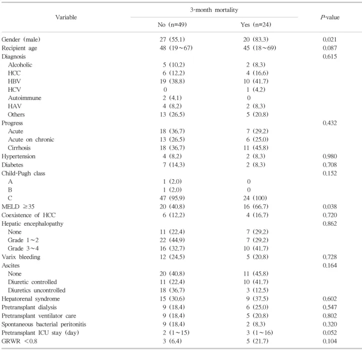
=0.025) was closely associated with 3-month mortality in patients in the pretransplant ICU after LDLT (Tables 4, 5).Table 4. Risk factor for 3-month mortality after living donor liver transplantation in patients requiring pretransplant intensive care
Variable
3-month mortality
P -value
No (n=49) Yes (n=24)
Gender (male) 27 (55.1) 20 (83.3) 0.021
Recipient age 48 (19∼67) 45 (18∼69) 0.087
Diagnosis Alcoholic HCC HBV HCV Autoimmune HAV Others
5 (10.2) 6 (12.2) 19 (38.8)
0 2 (4.1) 4 (8.2) 13 (26.5)
2 (8.3) 4 (16.6) 10 (41.7) 1 (4.2)
0 2 (8.3) 5 (20.8)
0.615
Progress Acute
Acute on chronic Cirrhosis
18 (36.7) 13 (26.5) 18 (36.7)
7 (29.2) 6 (25.0) 11 (45.8)
0.432
Hypertension 4 (8.2) 2 (8.3) 0.980
Diabetes 7 (14.3) 2 (8.3) 0.708
Child-Pugh class A
B C
1 (2.0) 1 (2.0) 47 (95.9)
0 0 24 (100)
0.152
MELD ≥35 20 (40.8) 16 (66.7) 0.038
Coexistence of HCC 6 (12.2) 4 (16.7) 0.720
Hepatic encephalopathy None
Grade 1∼2 Grade 3∼4
11 (22.4) 22 (44.9) 16 (32.7)
7 (29.2) 7 (29.2) 10 (41.7)
0.862
Varix bleeding 12 (24.5) 5 (20.8) 0.728
Ascites None
Diuretic controlled Diuretics uncontrolled
20 (40.8) 11 (22.4) 18 (36.7)
11 (45.8) 10 (41.7) 3 (12.5)
0.164
Hepatorenal syndrome 15 (30.6) 9 (37.5) 0.602
Pretransplant dialysis 9 (18.4) 6 (25.0) 0.547
Pretransplant ventilator care 9 (18.4) 5 (20.8) 0.802
Spontaneous bacterial peritonitis 9 (18.4) 2 (8.3) 0.320
Pretransplant ICU stay (day) 2 (1∼15) 3 (1∼16) 0.052
GRWR <0.8 3 (6.4) 5 (21.7) 0.104
Data are presented as number (%) or median (range).
Abbreviations: HCC, hepatocellular carcinoma; HBV, hepatitis B virus; HCV, hepatitis C virus; HAV, hepatitis A virus; MELD, model for end-stage liver disease; ICU, intensive care unit; GRWR, graft-to-recipient weight ratio.
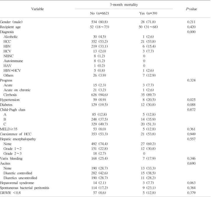
In patients in general ward, the presence of hypertension in patients who died before 3 months after transplantation was higher than survived patients. However, multivariate analysis showed that cirrhosis in disease progression (OR, 0.165; 95% CI, 0.040∼0.681;
P
=0.013) was an independent factor predicting 3-month mortality after LDLT.3. Outcomes for patients with pretransplant ICU care and high MELD score
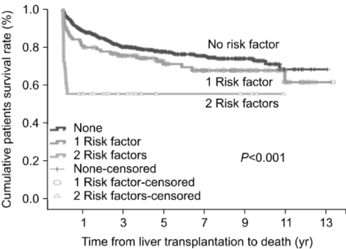
Ninety-five patients (12.3%) had at least one risk factor of those two risk factors analyzed in multivariate analysis.
and 36 patients (4.7%) had two risk factors, pretransplant ICU stay and high MELD score (≥35) both. Most patients (n=643, 83.1%) did not have any risk factors. Those with
Table 5. Risk factors for 3-month mortality after living donor liver transplantation in patients treated in the general ward pretransplant
Variable
3-month mortality
P -value
No (n=662) Yes (n=39)
Gender (male) 534 (80.8) 28 (71.8) 0.211
Recipient age 52 (18∼73) 50 (31∼68) 0.420
Diagnosis Alcoholic HCC HBV HCV NBNC Autoimmune HAV HBV+HCV Others
30 (4.5) 352 (53.2) 219 (33.1) 13 (2.0)
8 (1.2) 8 (1.2) 1 (0.2) 5 (0.8) 26 (3.9)
1 (2.6) 21 (53.8)
6 (15.4) 3 (7.7)
0 0 0 1 (2.6) 7 (12.9)
0.000
Progress Acute
Acute on chronic Cirrhosis
15 (2.3) 21 (3.2) 626 (94.6)
3 (7.7) 1 (2.6) 35 (89.7)
0.324
Hypertension 59 (8.9) 8 (20.5) 0.025
Diabetes 129 (19.5) 12 (30.8) 0.088
Child-Pugh class A
B C
85 (12.8) 248 (37.5) 329 (49.7)
5 (12.8) 14 (35.9) 20 (51.3)
0.872
MELD≥35 53 (8.0) 5 (12.8) 0.361
Coexistence of HCC 353 (53.3) 21 (53.8) 0.949
Hepatic encephalopathy None
Grade 1∼2 Grade 2∼3
492 (74.4) 151 (22.8) 18 (2.7)
27 (69.2) 12 (30.8)
0
0.557
Varix bleeding 168 (25.4) 7 (17.9) 0.346
Ascites None
Diuretic controlled Diuretics uncontrolled
190 (28.7) 282 (42.6) 190 (28.7)
13 (33.3) 15 (38.5) 11 (28.2)
0.690
Hepatorenal syndrome 14 (2.1) 3 (7.7) 0.063
Spontaneous bacterial peritonitis 114 (17.2) 9 (23.1) 0.384
GRWR <0.8 57 (8.6) 5 (12.8) 0.379
Data are presented as number (%) or median (range).
Abbreviations: HCC, hepatocellular carcinoma; HBV, hepatitis B virus; HCV, hepatitis C virus; NBNC, non B non C; HAV, hepatitis A virus; MELD, model for end-stage liver disease; GRWR, graft-to-recipient weight ratio.
no risk factors had 95.5% 3-month patient survival rate respectively. In contrast, patients with at least one risk fac- tor had 88.4% 3-month patient survival rates respectively, while patients with two risk factors had 55.6% 3-month pa- tient survival respectively (Fig. 2). These differences in pa- tient survival rates were statistically significant (
P
<0.001).DISCUSSION
In the present study, we aimed to determine character- istics of transplant recipients that might be useful for pre- dicting 3-month mortality after LDLT and that would im- pact the selection of patients for LDLT. Our results suggest that MELD scores can predict postoperative survival in pa-
Fig. 2. Patient survival with two risk factors which were high End-stage liver disease score (≥35) and pretransplant intensive care unit care when compared with those who had at least one risk factor or no risk factor.
tients with pretransplant ICU stays. Patients with two risk factors, such as ICU care before LDLT and MELD scores
≥35, had extremely poor 3-month survival. However, MELD scores were not associated with poor outcomes in pa- tients who were in the general ward before LDLT.
Using preoperative MELD scores to predict posttransplant outcomes after LDLT is controversial. Some studies have suggested that high MELD scores are associated with poor outcomes, whereas others reported that MELD scores had no prognostic value(8,13). While it is unclear if MELD scores are useful for predicting liver transplantation out- comes, the MELD score relies on objective laboratory data that reflects severity of illness in patients with liver disease.
It is generally accepted that live liver donation should be prohibited in recipients with MELD scores over 25(8,14).
However, some studies have raised questions about the im- pact of MELD scores on short-term outcomes after right lobe LDLT(8,13,15).
One study reported that high MELD scores (≥25) were associated with prolonged postoperative ICU stays and in- creased hospital costs but not with postoperative mortal- ity(13). Another showed that recipients of liver donor liver grafts with high MELD scores (≥25) had excellent outcomes. They had increased rates of postoperative pulmo- nary infections, but similar rates of graft function, post- operative graft injury, overall postoperative complications, length of hospital stay, short-term and long-term graft sur-
vival, and patient survival(8).
Most of these studies divide the patient population into two groups according to MELD score (< or >25), and sug- gest that MELD scores lack predictive power for short-term outcomes after LDLT(8,13). We also found that MELD scores ≥25 had no impact on 3-month mortality in patients who underwent right lobe LDLT. In the present study, the cutoff point on ROC curve was move to 35 from 25.
We found that MELD scores ≥35 were useful for pre- dicting the 3-month mortality following LDLT. However, high MELD scores above than or equal to 35 alone should not be an absolute contraindication of LDLT. Another pre- vious study reported that patients with high MELD scores had significantly more early postoperative complications, but comparable hospital mortality, graft survival, and overall survival compared to patients with low MELD scores(16).
A recent study reported that high MELD scores and ad- vanced donor age were associated with graft survival. In ad- dition, these variables had the highest sensitivity for predicting in-hospital mortality(17). However, our results did not sup- port an association between 3-month mortality and donor age.
High MELD score patients may not be suitable candidates for LDLT because of the need for greater liver mass and low tolerance to postoperative complications(18). DDLT may be indicated for recipients in very poor condition, elim- inating concerns about risk to the donor. LDLT for sicker patients is controversial due to ethical issues, and DDLT with whole liver graft transplantation is recommended as the best option for sicker patients compared with split or LDLT(19). Nevertheless, LDLT should be considered for patients with high MELD scores or those requiring intensive care to survival in situations when the use of liver grafts from deceased donor is limited. The decision to undertake LDLT can be difficult when available living-donor grafts are marginal and the recipient is judged to be at high risk of complications. LDLT should be performed only if the risk to the donor is justified by the expectation of an ac- ceptable outcome for the recipient(17,20). The balance of ethical issues needs to be considered in LDLT.
Preoperative renal dysfunction in patients with end-stage cirrhosisis common and ranges from 10%∼20% among pa- tients who undergo liver transplantation(21). Previous stud- ies suggested that preoperative renal dysfunction was asso-
ciated with a high incidence of infection, long ICU stay, and long hospital stay(22). However, in the present study, hep- atorenal syndrome or dialysis in the pretransplant patient was associated with 3-month mortality, but not with 3-month mortality according to multivariate analysis.
Small-for-size grafts was not a risk factor for 3-month mortality in the present study. All institutions at which LDLT is performed have lower limits for donated GV that include the following safety margin: GRWR >0.6 to 0.8 for transplanted GV(11). However, patients with small-for- size grafts may have poor outcomes in patients when they have high MELD scores(23).
This study includes the largest series of adult LDLT re- cipients with high MELD scores ever examined, and our re- sults suggest that the optimal cutoff point on ROC curve for MELD scores should move to 35 from 25. We identified high MELD scores (≥35) and preoperative ICU care as risk factors. High MELD scores should not be an absolute con- traindication for LDLT, but when patients with MELD scores above than or equal to 35 required ICU care, DDLT should be considered prior to LDLT as a treatment strategy.
The preoperative ICU management is one of the major risk factors in this study; however, other several risk factors as hepatic encephalopathy, hepatorenal syndrome, dialysis, and ventilator care have associated with pretransplant con- dition which demand on ICU management. Therefore, our study should be considered of these issues. Further studies are needed to reevaluate the risk factors associated with seri- ous patient condition after divide into two subgroups for MELD scores above than or equal to 35, and lower than 35.
In addition, in this study we analyzed 3-month mortality as a single end-point; however, further studies are needed to collect data on multiple end point. For example, the mor- tality related to pretransplant condition (sepsis and brain death), to procedure (presence of hepatic artery thrombosis, portal vein thrombosis, and bleeding), and to extrahepatic causes (heart failure, cerebrovascular event).
CONCLUSION
In conclusion, high MELD score alone is not an absolute contraindication for LDLT. We identified MELD score greater than or equal to 35 and preoperative ICU stay as
clinical risk factors for short-term mortality within 90 days after LDLT. We found that patients with both risk factors have extremely poor 3-month survival and should therefore be cautiously considered as candidates for LDLT.