• 검색 결과가 없습니다.

Non-Odontogenic Toothache Caused by the Fungal Ball of Maxillary Sinus: Case Reports JOMP

N/A
N/A
Protected

Academic year: 2021

Share "Non-Odontogenic Toothache Caused by the Fungal Ball of Maxillary Sinus: Case Reports JOMP"

Copied!
5
0
0

로드 중.... (전체 텍스트 보기)

전체 글

(1)

pISSN 2288-9272 eISSN 2383-8493 J Oral Med Pain 2019;44(4):174-178 https://doi.org/10.14476/jomp.2019.44.4.174

Non-Odontogenic Toothache Caused by the Fungal Ball of Maxillary Sinus: Case Reports

Ji-Woo Ha

1

, Won Jung

2

, Kyung-Eun Lee

2

, Bong-Jik Suh

2

1

Department of Oral Medicine, School of Dentistry, Jeonbuk National University, Jeonju, Korea

2

Department of Oral Medicine, Institute of Oral Bioscience, School of Dentistry, Jeonbuk National University, Jeonju, Korea

Received November 23, 2019 Revised December 10, 2019 Accepted December 10, 2019

A fungal ball (FB) of the paranasal sinuses is a chronic, non-invasive fungal sinusitis defined as the accumulation of dense aggregation of fungal hyphae in a sinus cavity. A patient with FB infection in a sinus cavity has usually non-specific symptoms such as post-nasal drip, nasal congestion, headache. However, facial pain and toothache can be developed if FB infection is in maxillary sinus. The aim of this case report is to present two cases of FB of the maxillary sinus which caused toothache in the upper molar region. It is also to make dental practitioners consider the non-odontogenic origins of toothache and to pay special attention to avoid unnecessary dental treatment.

Key Words: Chronic sinusitis; Fungal ball; Non-odontogenic toothache

Correspondence to:

Bong-Jik Suh

Department of Oral Medicine, School of Dentistry, Jeonbuk National University, 567 Baekje-daero, Deokjin-gu, Jeonju 54896, Korea

Tel: +82-63-250-2060 Fax: +82-63-250-2058 E-mail: [email protected]

https://orcid.org/0000-0002-1817-4645

JOMP

Journal of Oral Medicine and Pain

Copyright

2019 Korean Academy of Orofacial Pain and Oral Medicine. All rights reserved.

CC
This is an open-access article distributed under the terms of the Creative Commons Attribution Non-Commercial License (http://creativecommons.org/licenses/by-nc/4.0/), which permits unrestricted non-commercial use, distribution, and reproduction in any medium, provided the original work is properly cited.

INTRODUCTION

Orofacial pain can be a common clinical problem [1].

A total of 42% of the general population experienced one or more types of orofacial pain in the past 6 months.

Toothache was the most common pain which was reported by 26.8% of the population experienced orofacial pain [2].

However, orofacial pain can be a diagnostic dilemma for dental practitioners in clinical practice because most of the pain in the orofacial region mimics odontogenic pain and sometimes the site of pain might not be coincident with the origin of pain. In these regards, appropriate diagnosis and management were challenges for dentists [1,3].

Most toothaches are originated from pulpal or adjacent periodontal tissues. The orofacial pain from dental origin was termed “odontogenic toothache” [4]. “Non-odontogenic toothache” defined as the pain which is perceived on pulpal and adjacent structure but not originated from the tooth or periodontal tissues [4]. If clinical and radiographic exami- nations did not reveal any pathologic findings in teeth or

periodontal structures, nonodontogenic origin should be considered [5,6]. Nonodontogenic toothache can be caused by a variety of causes such as muscle disorders, sinus and nasal mucosal problems, neuropathic pain, neurovascu- lar pain, psychogenic problem, or cardiogenic disease [5].

According to Linn et al. [7], 44 out of 100 patients who suf- fered from non-odontogenic orofacial pain had either tooth extraction or root canal treatment in an attempt to resolve their pain. If the origin of toothache was misdiagnosed, un- necessary dental treatment such as root canal treatment or extractions could be performed and the patient’s pain would not be alleviated through such treatment. Therefore, the origin of toothache should be found correctly before dental treatment is performed [5]. Further tests and consul- tation with other medical specialties have to be performed to find the origin of a toothache.

The objective of this case report is to present uncommon

cases of two patients who had the nonodontogenic tooth-

ache caused by fungal ball (FB) of the maxillary sinus.

(2)

CASE REPORT

1. Case 1

A 41-year-old female referred from the department of conservative dentistry at the Jeonbuk National University Hospital with a chief complaint of persisted toothache on her maxillary left molar area. Her past history revealed that her toothache started about two years ago. She visited a dental clinic and dental caries was found on the first and second premolar of left upper dentition.

Although root canal treatment was done in a dental clin- ic, the symptoms were not improved. She visited the depart- ment of conservative dentistry for the surgical treatment on the first and second premolar of left maxillary dentition.

However, toothache on the molar area was not resolved al- though apicoectomy was finished.

Clinical examination was performed and findings were including tenderness to palpation on multiple sites with both sides of face, neck, and shoulder. There was no mouth opening limitation. Panoramic and periapical radiograph showed no specific findings (Fig. 1).

She was diagnosed with myofascial pain with referral and toothache was considered as referred pain from myofas- cial pain. The patient was treated with conservative treat- ment including patient education, physical therapy, and oral appliance. The symptoms of myofascial pain had been improved about a year later, but toothache had been re- mained. Other reasons for toothache should be considered.

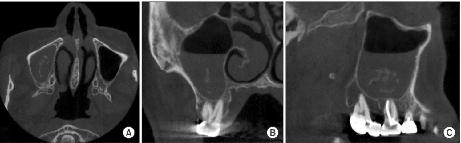
Cone beam computed tomography (CBCT) taken before she visited our department, revealed moderate mucosal thickening including small calcified bodies of the left max- illary sinus (Fig. 2). She was referred to the department of otorhinolaryngology for the treatment of the left maxillary sinus. It was diagnosed with FB from endoscopic features and histopathological findings after surgical removal under general anesthesia. Her toothache was disappeared about two months later.

2. Case 2

A 62-year-old female with a chief complaint of toothache in the right upper molar area was referred to our depart- ment from the department of conservative dentistry.

She had toothache in the right upper molar area about five months ago. Root canal re-treatment was treated on first premolar, second premolar and first molar of right

Fig. 1. Panoramic view shows no remarkable findings.

Fig. 2. Cone beam computed tomography view shows moderate mucosal thickening including small calcified bodies of the left maxillary sinus.

(A) Axial view, (B) coronal view, (C) sagittal view.

A B C

(3)

upper dentition at a dental clinic. However, toothache was not disappeared. She referred to the department of conser- vative dentistry at Jeonbuk National University Hospital.

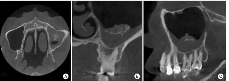
Although panoramic and periapical radiograph showed no specific lesions and findings, CBCT showed fungal sinus- itis in the right maxillary sinus (Fig. 3, 4). She was referred to the department of otorhinolaryngology for treatments of the right maxillary sinus. It was also diagnosed to FB in- fection from nasal endoscopic features and histopathologi- cal findings after surgical removal under general anesthesia (Fig. 5).

She visited our department about 3 weeks later after the surgery because of persisting toothache on the right upper molar area during mastication and uncomfortable sensation on the right middle face area.

Clinical examination showed palpation pain on both masseter muscles and first premolar, second premolar and first molar of right upper dentition sensitivity to percussion.

The patient was diagnosed as local myalgia and me dication,

physical therapy and patient education were given. We had planned to monitor the progress of the main symptoms over time. Her toothache got gradually declined two months lat- er after the surgery.

DISCUSSION

FB of the paranasal sinuses is a non-invasive form of fungal sinusitis defined as the accumulation of dense con- glomeration of fungal hyphae in sinus cavities [8,9].

Although it was considered relatively rare in the past, the number of patients has increased over the past two decades [9].

It occurs predominantly in older people and more fre- quently observed in females. The mean age of the patients

Fig. 3. Panoramic view shows no remarkable findings.

A B C

Fig. 4. Cone beam computed tomography view shows fungal sinusitis in the right maxillary sinus. (A) Axial view, (B) coronal view, (C) sagittal view.

Fig. 5. Aggregates of highly packed hyphae are observed in micro-

scopic examination (H&E staining, ×400).

(4)

was 53.6 and the ratio of female patients was 62.5% [10].

Klossek et al. [11] reported that most of FB can be found in a single sinus of patients. FB is most frequently occurred in the maxillary sinus followed by the sphenoid sinus [10].

The pathogenesis of FB remains unexplained. No predis- posing environmental or local anatomical factors have been demonstrated although most of the patients are usually immunocompetent [8]. However, two pathways of fungal entry have been suggested. An aerogenic pathway theory explains that high quantities of the airborne spores can en- ter the sinus and turn to be pathogenic when the sinus be- came anaerobic. Another pathway of fungal colonization in which occurs in the maxillary sinus is the iatrogenic oro- antral communication caused by dental extraction or root canal treatment [8].

FB is known to be asymptomatic in the early stage and developed slowly. It can be diagnosed incidentally in as- ymptomatic patients by computed tomography or magnetic resonance imaging for head examination in a part of the diagnosis of other diseases [8,12]. At the later stages of the disease, it causes non-specific clinical symptoms of chronic rhinosinusitis including post-nasal drip, nasal congestion, facial pain, headache, toothache, and retro-orbital pain [12].

In our cases, the patients complained of toothache in the upper molar area. Fungal sinusitis of maxillary sinus was incidentally found from CBCT images performed to find odontogenic causes.

The radiological findings of FB are quite specific.

Heterogenic densities with focal dense calcification could be found in conventional tomography (CT). The calcified bod- ies of the FB observed at CT are caused by calcium deposits from metabolic reactions of fungal mass [10,13].

Yoon et al. [14], reported that 21 of 39 patients with fun- gal sinusitis had intra-sinus calcifications, while 16 of 471 patients with nonfungal sinusitis had it. The location of cal- cification was mainly central of the maxillary sinus (95%) in fungal sinusitis and the calcification had irregular mar- gins with fine punctate, linear or nodular form. Smooth- margined calcification was only seen in nonfungal sinusitis [14].

Histopathological and mycological examinations are also needed to establish the diagnosis [13]. Microscopically, ag- gregates of highly packed hyphae can be observed and the

sinus mucosa shows non-specific inflammation [8]. It has to be confirmed no invasion of the sinus mucosa or bone by fungi in order to have a positive diagnosis of the FB [13].

Fungal cultures show a relatively poor yield. However, it is recommended to perform in every case to identify the causative fungal species, as complementary to special stain- ing techniques [8].

Functional endoscopic sinus surgery with a canine fossa approach is the treatment of choice [8,9]. Most patients may not need further antifungal treatment and rare recurrences had been reported [8,15].

In these present cases, toothache was alleviated after FB removal surgery. As mentioned earlier, it could cause non- specific clinical symptoms including facial pain, and tooth- ache at the later stages of the disease [12].

Similar to cases we reported, Yoon et al. [9] reported that 6.1% of patients (29 out of 477) had toothache caused by the FB of the maxillary sinus. Ahn et al. [12] reported that toothache was found in 3.8% of patients (1 out of 26) in the bilateral FB group and 3.9% of patients (16 out of 408) in the unilateral FB group. Sinusitis caused by FB is thought to induce toothache [8,9].

The exact mechanism of toothache caused by sinusitis was not fully understood, in so far. Toothache frequent- ly occurs in the maxillary premolar and molar regions in acute sinusitis, whereas chronic sinusitis usually does not accompany toothache [16,17]. In contrary to general be- lief, chronic sinusitis seldom induces facial pain or head- ache. Sinus lining mucosa is known to be nearly insensible to pain. Referred spontaneous toothache does not arise as a result of sinus inflammation. A condition that causes tooth- ache in sinusitis was suggested that neuritic neuralgia could occur by inflammation of superior alveolar plexus [18]. The neuritic symptoms occur in the peripheral distribution of the affected nerve branches because the dental nerve often lies just below the lining mucosa or are separated by a very thin bone. These conditions may cause pain in the ipsilat- eral maxillary teeth [18].

As we mentioned before, nonodontogenic toothache

could be caused by a variety of causes, including fungal si-

nusitis of the maxillary sinus. Therefore, dental practitioners

should consider the nonodontogenic toothache and avoid

performing unnecessary dental treatment such as root canal

(5)

treatment or extractions when clinical and radiographic findings did not reveal dental origin.

CONFLICT OF INTEREST

No potential conflict of interest relevant to this article was reported.

ORCID

Ji-Woo Ha

https://orcid.org/0000-0002-6708-796X Won Jung

https://orcid.org/0000-0002-8728-8013 Kyung-Eun Lee

https://orcid.org/0000-0001-8923-1478 Bong-Jik Suh

https://orcid.org/0000-0002-1817-4645

REFERENCES

1. Soni A. Toothache of non-dental origin: a review of its mecha- nism and clinical characteristics. Int J Sci Study 2018;6:26-35.

2. Chung JW, Kim JH, Kim HD, Kho HS, Kim YK, Chung SC. Chron- ic orofacial pain among Korean elders: prevalence, and impact using the graded chronic pain scale. Pain 2004;112:164-170.

3. Balasubramaniam R, Turner LN, Fischer D, Klasser GD, Oke- son JP. Non-odontogenic toothache revisited. Open J Stomatol 2011;1:92-102.

4. Kim KM, Byun JS, Jung JK, Choi JK. Non-odontogenic toothache caused by acute maxillary sinusitis: a case report. J Oral Med Pain 2016;41:80-84.

5. Kang JK, Ryu JW. Nonodontogenic toothache. Oral Biol Res

2018;42:241-247.

6. Mascia P, Brown BR, Friedman S. Toothache of nonodontogenic origin: a case report. J Endod 2003;29:608-610.

7. Linn J, Trantor I, Teo N, Thanigaivel R, Goss AN. The differential diagnosis of toothache from other orofacial pains in clinical prac- tice. Aust Dent J 2007;52(1 Suppl):S100-S104.

8. Grosjean P, Weber R. Fungus balls of the paranasal sinuses: a re- view. Eur Arch Otorhinolaryngol 2007;264:461-470.

9. Yoon YH, Xu J, Park SK, Heo JH, Kim YM, Rha KS. A retrospec- tive analysis of 538 sinonasal fungus ball cases treated at a single tertiary medical center in Korea (1996-2015). Int Forum Allergy Rhinol 2017;7:1070-1075.

10. Pinar E, Imre A, Ece AA, et al. Paranasal sinus fungus ball:

analysis of clinical characteristics and surgical outcomes. ENT Updates 2015;5:124-127.

11. Klossek JM, Serrano E, Péloquin L, Percodani J, Fontanel JP, Pessey JJ. Functional endoscopic sinus surgery and 109 myceto- mas of paranasal sinuses. Laryngoscope 1997;107:112-117.

12. Ahn SH, Lee EJ, Hong MP, Shin GC, Kim KS. Comparison of the clinical characteristics of bilateral and unilateral fungal balls in Korea. Eur Arch Otorhinolaryngol 2019;276:1975-1980.

13. Patrascu E, Manea C, Sarafoleanu C. Difficulties in the diag- nosis of fungal rhinosinusitis – literature review. Rom J Rhinol 2016;6:11-17.

14. Yoon JH, Na DG, Byun HS, Koh YH, Chung SK, Dong HJ. Cal- cification in chronic maxillary sinusitis: comparison of CT findings with histopathologic results. AJNR Am J Neuroradiol 1999;20:571-574.

15. Aribandi M, McCoy VA, Bazan C 3rd. Imaging features of inva- sive and noninvasive fungal sinusitis: a review. Radiographics 2007;27:1283-1296.

16. Yatani H, Komiyama O, Matsuka Y, et al. Systematic review and recommendations for nonodontogenic toothache. J Oral Rehabil 2014;41:843-852.

17. Hansen JG, Højbjerg T, Rosborg J. Symptoms and signs in cul- ture-proven acute maxillary sinusitis in a general practice popu- lation. APMIS 2009;117:724-729.

18. Okeson JP. Bell’s oral and facial pain. 7th ed. Chicago: Quintes-

sence Publishing Co, Inc; 2014. pp. 384, 458-459

수치

Fig. 2. Cone beam computed tomography view shows moderate mucosal thickening including small calcified bodies of the left maxillary sinus
Fig. 5. Aggregates of highly packed hyphae are observed in micro- micro-scopic examination (H&E staining, ×400).

참조

관련 문서

_____ culture appears to be attractive (도시의) to the

After first field tests, we expect electric passenger drones or eVTOL aircraft (short for electric vertical take-off and landing) to start providing commercial mobility

1 John Owen, Justification by Faith Alone, in The Works of John Owen, ed. John Bolt, trans. Scott Clark, "Do This and Live: Christ's Active Obedience as the

The thickness of the maxillary sinus lateral wall according to tooth site and measurement level was measured by using image-processing software and its histologic

With the increase of the dental implant procedure, there are many cases of Maxillary Sinus Floor Elevation Surgery(MSFES). Additionally, its side effect has

Methods to overcome insufficient bone due to poor bone quality, the pneumatization of a maxillary sinus and other anatomical limitations of implant placement

Thirdly, it appeared that social support had meaningful effect on self-efficacy and the quality of life, and in the case of the difference analysis by

The OSFE (osteotome sinus floor elevation) technique has been used for maxillary sinus augmentation.. The implants were clinically and radiographically followed