http://dx.doi.org/10.5090/kjtcs.2014.47.4.358 ISSN: 2233-601X (Print) ISSN: 2093-6516 (Online)
1
Department of Thoracic and Cardiovascular Surgery, Chonbuk National University Medical School,
2Research Institute of Clinical Medicine of Chonbuk National University and Biomedical Research Institute of Chonbuk National University Hospital
†This article was presented at the 45th Autumn Academic Meeting of the Korean Society for Thoracic and Cardiovascular Surgery.
Received: November 25, 2013, Revised: February 11, 2014, Accepted: February 17, 2014, Published online: August 5, 2014
Corresponding author: Jong Hun Kim, Department of Thoracic and Cardiovascular Surgery, Chonbuk National University Medical School, 567 Baekje-daero, Deokjin-gu, Jeonju 561-756, Korea
(Tel) 82-63-250-1485 (Fax) 82-63-250-1480 (E-mail) [email protected]
C
The Korean Society for Thoracic and Cardiovascular Surgery. 2014. All right reserved.
CC
This is an open access article distributed under the terms of the Creative Commons Attribution Non-Commercial License (http://creative- commons.org/licenses/by-nc/3.0) which permits unrestricted non-commercial use, distribution, and reproduction in any medium, provided the original work is properly cited.
Outcome of Concomitant Cox Maze Procedure with Narrow Mazes and Left Atrial Volume Reduction
Jong Bum Choi, M.D.
1,2, Jong Hun Kim, M.D.
1,2, Byong Ki Cha, M.D.
1,2Background: To improve sinus rhythm conversion, the Cox maze III procedure with narrow mazes (width: ≤3.0 cm) was performed in combination with left atrial volume reduction. Methods: From October 2007 to April 2013, 87 patients with atrial fibrillation (paroxysmal in 3, persistent in 14, and permanent in 70) underwent the Cox maze procedure concomitant with another cardiac procedure. They were followed-up with serial electrocardiographic and echocardiographic studies. We used 24-hour Holter monitoring tests to evaluate postoperatively symptomatic patients.
Results: At the mean follow-up time of 36.4 months, 81 patients (94.2%) had sinus rhythm and two were on an- ti-arrhythmic medication (one on a beta-blocker and the other on amiodarone). Five patients (5.8%) with post- operative recurrent and persistent atrial fibrillation never experienced sinus rhythm conversion; however, they did not require any medication for rate control. On postoperative echocardiography, the left atrial A waves were more fre- quently observed after concomitant mitral valve repair than after concomitant mitral valve replacement (82.4% vs.
40.4%, respectively; p<0.001). Conclusion: For the Cox maze procedure, narrow mazes and atrial volume reduc- tion resulted in excellent sinus rhythm conversion without the preventive use of anti-arrhythmic drugs, and they did not affect the presence of the left atrial A waves on echocardiography.
Key words: 1. Arrhythmia surgery 2. Technique
3. Outcome assessment
INTRODUCTION
Atrial fibrillation (AF) is present in up to 50% of the pa- tients undergoing mitral valve surgery and in 1% to 6% of the patients undergoing coronary artery bypass grafting or aortic valve surgery [1-3]. Since the introduction of the maze procedure for the treatment of AF in 1987 [4], it has been simplified by replacing the maze incisions with cryoablation and radiofrequency ablation [5]. Although some randomized trials have demonstrated an excellent conversion rate of sinus
rhythm with radiofrequency ablation [6], 20% to 30% of the patients undergoing the maze procedure have exhibited re- current AF during the follow-up period [7].
Among the maze procedures that were performed in the
same fashion, the dimensions of the mazes differ widely in
accordance with the left atrial sizes. However, maze di-
mensions are an important factor for successful sinus rhythm
conversion, interrupting reentrant circuits in the left atrium
(LA). We set the maze dimension at ≤3.0 cm in all patients
requiring the maze procedure. The enlarged LA was reduced
Paroxysmal/persistent/permanent AF Left ventricular ejection fraction (%) Previous cerebro-vascular accident Redo-valve surgery
Mitral valve lesion MS dominant MR dominant
Double- and triple-valve disease Non-valve lesion
3/14/70 (3.4/16.1/80.5) 55.8±6.7
5 (5.7) 3 (3.4) 78 (89.5) 44 (56.4) 34 (43.6) 25 (28.7) 3 (3.4)
Values are presented as mean±standard deviation, number (%), or median (range).
AF, atrial fibrillation; MS, mitral stenosis; MR, mitral regurgi- tation.
a)
Left atrial dimension>60 mm.
Atrial septal defect Ventricular septal defect No. of procedures
2 3 4 5
Left atrial reduction plasty Left atrial appendage excision Isthmus resection
Left atrial lateral wall plication
1 (1.1) 1 (1.1)
11 (12.6) 48 (55.5) 25 (28.7) 3 (3.4)
87 (100.0) 34 (39.1) 9 (10.3) Values are presented as number (%).
by the resection of the redundant inferior wall of the LA with and without plication of the posterior walls, left lateral wall, or superior walls of the LA. The aim of this study is to evaluate the efficacy of narrow mazes and left atrial reduction in the Cox maze procedure.
METHODS
This study was approved by Chonbuk National University Hospital institutional review board. Each patient included in these analyses provided informed consent.
1) Patients and combined surgery
From October 2007 to April 2013, 87 patients (34 men and 53 women; mean age, 59.5±10.0 years) with paroxysmal (n=3.4%), persistent (n=14, 16.1%), or permanent AF (n=70, 80.5%) underwent the Cox maze procedure concomitant with another cardiac operation (Table 1). The primary surgical in- dication was structural heart disease in all patients. Mitral valve disease was observed in 78 patients (89.5%); 41
(47.5%) underwent mitral valve replacement, and 37 (42.5%) underwent mitral valve repair. Non-valve cardiac disease was present in three patients (3.4%); one each with myxoma, a huge left atrial thrombus, and an ascending aortic aneurysm.
Twenty-five patients (28.7%) underwent multi-valve surgery.
The mean duration of AF was 58.1±89.2 months (range, 2 to 500 months).
The mean atrial dimension was 55.8±9.7 mm (range, 31 to 85 mm) in the two-dimensional parasternal long-axis view.
Left atrial reduction plasty by an isthmus excision was per-
formed in 34 patients (39.1%); nine (10.3%) required addi-
tional plication of the posterior wall, the left lateral wall, and
the superior wall of the enlarged LA (Table 2). Sixty-nine
patients (79.3%) underwent tricuspid valve repair. Of these,
three (3.4%) underwent isolated tricuspid valve repair and
right atrial reduction plasty. All data were retrospectively ana-
lyzed from the medical records, including the operative re-
cords, standard 12-lead electrocardiograms, and transthoracic
and transesophageal echocardiograms.
Fig. 1. The Cox maze procedure. Epicardial view of the atria and endocardial view of the right atrium. Both atrial appendages were amputated and narrow mazes (maze width<3.0 cm) were made in both atria. Thick single-dotted lines represent ‘cut-and-sew’
lesions; thick double-dotted lines represent cryoablation lines; and fine single-dotted lines represent ablation lines that were made us- ing a bipolar radiofrequency electrode. MV, mitral valve; TV, tri- cuspid valve; LAA, left atrial appendage; RAA, right atrial append- age; SVC, superior vena cava; IVC, inferior vena cava; FO, fora- men ovale.
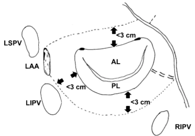
Fig. 2. A narrow maze in the left atrium. The ablation lines cir- cumscribing the pulmonary veins were made ≤3.0 cm away from the mitral valve annulus in all directions. LSPV, left superior pul- monary vein; LAA, left atrial appendage; LIPV, left inferior pulmo- nary vein; AL, anterior leaflet; PL, posterior leaflet; RIPV, right in- ferior pulmonary vin.
2) Surgical procedures
(1) Maze procedure: We performed the Cox maze proce- dure for all the study patients by using a combination of ra- diofrequency and cryoablation lesions. To make precise abla- tion lesions, a bipolar radiofrequency electrode (Medtronic Inc., Minneapolis, MN, USA) was applied for an additional 40 seconds after each lesion did not conduct electricity. A cryoprobe (Cooper Surgical, Trumbull, CT, USA) was applied for 90 seconds per lesion (Fig. 1) [8]. A left atriotomy in- cision was made through Sondergaard’s groove, and a vertical right atriotomy was made at the bottom 1/3 of the right atrium. After both atrial appendages were amputated, a box lesion circumscribing four pulmonary veins was made through the left atriotomy and the left atrial appendage opening, using a bipolar radiofrequency electrode, and four more radio- frequency lesions were made in the right atrial wall, the pos- terior septal wall, and the superior and inferior sinuses. Two cryoablation lesions were made on the right atrial wall: one between the right atriotomy and the tricuspid annulus, and the other between the right atrial appendage opening and the tri- cuspid annulus. Two additional cryoablation lesions were
made on the inner and the outer LA walls between the in- ferior end of the left atriotomy and the mitral annulus, in- cluding the coronary sinus. The left atrial appendage opening was externally closed with a continuous 4-0 polypropylene suture and reinforced with 3 to 4 interrupted pledgeted 4-0 polypropylene sutures.
The radiofrequency box lesions circumscribing four pulmo- nary veins were made within 3.0 cm of the mitral annulus in all directions (Fig. 2). In the right atrial wall and the in- teratrial septum, the distances between the ablation lines were also ≤3.0 cm.
(2) Atrial reduction plasty: The redundant inferior wall of
the enlarged LA (LA dimensions: >50–55 mm) was excised
just above the coronary sinus and then re-sutured using con-
tinuous 4-0 polypropylene sutures. For a markedly enlarged
LA with a redundant thin atrial wall in all directions, plica-
tion of the left lateral wall, the superior wall, and the posteri-
or walls of the LA was performed if necessary, as well as
the excision of the redundant inferior wall. The left atrial pli-
cation was performed using two rows of continuous 4-0 poly-
propylene sutures [9].
At the last follow-up
Mean clinical follow-up period (mo)
Mean echocardiographic follow-up period (mo)
81/86 (94.2) 36.4±17.4 17.8±17.2 Values are presented as mean±standard deviation or number (%).
3) Echocardiography
Transthoracic echocardiographic images were obtained with commercially available echocardiography machines (Vivid 7 system; GE Healthcare, Milwaukee, WI, USA) equipped with 2.5MHz transducers. LA dimensions were measured on an M-mode tracing taken from a parasternal long-axis view. At discharge from the hospital and after more than 6 post- operative months, left atrial ‘A’ waves, suggesting the me- chanical contraction of LA, were assessed in four-chamber views and the E/A ratios were measured.
4) Postoperative follow-up
Patients who experienced postoperative sinus rhythm con- version were temporarily prescribed anticoagulation therapy and either digoxin or a beta-blocker. Class I or III antiar- rhythmic agents were not used. In cases of recurrent or per- sistent AF after surgery, a class III antiarrhythmic drug (amiodarone) was held for three months until the sinus rhythm was restored; electrical cardioversion was not performed. Anticoagulation therapy was discontinued at 3 postoperative months for patients without mechanical valve replacement and another indication for anticoagulation medication. Continuous treatment of warfarin was employed for one patient with a huge left atrial thrombus and another with a questionable left ventricular thrombus. All other pa- tients received continuous aspirin (100 mg per a day).
Fisher’s exact tests. The Mann-Whitney U-test was used to compare the nonparametric samples. A logistic regression analysis was performed to determine predictive factors regard- ing the maintenance of the sinus rhythm, and a stratified analysis using the Mantel-Haenszel chi-square test was per- formed to assess the impacts of rheumatic valve disease and mitral valve surgery on the presence of the left atrial A waves. The PASW SPSS ver. 18.0 (SPSS Inc., Chicago, IL, USA) program was used for all the statistical calculations. A confidence level of 95% was considered statistically sign- ificant.
RESULTS 1) Postoperative data
In the operating room after surgery, 22 patients (25.6%) re- quired temporary ventricular pacing due to bradycardia: the remaining 64 (74.4%) showed sinus rhythm (Table 3).
Twenty-two patients (26.5%) experienced an AF relapse dur- ing their hospital stay: 17 of them experienced return to sinus rhythm before discharge.
During a mean follow-up of 38.4±17.4 months (range, 6 to
69 months), 86 patients were completely followed-up, while
one died of pneumonia at postoperative day 82. At discharge,
80 (93.0%) of the patients exhibited sinus rhythm, and the re-
maining 6 (7.0%) were prescribed amiodarone. Of the 80 pa-
tients who obtained sinus rhythm conversion, 73 (84.9%) had
no anti-arrhythmic medication at discharge and seven (8.1%)
were prescribed a beta-blocker or diltiazem for rate control
due to sinus tachycardia. At 6 months, 80 patients (93.0%)
had sinus rhythm recovery without antiarrhythmic medication,
while one patient returned to sinus rhythm and another to re-
current AF. At the last follow-up, 81 patients (94.2%) had si-
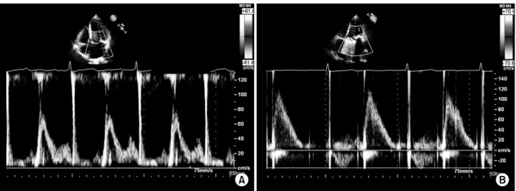
Fig. 3. Sample echocardiographic examinations of two patients after the Cox maze procedure. Examples of echocardiograms from a patient (A) with and (B) without the left atrial A waves, suggesting mechanically active and inactive atria, respectively.
nus rhythm (Table 3) and two (2.3%) were taking amiodar- one due to the intermittent recurrence of AF. Two patients (2.3%) had permanent pacemaker insertion for sinus node dysfunction at 4 and 6 months, respectively.
A postoperative left atrial thrombus developed after the dis- continuation of warfarin in one woman with an LA 60 mm in size that would not be diminished due to the LA wall calcification. It disappeared quickly with warfarin re-medi- cation.
2) Factors predicting sinus rhythm conversion after the maze procedure
The presence of sinus rhythm at postoperative month 6 was the only factor predicting the sinus rhythm at the last follow-up (odds ratio, 158.0; 95% confidence interval, 11.7 to 2,131; p<0.001). Sex, age, underlying cardiac lesions, num- ber of procedures, duration of AF, presence and type of mi- tral valve disease, preoperative and postoperative left atrial di- mensions, and left ventricular ejection fraction were not risk factors for the failure of the sinus rhythm conversion.
3) Left atrial dimensions and left atrial reduction plasty
Although preoperative LA dimensions were greater in the patients (35 patients, 40.2%) with LA reduction plasty than in those (52 patients, 59.8%) without (59.5±11.6 cm vs.
53.4±7.3 cm; p=0.008), postoperative LA dimensions did not
differ between the two groups (49.9±7.0 cm vs. 49.2±6.7 cm;
p=0.652).
Even without reduction plasty, the 52 patients experienced a significant decrease in the LA dimensions with the removal of the left atrial appendage and mitral valve surgery or other concomitant cardiac surgery (53.4±7.3 cm preoperatively vs.
49.2±6.7 cm postoperatively; p<0.0001). Thirty-five patients who underwent reduction plasty also showed marked reduc- tion in the LA dimensions (preoperatively vs. postoperatively, 59.5±11.6 cm vs. 49.9±7.0 cm; p<0.0001). A giant LA (>
6.0 cm) was observed in 24 patients (27.6%) preoperatively and in four (4.6%) postoperatively. All three patients who had isolated tricuspid valve repair underwent concomitant right atrial reduction plasty due to the marked dilatation of the right atrium.
4) Impact of mitral valve surgery and rheumatic valve disease on left atrial A wave
Of the 78 patients undergoing mitral valve surgery, 73 (n=34, mitral valve repair; n=39, mitral valve replacement) revealed sinus rhythm conversion at a mean echocardio- graphic follow-up time of 17.8±17.2 months. Of the 73 pa- tients with sinus rhythm conversion, 44 (60.3%) showed left atrial A waves (Fig. 3), suggesting mechanically active atria.
A larger number of rheumatic valve patients with sinus con-
version (total n=44) underwent mitral valve replacement than
Non-rheumatic (n=29) Rheumatic (n=44)
24 (82.8) 20 (45.5)
5 (17.2)
24 (54.5) 0.004 Values are presented as number (%).
a)