J Korean Ophthalmol Soc 2019;60(12):1169-1175 ISSN 0378-6471 (Print)⋅ISSN 2092-9374 (Online)
https://doi.org/10.3341/jkos.2019.60.12.1169
Original Article
안구건조증에서 자가 혈소판 풍부 혈장이 안구표면에 미치는 영향:
임상 및 실험 분석
Effects of Platelet-rich Plasma on Ocular Surface in Patients with Dry Eye Syndrome: Clinico-experimental Analysis
정재욱1⋅이상희2⋅김홍균1
Jae Uk Jung, MD1, Sang Hee Lee, MD, PhD2, Hong Kyun Kim, MD, PhD1
경북대학교 의과대학 안과학교실1, 제일안과병원2
Department of Ophthalmology, School of Medicine, Kyungpook National University1, Daegu, Korea Cheil Eye Hospital2, Daegu, Korea
Purpose: To evaluate the effect of platelet rich plasma (PRP) on ocular surface damage caused by hyperosmotic conditions us- ing retrospective clinical and experimental analyses.
Methods: Eighty eyes of moderate dry eye syndrome patients who had no responses using conventional treatments were in- cluded in the study. Before and 1, 3, and 6 months after the use of autologous PRP, the visual acuity, intraocular pressure, tear break-up time (TBUT), ocular staining score (OSS), and ocular surface disease index (OSDI) were compared. The changes in in- flammatory factors of ocular surface cells were analyzed using a corneo-limbal epithelial cell culture and a hyperosmotic stress experimental model.
Results: Using retrospective clinical analyses, in 64 eyes (80%) after the use of autologous PRP, the symptom scores and symp- toms were significantly reduced in the OSDI questionnaire when compared with the symptom scores and symptoms before treatment. The TBUT and OSS, which were objective indicators showed a significant increase of TBUT and significant decrease of OSS in 68 eyes (85%) and 72 eyes (90%), respectively. The expression of inflammatory factors such as interleukin-1, tumor necrosis factor-α, metalloproteinase (MMP)-1, and MMP-3 decreased in corneo-limbal epithelial cells under hyperosmotic con- ditions when PRP was added.
Conclusions: The use of autologous PRP showed significant improvement before and after treatment in the TBUT, OSS, symp- tom scores and symptoms, and OSDI. In addition, anti-inflammatory effects were demonstrated in hyperosmotic models simulat- ing dry eye syndrome. Therefore, autologous PRP could be used effectively for the treatment of moderate dry eye syndrome.
J Korean Ophthalmol Soc 2019;60(12):1169-1175
Keywords: Dry eye syndromes, Inflammatory factors, Platelet-rich plasma
■Received: 2019. 7. 12. ■ Revised: 2019. 8. 27.
■Accepted: 2019. 12. 6.
■Address reprint requests to Hong Kyun Kim, MD, PhD Department of Ophthalmology, School of Medicine, Kyungpook National University, #130 Dongdeok-ro, Jung-gu, Daegu 41944, Korea
Tel: 82-53-420-5816, Fax: 82-53-426-6552 E-mail: [email protected]
*Conflicts of Interest: The authors have no conflicts to disclose.
ⓒ2019 The Korean Ophthalmological Society
This is an Open Access article distributed under the terms of the Creative Commons Attribution Non-Commercial License (http://creativecommons.org/licenses/by-nc/3.0/) which permits unrestricted non-commercial use, distribution, and reproduction in any medium, provided the original work is properly cited.
안구건조증은 매우 흔한 안과적 질환으로 눈에 다양한 자극 증상을 일으키며, 매우 경한 경우에서 시력상실을 일 으킬 수 있을 정도의 중증까지 다양한 임상양상을 가진다. 또한 안구건조증은 삶의 질에 중요한 영향을 미치는 업무, 독서, 운전 및 사회 활동과 같은 일상 생활에 영향을 준 다.1,2 안구표면의 항상성 유지를 위해서는 정상 눈물막의 생성, 건강한 안부속기, 눈꺼풀테, 적절한 눈깜빡임 등의 상호 긴밀한 관계가 요구된다. 그중에서 눈물에는 상피 성장인
자(epidermal growth factor, EGF), 파이브로넥틴(fibronectin), 비타민 A, 싸이토카인(cytokine) 등 여러 영양인자를 포함 하고 있어 각결막상피의 재생, 이주, 분화에 중요한 역할을 함으로써 안구표면 상피의 안정성을 유지하는 데 필수적인 역할을 한다.3-5 이러한 인자들이 부족하면 안구표면이 지속 적으로 손상되어 심각한 질병을 유발할 수 있다.6-11 따라서, 안구표면 질환에서의 창상 치유를 위해서 성장인자의 치료 적 공급이 매우 중요하다.
혈소판 풍부 혈장(platelet rich plasma, PRP)은 혈소판 농 도가 높고 각막과 결막상피의 성장 및 유지에 필수적인 더 많은 인자를 포함한다. 혈소판은 상처 치유 과정에서 매우 중요한 역할을 하는 것으로 알려져 있다. 혈소판은 창상면 으로 빠르게 이동하여 손상된 조직에 부착하고 다양한 사 이토카인과 성장인자들을 분비하여 상처 치유를 유도한 다.12 혈소판 내의 α-granule은 platelet-derived growth fac- tor (PDGF), EGF, platelet factor IV, transforming growth factor-β (TGF-β) 등을 분비한다. 따라서 PRP는 고농축 성 장인자를 보유하고 효과적으로 상처 치유를 유도할 수 있 다고 알려져 있다.13,14 이러한 특성으로 PRP가 다양한 안구 표면 질환에 효과적으로 사용될 수 있다. Alio et al15-17은 안과적 수술 후 발생한 난치성 각막 궤양과 안구건조증에 서 자가 PRP를 사용하여 성공적으로 치료할 수 있었다고 보고하였다. 우리는 이전에 전염성 각막염 이후 지속적인 상피 결손이 있는 환자와 반복각막미란환자에서 자가 PRP 의 효과를 보고하였으며, 여러 성장인자의 농도를 비교 분 석하였다.18,19 따라서 저자들은 이러한 관찰을 바탕으로 안 구건조증의 치료에서 자가 PRP의 효과와 눈물막 및 안구 표면의 변화를 알아보고자 하였으며, PRP가 안구표면에 미 치는 영향을 안구건조증 환경을 시뮬레이션한 고삼투압 스 트레스 실험모델을 이용하여 연구해 보고자 하였다.
대상과 방법
본 연구는 본원 IRB 승인을 받았으며(승인 번호: 2016-03- 019-004), 헬싱키선언을 준수하였다. 2016년 1월부터 2018년 1월까지 본원 안과에 내원한 환자들 중 중등도 안구건조증 으로 진단받고 기존의 치료에 효과가 없었던 56명의 80안 을 대상으로 최소 6개월 이상 경과 관찰이 가능했던 환자 를 대상으로 의무기록을 토대로 후향적으로 분석하였다.
안구건조증의 중증도 분류는 2007년 International Dry Eye Workshop (DEWS)에서 제시한 중증도 체계에 따라 level 1은 경도, level 2-3는 중등도, level 4는 중증으로 분류하였다.
경도 및 중증 안구건조증, 스테로이드 점안액을 사용한 경 우, 안구 수술을 받은 경우, 안구 알레르기가 있는 경우, 기
타 안구의 급성 감염 또는 염증이 있는 경우 그리고 눈꺼풀 및 속눈썹에 이상이 있는 경우는 대상에서 제외하였다.
자가 PRP의 제조를 위해 환자로부터 동의를 구한 후 50 mL 전혈을 채취하여 1.4 mL anticoagulant citrate dextrose sol- ution 용액이 들어있는 진공 채혈관에 넣고 10분간 200 g으 로 원심 분리하였다. 원심 분리된 혈액의 상층액을 무균적 으로 분리하고 생리식염수에 섞어 20%로 희석하였다. 자 외선에 의한 비타민 A의 분해를 막기위해 알루미늄 호일로 둘러 싼 소독된 5 mL 병들에 완성된 자가 PRP를 나누어 담았다. 사용 전까지 -20°C 냉동 보관하였고 사용 시 해동 하여 쓰고 4°C 냉장고에 보관하도록 하였다.
모든 환자는 기존에 사용하던 점안액과 더불어 자가 PRP를 1일 6회 점안하였다. 자가 PRP 사용 후의 안구표면 과 눈물막의 변화를 알아보기 위해 모든 환자는 자가 PRP 사용 전과 사용 후 1, 3, 6개월의 시력, 안압, 눈물막파괴시 간(tear break-up time, TBUT), 각결막 표면 형광염색 점수 (ocular staining score, OSS) 및 안구표면질환지수(ocular surface disease index, OSDI) 설문조사 결과를 비교 분석하 였다.
고삼투압 환경에서의 각막윤부상피세포에 대한 PRP의 효과를 알아보기 위해 본원 안은행에서 기증받은 기증자 각막의 이식 후 남은 각막윤부조직을 Phosphate buffered saline (PBS)에서 2번 헹궈준 다음 0.3 cm 정도의 크기로 작게 자른 후 조직은 collagease/dispase (0.1 U/mL collage- nase; 0.8 U/mL dispase, Roche, Mannheim, Germany)에 담 가서 상온에 두었다. 그 다음 분리된 표피를 PBS에 헹군 후 Trypsin-EDTA (0.25% Trypsin with EDTA 4Na) (Gibco BRL, Gaithersburg, MD, USA)에 넣고 37℃에서 20분 동안 배양하여 fetal bovine serum를 넣고 중화시킨 후 15분 동안 약하게 진동을 주었다. Cell stainer (40 μm; BD Falcon TM, Bedford, MA, USA)에서 용액을 걸러 준 다음 원심분리시 켜 배양된 세포를 가라앉혔다. 모아진 세포는 defined kera- tinocyte-SFM (serum-free keratinocyte medium) (Gibco in- vitrogen, Carlsbad, CA, USA)에 풀어서 type 1 bovine col- lagen이 코팅된 plastic dish (Iwaki Glass, Tokyo, Japan)에 서 배양했다.
배양된 각막윤부상피세포들(growth for 12-14 days, about 4-5×10⁵ cells/well)은 defined keratinocyte-SFM에서 배양 하여 스트레스를 주기 전에 type 1 bovine collagen이 코팅 된 6-well plate에 깔았다. 3개 각각의 배지들 중 1개는 대 조군(defined keratinocyte-SFM)으로 하고 나머지 2개에 10% PRP을 준 후 40분 뒤에 1개의 배지에 90 mM sodium chloride를 주어 312-500 mOsM 범위의 hyperosmolar stress 를 만들어 주었다. 이와 같은 조건으로 24시간 동안 배양한
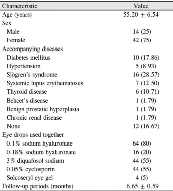
Characteristic Value
Age (years) 55.20 ± 6.54
Sex
Male 14 (25)
Female 42 (75)
Accompanying diseases
Diabetes mellitus 10 (17.86)
Hypertension 5 (8.93)
Sjögren’s syndrome 16 (28.57)
Systemic lupus erythematosus 7 (12.50)
Thyroid disease 6 (10.71)
Behcet's disease 1 (1.79)
Benign prostatic hyperplasia 1 (1.79)
Chronic renal disease 1 (1.79)
None 12 (16.67)
Eye drops used together
0.1% sodium hyaluronate 64 (80)
0.18% sodium hyaluronate 16 (20)
3% diquafosol sodium 44 (55)
0.05% cyclosporin 44 (55)
Solcoseryl eye gel 4 (5)
Follow-up periods (months) 6.65 ± 0.59
Values are presented as mean ± standard deviation or number (%).
Table 1. Demographic and clinical data of patients 다음, 배지는 제거하고 세포에서는 RNA를 분리했다.
각각 다른 hyperosmolar stress을 받은 각막윤부상피세포 들의 total RNA는 trizol (Invitrogen, Carlsbad, CA, USA)을 사용하여 분리하였다. 먼저 배양된 세포에서 배양액을 제 거한 후 trizol을 넣어 세포막을 용해하였다. 그리고 trizol 양의 1/10만큼 1-bromo-3-chloropropane을 넣고 강하게 섞 어 층이 분리된 후 30분간 원심 분리(4°C, 13,000 rpm)한 다음 상층액을 새 tube로 옮겼다. Isopropanol을 상층액과 동일한 양으로 넣어준 후 4°C에서 overnight하여 30분간 원심 분리(4°C, 13,000 rpm)한 후 상층액은 버리고 침전 물은 75% ethanol로 헹궈준 후 말렸다. 마지막으로 DEPC (diethylpyrocarbonate)-H2O에 녹인 후 NanoDrop ND-1000 (Thermo, Waltham, MA, USA)으로 정량하였다.
역전사 중합효소연쇄반응(Reverse Transcription-Polymerase Chain Reaction)은 total RNA 1 μg, Oligo dT 2 μL와 총 vol- ume이 20 μL가 되게 물(pure water)을 섞은 후 70°C에서 10분 정도 둔다. 모든 cDNA의 역전사를 확인하기 위하여 β-actin primer로 PCR을 하였고 이 PCR은 cDNA 산물 1 μL, 5Xbuffer 5 μL, MgCl2 (25 mM) 2 μL, dNTPs (2.5 mM) 2 μL, primer pair 0.5 μL (forward 0.25 + reverse 0.25), Taq DNA polymerase 0.125 μL, 물(pure water) 14.375 μL 를 섞어서 수행하였다. 각 단계의 온도는 94°C-5분, 94°C-1분, primer 반응 온도-1분, 72°C-1분, 72°C-10분으로 진행하였 다. 그리고 각 primer에서 β-actin은 59°C에서 29 cycles을 시행하였고, interleukin-1 (IL-1)은 56°C에서 25 cycles, tu- mor necrosis factor-α (TNF-α)는 56°C에서 35 cycles, ma- trix metalloproteinase (MMP)-1, -3은 55°C에서 35 cycles 로 시행하였다.
치료 전후 효과를 비교하기 위하여 paired t-test를 이용하 였으며, p-value가 0.05 미만인 값을 통계적으로 유의하다 고 간주하였다. 자료에 대한 통계적인 분석을 위해 SPSS version 20.0 (IBM Corp., Armonk, NY, USA)을 이용하였다.
결 과
본 연구는 56명의 환자(남자 14명, 여자 42명) 80안을 대 상으로 하였고, 평균 연령은 55.2 ± 6.54세였으며, 평균 추 적 관찰기간은 6.65 ± 0.59개월이었다. 기존에 사용 중이었 던 점안액은 16안에서 0.1% sodium hyaluronate (Hyalein mini®; Santen Pharmaceutical Co., Ltd., Osaka, Japan), 0.05%
cyclosporine (Restasis®; Allergan Inc., Irvine, CA, USA), 3%
Diquafosol sodium (Diquas®; Santen Pharmaceutical Co., Ltd.)을, 16안에서 0.1% sodium hyaluronate, 3% Diquafosol sodium을, 16안에서 0.1% sodium hyaluronate, 0.05% cy-
closporine을, 12안에서 0.1% sodium hyaluronate을 단독으 로, 8안에서 0.18% sodium hyaluronate (Kynex2®; Alcon Korea, Seoul, Korea), 0.05% cyclosporine을, 4안에서 0.18% sodium hyaluronate, 0.05% cyclosporine, 3% Diquafosol sodium을, 4안에서 0.18% sodium hyaluronate, 3% Diquafosol sodium 을, 4안에서 0.1% sodium hyaluronate, 3% Diquafosol so- dium, Solcoseryl eye gel (Solcorin ophthalmic gel®; Hanlim Pharm. Co., Seoul, Korea)을 함께 점안 중이었다(Table 1).
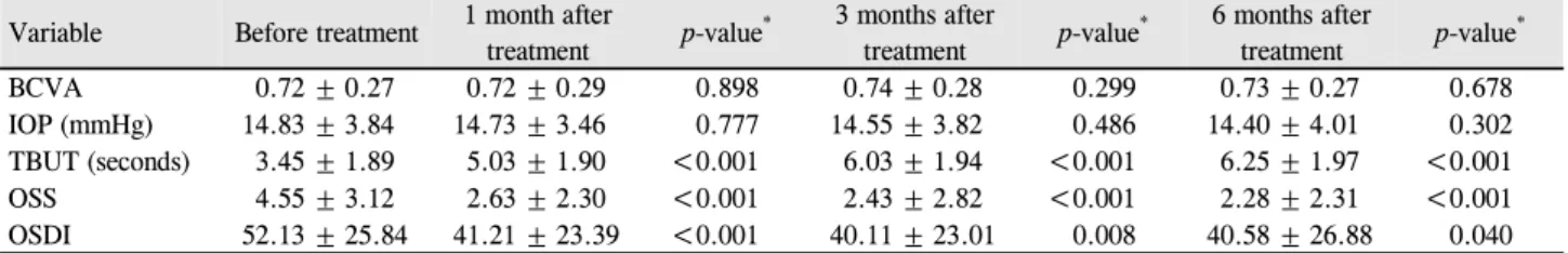
자가 PRP 사용 전후의 시력, 안압, TBUT, OSS 및 OSDI 점수는 Table 2에 나타내었다. 시력과 안압은 치료 전후 유 의한 차이를 보이지 않았다(p>0.05). TBUT는 85% (80안 중 68안)에서 증가하였으며, 치료 전 3.45 ± 1.89초에서 치 료 1개월 후 5.03 ± 1.90초(p<0.001), 치료 3개월 후 6.03 ± 1.94초(p<0.001), 치료 6개월 후 6.25 ± 1.97초(p<0.001)로 의미 있게 증가하였다. OSS는 90% (80안 중 72안)에서 감 소하였으며, 치료 전 4.55 ± 3.12에서 치료 1개월 후 2.63
± 2.30 (p<0.001), 치료 3개월 후 2.43 ± 2.82 (p<0.001), 치 료 6개월 후 2.28 ± 2.31 (p<0.001)로 의미 있게 감소하였 다. OSDI 점수는 80% (80안 중 64안)에서 감소하였으며, 치료 전 52.13 ± 25.84에서 치료 1개월 후 41.21 ± 23.39 (p<0.001), 치료 3개월 후 40.11 ± 23.01 (p=0.008), 치료 6개 월 후 40.58 ± 26.88 (p=0.040)로 의미 있게 감소하였다. 증 상이 악화된 환자는 없었으며, 모든 환자에서 PRP와 관련 된 합병증은 관찰되지 않았다.
Variable Before treatment 1 month after
treatment p-value* 3 months after
treatment p-value* 6 months after
treatment p-value*
BCVA 0.72 ± 0.27 0.72 ± 0.29 0.898 0.74 ± 0.28 0.299 0.73 ± 0.27 0.678
IOP (mmHg) 14.83 ± 3.84 14.73 ± 3.46 0.777 14.55 ± 3.82 0.486 14.40 ± 4.01 0.302
TBUT (seconds) 3.45 ± 1.89 5.03 ± 1.90 <0.001 6.03 ± 1.94 <0.001 6.25 ± 1.97 <0.001
OSS 4.55 ± 3.12 2.63 ± 2.30 <0.001 2.43 ± 2.82 <0.001 2.28 ± 2.31 <0.001
OSDI 52.13 ± 25.84 41.21 ± 23.39 <0.001 40.11 ± 23.01 0.008 40.58 ± 26.88 0.040
Values are presented as mean ± standard deviation.
BCVA = best corrected visual acuity; IOP = intraocular pressure; TBUT = tear break up time; OSS = ocular staining score; OSDI = Ocular Surface Disease Index.
*Paired t-test.
Table 2. Changes of tear film, cornea and conjunctival fluorescein stain, and symptom after autologous platelet-rich plasma therapy in all patients with dry eye syndrome
Figure 1. The relative gene expression of inflammatory mark- ers: IL-1, MMP-1, -3, TNF-α in three different groups of hu- man limbal epithelial cell. The second group with adding 10%
PRP was showed significant increase of inflammatory markers IL-1 and TNF-α. However, in the third group under hyper- osmolar condition with 10% PRP, the gene expression of in- flammatory marker was decreased into below or normal level.
IL-1 = interleukin-1; MMP = matrix metalloproteinase; TNF-α
= tumor necrosis factor-α; PRP = platelet rich plasma. *p <
0.05, Mann-W hitney U test.
각막윤부상피세포에 대한 PRP의 효과를 확인한 결과 정 상 삼투압하에서 10% PRP가 배양액에 첨가된 각막윤부상 피세포에서 IL-1, TNF-α, MMP-1, MMP-3의 모든 변수에 서 발현이 증가되었다. 일반 배양 조건하인 대조군을 1로 보았을 때 비해 IL-1은 4.35 ± 0.87로 발현이 증가되었으며 MMP-1은 1.85 ± 0.39, MMP-3은 1.84 ± 0.7, TNF-α는 2.19
± 0.43로 각각 발현이 증가되었다. 하지만 10% PRP가 첨 가된 고삼투압하에서의 각막윤부상피세포는 대조군에 비 해 IL-1은 0.29 ± 0.08로 발현이 감소되었으며, MMP-1은 1.39 ± 0.67, MMP-3은 1.61 ± 0.54, TNF-α는 1.44 ± 0.2로 각각 발현이 정상 삼투압하의 환경에 비해 이들의 발현이 감소하거나 유사하였다. 특히 IL-1는 정상 대조군에 비해서 약 30% 수준으로 발현이 감소되었다(Fig. 1).
고 찰
최근 안구건조증의 연구가 발달함에 따라 개정된 DEWS II에서 안구건조증은 안구표면의 다요인성 질병으로 눈물 막의 항상성을 상실하고 안구증상이 동반되는 특징을 가지 는데, 눈물막의 불안정과 고삼투압, 안구표면의 염증 및 손 상, 신경감각 이상이 원인적인 역할을 하는 것으로 정의되 었다.20 이는 눈물의 항상성 상실이 주된 원인으로 설명하 고 있다. 또한 다양한 치료가 시도되고 있지만 여러 원인과 다양한 종류의 안구건조증 때문에 명확한 치료법이 확립되 지 않았으며 치료가 항상 성공적인 것은 아니다.
각결막상피의 성장과 유지에는 EGF, TGF-β, 파이브로넥 틴, 비타민 A와 같은 성장인자와 싸이토카인이 필수적이며 안구표면 질환이 있는 경우에는 이러한 성장인자와 싸이토 카인을 공급하여주는 치료가 유용하다. 자가혈청 점안액은 윤활 작용을 도와줄 뿐만 아니라, 성장인자에 대한 공급원 으로 이용되어 임상적으로 지속된 각막상피 결손, 상윤부 각결막염, 반복각막미란 등의 안구표면 질환의 치료에 유
용한 효과가 보고되었다.21-25 또한, 안구건조증의 치료에 있 어서도 혈액성분이 유용하게 사용될 수 있는데 이것은 혈 액성분의 사용이 윤활제 역할을 할 수 있도록 하거나, 혈액 내에 함유된 성장인자에 의해 안구건조증에 의해 손상된 안구표면 상피세포의 치유 효과를 기대하는 것이다. 여러 연구에서 중증의 안구건조증에 대한 자가혈청의 효과가 보 고되었다. Tsubota et al26은 쇼그렌증후군에 의한 중증의 안구건조환자에서 4주간의 자가혈청의 사용으로 플루오레 신 염색 지수가 향상되고 mucin 발현이 증가되었음을 보고 하였고, 안구건조증환자에 대한 무작위 비교 임상실험에서 도 자가혈청의 효과가 입증되었다.24 이와 같이, 자가혈청
은 안구표면 질환에서 상피증식성 인자들에 대한 중요한 공급원으로 이용할 수 있으며, 항원성이 없고 다른 부가적 인 독성이 없다는 장점이 있다.
그러나 혈액 내에 존재하는 상피증식성 성장인자와 싸이 토카인은 혈청이 아닌 다른 구성성분에도 존재한다. 특히 혈소판에 다량으로 성장인자가 검출된다. 혈소판은 혈관 손상부위에서 손상된 내피에 부착하고 트롬빈(thrombin) 생성 및 피브린(fibrin) 형성을 유도하여 혈액 손실을 예방 하는 데 도움이 된다. 또한 혈소판은 손상된 조직 회복을 촉진하고 혈관신생 및 염증에서 혈관 및 다른 혈액 세포의 반응에 영향을 주는 물질을 방출한다. 혈소판 내의 α-gra- nule은 EGF, PDGF, TGF-β, fibriblast growth factor, vas- cular endothelial growth factor와 같은 성장인자뿐만 아니 라 다양한 사이토카인을 방출한다.27-29 특히 EGF는 각막상 피의 증식과 이동과 분화에 중요한 역할을 하며, TGF-β는 각막상피의 증식을 조절하고 미분화된 상태로 세포를 유지 할 수 있다.30-34 즉, 혈소판이 성장인자와 다양한 대사물질 을 분비한다는 것은 PRP가 신속한 치유와 조직재생이 필 요한 임상상황에서 긍정적인 효과를 나타낼 수 있음을 의 미한다.
따라서 PRP는 구강 악안면수술, 심혈관수술, 정형외과, 성형외과와 같은 여러 임상에서 사용되어 왔다.35-38 안과에 서는 Hartwig et al12은 혈소판 분비물이 각막상피세포의 이 동과 분화 및 세포성장에 영향을 미친다고 보고하였다.
Alio et al15-17은 안과적 수술 후 발생한 난치성 각막궤양과 안구표면증후군, 안구건조증에서 자가 PRP를 사용하여 부 작용 없이 성공적으로 치료할 수 있었다고 보고하였다. 우 리는 이전에 감염각막염 후 지속되는 상피결손환자를 대상 으로 자가혈청과 자가 PRP의 치료 효과를 비교하였으며, 자가혈청과 자가 PRP에서 여러 성장인자의 정량적 비교를 하였다. 비교 결과 자가 PRP 치료군이 자가혈청 치료군보 다 평균 치유율은 유의하게 높았으며, PRP의 평균 혈소판 농도는 전혈에서보다 약 1.5배 유의하게 높았다. 또한 PRP 에서 EGF의 평균 농도는 자가혈청에서보다 약 2.5배 유의 하게 높았으며, TGF-β1의 평균 농도는 자가혈청에서보다 약 1.4배 유의하게 높았다.18
저자들은 이러한 PRP의 장점과 더불어 보다 객관적인 경험을 바탕으로 대표적인 안구표면 질환인 안구건조증환 자의 치료에 있어 자가 PRP의 임상효과를 알아보기 위해 치료 후의 증상 변화와 더불어 눈물막과 안구표면의 변화 에 대해 조사하였다. 그 결과 자가 PRP를 사용한 80안에서 TBUT, OSS 및 OSDI 점수에서 치료 전후 통계학적으로 의미 있는 호전을 보였다.
또한 PRP가 안구표면 상피세포에 어떠한 효과로 의미
있는 호전을 보였는지 확인하기 위하여, 인간 윤부상피세 포를 일차 배양하여 PRP에 대한 반응을 정상삼투압과 안 구건조증 환경을 시뮬레이션한 고삼투압하의 환경에서 서 로 비교하였다. Li et al39,40은 정상삼투압에 비해 고삼투압 하에서 스트레스를 받은 윤부상피세포는 IL-1β, TNF-α, IL-8과 같은 염증성 싸이토카인의 분비가 증대되며 또한 MMP-1, -3, -9, -13과 같은 단백분해효소도 증가된다고 보 고하였다. 고삼투압에 대한 세포의 반응은 다양한 경로를 통해 나타나며, Li et al39,40의 연구에서는 고삼투압 환경하 에 있는 윤부상피세포가 c-Jun N-terminal kinases (JNK)와 extracellular regulated kinase (ERK) 경로를 통해서 염증성 싸이토카인의 유도가 일어나는 것을 실험으로 증명하였다. 본 연구에서는 Li et al39,40의 실험 모델을 이용하여 PRP에 대해 윤부상피세포에서 IL-1, TNF-α, MMP-1, -3가 어떻게 반응하는지를 보았는데, 정상 삼투압에서 윤부상피세포는 정상 대조군에 비해 모든 변수에서 증가된 유전자 발현을 보였다. 그러나 고삼투압 환경하에서는 IL-1는 정상 대조군 보다 더 감소되었으며 TNF-α, MMP-1, -3는 정상 수준으로 감소하였다. 혈소판에 함유된 풍부한 성장인자는 세포에 자극을 전달하는 충분한 신호를 보내며, 특히 EGF와 PDGF 등은 ERK 경로를 강하게 활성화한다.41-43 PRP가 첨가된 윤부상피세포는 이러한 성장인자를 매개로 하여 염증성인 자들이 유도된 것으로 보인다. 그러나 본 연구에서 고삼투 압 환경하에 놓인 윤부상피세포들은 PRP가 첨가된 경우에 염증성 인자들의 발현이 감소하였고, 거의 정상 수준으로 환원됨을 알 수 있었다. 이것은 이미 고삼투압에 의한 스트 레스에 의해 JNK와 ERK 경로가 모두 활성화된 환경에 놓 인 세포들에서는 PRP가 오히려 항염증 효과를 가지는 것 으로 생각되며, 이것은 PRP가 JNK와 ERK 세포 신호 경로 에 영향을 주어 이를 차단하는 효과가 있는 것으로 추정된 다. 이러한 실험의 결과로서 PRP는 안구표면 질환에서 상 피세포의 증식, 이동 및 분화를 돕는 효과가 있으며 안구건 조증과 같이 고삼투압 환경하에서 염증성 발현이 증가된 세포에서 염증을 억제할 수 있는 효과를 가지는 것으로 생 각된다. 즉, 자가 PRP는 고농축의 혈소판과 다양한 종류의 성장인자가 다량 포함되어있다. 또한 환자 자신의 혈액에 서 얻은 보존제가 없는 생물학적 제제이다. 따라서 손상된 안구표면에 대한 항염증 효과와 세포 성장과 치유 과정을 보다 효과적으로 촉진하는 것으로 생각된다.
한편, 고삼투압 환경이 아닌 안구표면 질환에서도 자가 PRP의 우수한 치료 효과가 앞선 여러 연구에서 입증되었 다. 하지만 우리는 이번 실험을 통해 고삼투압 환경이 아닌 상태에서는 오히려 염증성 싸이토카인이 증가함을 확인하 였다. 그러나 PRP에는 높은 농도의 성장인자들뿐만 아니라
항 염증인자들도 높은 농도로 존재하여 증가된 염증성 싸 이토카인의 영향을 상쇄시키는 것으로 추정된다. 이러한 관점에서 PRP의 효과와 관련된 명확한 기전을 확인하기 위해 추가적인 논의 및 연구가 필요할 것이다.
또한 본 연구에서 환자들은 20%의 PRP를 사용했다는 점도 고려할 필요가 있는데, 현재까지 다양한 농도의 혈청 점안제의 효과들이 발표되고 있지만 치료에 가장 효과적인 농도에 관해서는 아직까지 확정된 바는 없다. 오히려 높은 농도의 성장인자가 유해한 영향을 줄 수 있으며, 고농도의 EGF는 각막 신생혈관 촉진인자로 보고된 바 있다.44 본 연 구에서 각막 신생혈관을 관찰할 수 없었지만 향후 적절한 농도를 결정하기 위해 여러 농도 PRP의 비교가 포함된 연 구가 필요하다고 생각된다. 결론적으로 자가혈청보다 많은 성장인자를 가진 자가 PRP는 기존의 치료에 반응하지 않 는 대표적인 안구표면 질환인 안구건조증에 효과적으로 사 용될 수 있을 것으로 기대된다.
REFERENCES
1) Miljanović B, Dana R, Sullivan DA, Schaumberg DA. Impact of dry eye syndrome on vision-related quality of life. Am J Ophthalmol 2007;143:409-15.
2) Tong L, Waduthantri S, Wong TY, et al. Impact of symptomatic dry eye on vision-related daily activities. the Singapore Malay Eye Study. Eye (Lond) 2010;24:1486-91.
3) Gupta A, Monroy D, Ji Z, et al. Transforming growth factor beta-1 and beta-2 in human tear fluid. Curr Eye Res 1996;15:605-14.
4) Nishida T, Nakamura M, Ofuji K, et al. Synergistic effects of sub- stance P with insulin-like growth factor-1 on epithelial migration of the cornea. J Cell Physiol 1996;169:159-66.
5) Watanabe K, Nakagawa S, Nishida T. Stimulatory effects of fi- bronectin and EGF on migration of corneal epithelial cells. Invest Ophthalmol Vis Sci 1987;28:205-11.
6) Barton K, Nava A, Monroy DC, Pflugfelder SC. Cytokines and tear function in ocular surface disease. Adv Exp Med Biol 1998;438:
461-9.
7) Fukuda M, Fullard RJ, Willcox MD, et al. Fibronectin in the tear film. Invest Ophthalmol Vis Sci 1996;37:459-67.
8) Lopez Bernal D, Ubels JL. Artificial tear composition and promo- tion of recovery of the damaged corneal epithelium. Cornea 1993;12:115-20.
9) Pancholi S, Tullo A, Khaliq A, et al. The effects of growth factors and conditioned media on the proliferation of human corneal epi- thelial cells and keratocytes. Graefes Arch Clin Exp Ophthalmol 1998;236:1-8.
10) Tsubota K, Xu KP, Fujihara T, et al. Decreased reflex tearing is as- sociated with lymphocytic infiltration in lacrimal glands. J Rheumatol 1996;23:313-20.
11) Wilson SE, Liang Q, Kim WJ. Lacrimal gland HGF, KGF, and EGF mRNA levels increase after corneal epithelial wounding.
Invest Ophthalmol Vis Sci 1999;40:2185-90.
12) Hartwig D, Harloff S, Liu L, et al. Epitheliotrophic capacity of a
growth factor preparation produced from platelet concentrates on corneal epithelial cells: a potential agent for the treatment of ocular surface defects? Transfusion 2004;44:1724-31.
13) Knighton DR, Ciresi K, Fiegel VD, et al. Stimulation of repair in chronic, nonhealing, cutaneous ulcers using platelet derived wound healing formula. Surg Gynecol Obstet 1990;170:56-9.
14) Lynch SE, Colvin RB, Antoniades HN. Growth factors in wound healing. Single and synergistic effects in partial thickness porcine skin wounds. J Clin Invest 1989;84:640-6.
15) Alio JL, Abad M, Artola A, et al. Use of autologous platelet-rich plasma in the treatment of dormant corneal ulcers. Ophthalmology 2007;114:1286-93.e1.
16) Alio JL, Pastor S, Ruiz-Colecha J, et al. Treatment of ocular sur- face syndrome after LASIK with autologous platelet-rich plasma. J Refract Surg 2007;23:617-19.
17) Alio JL, Colecha JR, Pastor S, et al. Symptomatic dry eye treatment with autologous platelet-rich plasma. Ophthalmic Res 2007;39:124-9.
18) Kim KM, Shin YT, Kim HK. Effect of autologous platelet-rich plasma on persistent corneal epithelial defect after infectious keratitis. Jpn J Ophthalmol 2012;56:544-50.
19) Lee JH, Kim MJ, Ha SW, Kim HK. Autologous platelet-rich plas- ma eye drops in the treatment of recurrent corneal erosions. Korean J Ophthalmol 2016;30:101-7.
20) Nelson JD, Craig JP, Akpek EK, et al. TFOS DEWS II introduction.
Ocul Surf 2017;15:269-75.
21) Tsubota K, Goto E, Shimmura S, Shimazaki J. Treatment of persis- tent epithelial defect by autologous serum application. Ophthalmology 1999;106:1984-9.
22) Goto E, Shimmura S, Shimazaki J, Tsubota K. Treatment of superi- or limbic keratoconjunctivitis by application of autologous serum.
Cornea 2001;20:807-10.
23) del Castillo JM, de la Casa JM, Sardiña RC, et al. Treatment of re- current corneal erosions using autologous serum. Cornea 2002;21:
781-3.
24) Tananuvat N, Daniell M, Suillivan LJ, et al. Controlled study of the use of autologous serum in dry eye patients. Cornea 2001;20:802-6.
25) Ogawa Y, Okamoto S, Mori T, et al. Autologous serum eyedrops for the treatment of severe dry eye in patients with chronic graft- versus-host disease. Bone Marrow Transplant 2003;31:579-83.
26) Tsubota K, Goto E, Fujita H, et al. Treatment of dry eye by autolo- gous serum application in Sjögren's syndrome. Br J Ophthalmol 1999;83:390-5.
27) Anitua E, Andia I, Ardanza B, et al. Autologous platelets as a source of proteins for healing and tissue regeneration. Thromb Haemost 2004;91:4-15.
28) Stassen JM, Arnout J, Deckmyn H. The hemostatic system. Curr Med Chem 2004;11:2245-60.
29) Klinger MH, Jelkmann W. Role of blood platelets in infection and inflammation. J Interferon Cytokine Res 2002;22:913-22.
30) Ho PC, Davis WH, Elliott JH, Cohen S. Kinetics of corneal epi- thelial regeneration and epidermal growth factor. Invest Ophthalmol 1974;13:804-9.
31) Gönül B, Koz M, Ersöz G, Kaplan B. Effect of EGF on the corneal wound healing of alloxan diabetic mice. Exp Eye Res 1992;54:
519-24.
32) Rodeck U, Jost M, Kari C, et al. EGF-R dependent regulation of keratinocyte survival. J Cell Sci 1997;110(Pt 2):113-32.
33) Yashino Y, Garg R, Monroy D, et al. Production and secretion of
= 국문초록 =
안구건조증에서 자가 혈소판 풍부 혈장이 안구표면에 미치는 영향:
임상 및 실험 분석
목적: 고삼투압성 눈물막에 의한 안구표면손상이 있는 환경에서 자가 혈소판 풍부 혈장이 어떠한 영향을 주는지 후향적 임상분석 및 실험 방법을 통하여 확인하고자 한다.
대상과 방법: 기존 치료에 효과가 없었던 중등도 안구건조증환자 80안을 대상으로 자가 혈소판 풍부 혈장 사용 전과 사용 후 1, 3, 6개월에 각각 시력, 안압, 눈물막파괴시간, 각결막 표면 형광염색점수, 안구표면질환지수를 비교 분석하였으며, 각막윤부상피세포 배 양과 고삼투압 스트레스 실험 모델을 이용하여 안구표면세포의 염증성 인자의 변화를 분석하였다.
결과: 후향적 임상분석에서 자가 혈소판 풍부 혈장을 사용 후 64안(80%)에서 안구표면질환지수 설문조사에서 치료 전과 비교하여 증상 호전 및 증상 점수의 유의한 감소를 보였다. 객관적 지표인 눈물막파괴시간 및 각결막 표면 형광염색 점수 역시 각각 68안(85%), 72안(90%)에서 눈물막파괴시간의 유의한 증가 및 각결막 표면 형광염색점수의 유의한 감소를 보였다. 고삼투압하에서의 각막윤부상 피세포는 혈소판 풍부 혈장을 추가한 경우 interleukin-1, tumor necrosis factor-α, metalloproteinase (MMP)-1, MMP-3과 같은 염증 성 인자들이 감소하였다.
결론: 자가 혈소판 풍부 혈장의 사용으로 눈물막파괴시간, 각결막 표면 형광염색 점수, 안구표면질환지수에서 치료 전후 통계학적으로 의미 있는 호전을 보였으며, 안구건조증을 시뮬레이션한 고삼투압모델에서 항염증 효과가 입증되어 중등도 안구건조증에 자가혈소판 풍부 혈장 투여가 효과적으로 사용될 수 있을 것으로 보인다.
<대한안과학회지 2019;60(12):1169-1175>
정재욱 / Jae Uk Jung
경북대학교 의과대학 안과학교실 Department of Ophthalmology,
School of Medicine, Kyungpook National University transforming growth factor beta (TGF-β) by the human lacrimal
gland. Curr Eye Res 1996;15:615-24.
34) Sammartino G, Tia M, Marenzi G, et al. Use of autologous plate- let-rich plasma (PRP) in periodontal defect treatment after ex- traction of impacted mandibular third molars. J Oral Maxillofac Surg 2006;63:766-70.
35) Choi BH, Im CJ, Huh JY, et al. Effect of platelet-rich plasma on bone regeneration in autogenous bone graft. Int J Oral Maxillofac Surg 2004;33:56-9.
36) Englert SJ, Estep TH, Ellis-Stoll CC. Autologous platelet gel appli- cations during cardiovascular surgery: effect on wound healing. J Extra Corpor Technol 2005;37:148-52.
37) Man D, Plosker H, Winland-Brown JE. The use of autologous pla- telet-rich plasma (platelet gel) and autologous platelet-poor plasma (fibrin glue) in cosmetic surgery. Plast Reconstr Surg 2001;107:
229-37; discussion 238-9.
38) Bhanot S, Alex JC. Current applications of platelet gels in facial plastic surgery. Facial Plast Surg 2002;18:27-33.
39) Li DQ, Luo L, Chen Z, et al. JNK and ERK MAP kinases meidate
induction of IL-1β, TNF-α and IL-8 following hyperosmolar stress in human limbal epithelial cells. Exp Eye Res 2006;82:588-96.
40) Li DQ, Chen X, Song XJ, et al. Stimulation of matrix metal- lopreteinases by hyperosmolarity via a JNK pathwasy in human corneal epithelial cells. Invest Ophthalmol Vis Sci 2004;45:4302-11.
41) Zeigler ME, Chi Y, Schmidt T, Varani J. Role of ERK and JNK pathways in regulating cell motility and matrix metalloproteinase 9 production in growth factor-stimulated human epidermal keratinocytes. J. Cell Physiol 1999;180:271-84.
42) Cho A, Graves J, Reidy MA. Mitogen-activated protein kinases mediate matrix metalloproteinase-9 expression in vascular smooth muscle cells. Arterioscler Thromb Vasc Biol 2000;20:2527-32.
43) Hayashida T, Decaestecker M, Schnaper HW. Cross-talk between ERK MAP kinase and Smad signaling pathways enhances TGF- beta dependent responses in human mesangial cells. FASEB J 2003;17:1576-8.
44) Gospodarowicz D, Bialecki H, Thakral TK. The angiogenic activ- ity of the fibroblast and epidermal growth factor. Exp Eye Res 1979;28:501-14.