pISSN: 0378-6471 eISSN: 2092-9374 http://dx.doi.org/10.3341/jkos.2013.54.4.581
= 증례보고 =
약물에 의한 동공 확장이 안구, 각막 및 안내 수차에 미치는 영향
유은주⋅강수연⋅김효명⋅송종석 고려대학교 의과대학 안과학교실
목적: 정상인을 대상으로 약물에 의한 동공 확장이 안구, 각막 및 안내의 수차에 미치는 영향을 알아보고자 하였다.
대상과 방법: 백내장 또는 시력교정수술 등의 안수술을 시행 받지 않은 62명 62안을 대상으로 산동 전 암실에서 KR-1W수차계(Topcon Corp., Tokyo, Japan)를 이용하여 안구, 각막 및 안내 구면수차를 측정한 뒤, 산동제(phenylephrine chloride 0.5% + tropicamide 0.5%) (Mydrin-P, Santen, Osaka, Japan)를 점안하여 산동한 후 다시 측정하여 동공 지름 4 mm와 6 mm 범위에서의 산동 전후 수차 변화를 대응표본 t-검정을 통해 비교하였다.
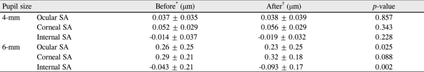
결과: 동공 지름 4 mm 범위에서 산동 전후의 안구, 각막 및 안내 구면수차 값은 차이를 보이지 않았으며, 6 mm 범위에서 산동 전후의 각막 구면수차 값은 차이가 없었으나 안구와 안내 구면수차 값은 통계적으로 유의한 차이를 나타내었다(p=0.025, p=0.002, respectively, paired t-test). 안내 구면수차 값은 산동 전 평균 -0.043 (±0.21) μm였으며, 산동 후 -0.093 (±0.17) μm로 산동 후에 음(-)의 방향으로 증가하였고, 안구 구면수차 또한 음(-)의 방향으로 변화하였다. 고위수차 및 난시값은 산동 전후에 차이를 보이지 않았다.
결론: 정상안에서 산동 후에 안구와 안내 구면수차 값은 음(-)의 방향으로 변하였고, 안구 구면수차 값의 변화는 안내 구면수차 값의 변화에 의한 것으로 생각한다.
<대한안과학회지 2013;54(4):581-586>
■ 접 수 일: 2012년 4월 30일 ■ 심사통과일: 2012년 7월 23일
■ 게재허가일: 2013년 2월 15일
■ 책 임 저 자: 송 종 석
서울특별시 구로구 구로동로 148 고려대학교 구로병원 안과
Tel: 02-2626-3178, Fax: 02-857-8580 E-mail: [email protected]
* 이 논문의 요지는 2011년 대한안과학회 제106회 학술대회에서 구연으로 발표되었음.
광학적오차를 교정하는 방법은 약 800년 전부터 시작된 것으로 알려졌으며, 구면 렌즈를 사용한 안경은 대략 13세 기부터, 난시를 교정하기 위한 원주렌즈는 19세기부터 쓰 이기 시작하였다.1이후 19세기 중반부터 고위수차의 개념 이 알려지게 되었고, 20세기에 고위광학수차를 제니케 다 항식(Zernike polynomials)으로 산술적으로 정량화하여 표 현하는 것이 가능해지면서다양한 수차계들이 개발되었다.2 현재 상용화된 수차계는 그 원리에 따라 Ray tracing, Automatic retinoscopy, Tscherning, Hartmann Shack 방 식으로 분류할 수 있다.3-8Hartmann Shack 방식의 수차계 는 객관적 병렬 측정을 하고 있으며, 후방 투사 방식을 채 택하고 있다. 후방 투사법은 황반부에 조사된 레이저가 반 사되어 눈을 통과하여 나올 때 발생하는 수차를 바둑판 모양 으로 정렬된 여러 개의 작은 렌즈군과 전하결합소자(charge
coupled device, CCD)에서 동시에 기록하여 측정한다.9 Hartmann Shack 방식 중 Topcon사에서 새로 개발된 KR-1W 수차계는 파면측정기(wavefront sensor)로 측정한 전체 안구값에서 각막형태검사기기(corneal topographer)로 측정한 각막값을 빼서 안구내값을 계산하는 방식으로 각막 과 렌즈의 정보를 따로 제시함으로써, 굴절 수술 전 각막 고위수차를 확인하고 백내장 수술 후 인공 수정체의 효과 를 알아보기 위해 사용할 수 있다.10
통상적으로 수차계의 사용 지침은 주관적인 굴절상태를 더 잘 반영하기 위해 조절마비제나 산동제를 사용하지 않 은 상태로 측정하기를 추천한다. 그러나 KR-1W 수차계는 8 lux의 중등도 조명환경에서 실제 동공 크기 범위의 데이 터를 측정한 후, 이때 얻은 자료로부터 동공 지름 4 mm와 6 mm 범위의 데이터를 따로 제시하는 방식이므로, 실제 동 공 지름이 6 mm 미만일 때에 6 mm 범위의 데이터가 산출 되지 않는 경우가 발생하게 된다. 이러한 경우에 제작사에 서는 산동 후 측정할 것을 권유하고 있는데, 이때 산동에 의해 데이터 값의 변화가 발생하지는 않는지 여부가 문제 될 수 있다. 따라서 본 연구에서는 정상인을 대상으로 KR-1W 수차계를 이용하여 산동 전후의 구면수차, 고위수 차 및 난시의 변화를 알아보고, 나이와 구면수차 간에 상관
Table 1. The average of ocular, corneal and internal spherical aberrations on 4-mm and 6-mm diameter zone before and after
mydriasis. The ocular and internal spherical aberrations on 6-mm diameter zone were statistically significantly shifted in a negative direction after mydriasisPupil size Before* (μm) After† (μm) p-value
4-mm Ocular SA Corneal SA Internal SA
0.037 ± 0.035 0.052 ± 0.029 -0.014 ± 0.037
0.038 ± 0.039 0.056 ± 0.029 -0.019 ± 0.032
0.857 0.343 0.228
6-mm Ocular SA 0.26 ± 0.25 0.23 ± 0.25 0.025
Corneal SA 0.29 ± 0.21 0.32 ± 0.18 0.088
Internal SA -0.043 ± 0.21 -0.093 ± 0.17 0.002
Values are presented as mean ± SD.
SA = spherical aberrations.
*Before mydriasis, at mesopic conditions, 8 lux; †After pharmacologically induced mydriasis.
Ocular
SA (4 mm) Cornea
SA (4 mm) Internal
SA (4 mm) Ocular
SA (6 mm) Corneal
SA (6 mm) Internal SA (6 mm) 0.35
0.3 0.25
0.2
0.15
0.1 0.05
0
-0.05
-0.1 -0.15
Before After
Figure 1. Comparison of spherical aberrations (SA) before and
after mydriasis (in micrometer). The ocular and internal spher- ical aberrations were statistically significantly different on 6-mm diameter zone (p = 0.025, p = 0.002, respectively, paired t-test). Before = before mydriasis, at mesopic conditions, 8 lux;After = after pharmacologically induced mydriasis.
관계가 있는지 알아보고자 하였다.
대상과 방법
2011년 7월부터 2012년 6월까지 본원 안과에 내원한 62명의 우안을 대상으로 하였다. 현성굴절검사를 시행한 후 세극등 검사상 각결막과 홍채 및 전안부의 병변이 없음 을 확인하고, 산동 전 암실에서 KR-1W 수차계(Topcon Corp., Tokyo, Japan)를 이용하여 구면수차, 고위수차, 난 시값을 측정한 뒤, 산동제(phenylephrine chloride 0.5% + tropicamide 0.5%) (Mydrin-P, Santen, Osaka, Japan)를 10분 간격으로 3회 점안하여 산동한 후 동일한 방법으로 다시 측정하였다. 산동 후에는 안저 검사를 시행하여 망막 의 이상 소견이 없음을 확인하였다. 그 중 산동 전 동공 지 름이 6 mm 미만이어서 데이터가 측정되지 않았던 15안을 제외한 나머지 47안을 대상으로 산동 전후의 변화를 비교 하였다.
KR-1W 수차계는 Hartmann Shack 방식의 파면 측정기 (wavefront aberrometry)와 Placido-disk 각막형태검사기 기(corneal topography), 표준 자동굴절검사기기(standard autorefraction)가 결합되어 안구의 광학적 성능을 분석할 수 있 는 기계이다. 각막지형도(topography), 수차계(aberrometry), 동공측정계(pupillometry), 각막곡률계(refractometry), 굴 절계(keratometry)의 기능을 수행할 수 있으며, 각막과 안 구를 거의 동시에 같은 중심축을 기준으로 측정하므로 여 러 기계를 이용하여 각막의 수차를 측정할 때 나타날 수 있 는 정렬오차(misalignment)를 줄여서 정확한 안내값을 얻 을 수 있는 것이 장점이다.
통계는 SPSS v12.0 for Windows (SPSS Inc., Chicago, IL) 프로그램의 대응표본 t-검정(paired t-test)으로 비교 분석하였다. 통계분석결과의 p값이 0.05 이하인 경우 통계 적으로 의미 있는 차이로 간주하였다.
결 과
62명의 우안이 연구에 포함되었으며 이들의 평균 연령은 48.28 (±18.41) (range, 25-79)세였고 남성이 28명, 여 성이 34명이었다. 연령 분포는 20대 21명, 30대 12명, 40 대 6명, 50대 8명 60대 이상 15명으로 구성되었다. 산동 전 평균 동공 지름은 8 lux의 중등도 조명환경에서 5.76 (±0.79) (range, 3.35-7.65) mm였다. 산동 후 평균 동공 지름은 7.66 (±0.56) (range, 6.36-9.07) mm로, 산동 전 과 약 1.9 mm 정도의 차이를 보였다. 이중 산동 전 동공 지 름이 6 mm 이하여서 데이터가 측정되지 않은 경우가 15안 (24%)이었으며, 나머지 47안을 대상으로 대응표본 t-검 정을 실시하였다. 산동 전 동공 지름이 6 mm 미만인 경우 는 60대 이상에서 8안으로 가장 많았다.
동공 지름 4 mm 범위에서는 산동 전후 안구, 각막 및 안 내 구면수차에 통계학적으로 유의한 변화가 없었으나, 6
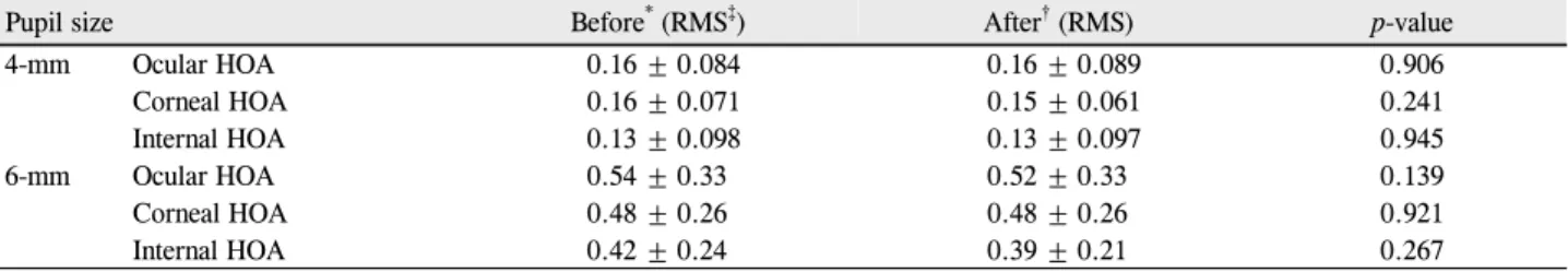
Table 2. The average of ocular, corneal and internal high order aberrations on 4-mm and 6-mm diameter zone before and after
mydriasis. The high order aberrations were not statistically significantly different after mydriasisPupil size Before* (RMS‡) After† (RMS) p-value
4-mm Ocular HOA 0.16 ± 0.084 0.16 ± 0.089 0.906
Corneal HOA 0.16 ± 0.071 0.15 ± 0.061 0.241
Internal HOA 0.13 ± 0.098 0.13 ± 0.097 0.945
6-mm Ocular HOA 0.54 ± 0.33 0.52 ± 0.33 0.139
Corneal HOA 0.48 ± 0.26 0.48 ± 0.26 0.921
Internal HOA 0.42 ± 0.24 0.39 ± 0.21 0.267
Values are presented as mean ± SD.
HOA = high order aberrations.
*Before mydriasis, at mesopic conditions, 8 lux; †After pharmacologically induced mydriasis; ‡Average root mean square.
Table 3. The average of ocular, corneal and internal astigmatism on 4-mm and 6-mm diameter zone before and after mydriasis. The
astigmatism was not statistically significantly different after mydriasisPupil size Before* (D) After† (D) p-value
4-mm Ocular Astig. -0.86 ± 0.49 -0.85 ± 0.46 0.936
Corneal Astig. -0.83 ± 0.54 -0.80 ± 0.54 0.409
Internal Astig. -0.65 ± 0.37 -0.70 ± 0.39 0.229
6-mm Ocular Astig. -0.92 ± 0.49 -0.95 ± 0.55 0.417
Corneal Astig. -0.65 ± 0.43 -0.63 ± 0.47 0.579
Internal Astig. -0.66 ± 0.39 -0.69 ± 0.43 0.427
Values are presented as mean ± SD.
D = Diopters; Astig. = Astigmatism.
*Before mydriasis, at mesopic conditions, 8 lux; †After pharmacologically induced mydriasis.
Ocular
HOA (4 mm) Corneal
HOA (4 mm) Internal
HOA (4 mm) Ocular
HOA (6 mm) Corneal
HOA (6 mm) Internal HOA (6 mm) 0.6
0.5
0.4
0.3
0.2
0.1
0
Before After
Figure 2. Comparison of high order aberrations (HOA) before
and after mydriasis (in average root mean square; RMS). The high order aberrations did not change significantly after mydriasis. Before = before mydriasis, at mesopic conditions, 8 lux; After = after pharmacologically induced mydriasis.0 -0.1 -0.2 -0.3 -0.4 -0.5 -0.6 -0.7 -0.8 -0.9 -1
Before After
Figure 3. Comparison of astigmatism (Astig.) before and after
mydriasis (in diopter). The astigmatism did not change sig- nificantly after mydriasis. Before = before mydriasis, at mes- opic conditions, 8 lux; After = after pharmacologically in- duced mydriasis.mm범위에서는안내 구면수차가 산동 전 -0.043 (±0.21) μm에서 산동 후 -0.093 (±0.17) μm로 통계적으로 유의 하게 음의 방향으로 증가하였다(p=0.002, paired t-test).
또한 동공 지름 6 mm 범위에서 각막 구면수차는 산동 전후 변 화가 없었으나, 안구 구면수차는 0.26 (±0.25) μm에서 0.23 (±0.25) μm로 유의하게 감소하였다(p=0.088, p=0.025, respectively, paired t-test) (Table 1) (Fig. 1).
고위수차의 경우에는 산동 전후 통계적으로 유의한 차이 를 보이지 않았으며(Table 2) (Fig. 2), 불규칙한 동공 확 장에 의해 안내 난시값의 변화가 있는지 알아보기 위해 안 구, 각막 및 안내 난시값을 비교해 보았으나 유의한 변화가 없었다(Table 3) (Fig. 3).
산동 전후 실제 동공 크기에서 측정한 구면대응치(spherical equivalent)는 산동 후에 평균 0.26 (±0.42)D 원시 쪽으로 이 행하였다(p=0.000, paired t-test).
고 찰
굴절교정수술을 시행하기 위해 수차를 측정할 때, 더 많 은 정보를 얻기 위해 산동을 하는 경우가 임상에서 자주 있 으므로, 산동제의 효과에 의한 수차의 변화를 연구한 논문 들이 여러 편 보고된 바 있다.11
2002년 Carkeet et al12이 31안을 대상으로 1% cyclo- pentolate hydrochloride와 2.5% phenylephrine hydro- chloride로 5일 이상의 간격을 두고 산동하여 Zywave 수차 계(Bausch&Lomb, New York, USA)로 동공 지름 6 mm 범위의 고위수차를 비교한 결과, cyclopentolate로 산동한 경우가 phenylephrine로 산동한 경우에 비해 유의하게 높 았다고 발표하였다. 2011년 Taneri et al13이 200 근시안을 대상으로 100안은 tropicamide 0.5% + phenylephrine 2.5%
로, 나머지 100안은 tropicamide 0.5%만으로 산동하여 Zywave 수차계(Technolas Perfect Vision, Munich, Germany)로 측정 한 결과 두 경우 모두에서 산동 전에 비해 산동 후 고위수차 가 유의하게 증가하였으며, predicted phoropter refraction (PPR)이 원시쪽으로 이행하였으나 두 군간에 차이는 없었 다고 보고하였다. 2002년 Giessler et al11이 50안을 대상 으로 중등도 조명환경 및 tropicamide, phenylephrine, cy- clopentolate를 이용한 산동 상태에서 수차계를 이용하여 굴절변화를 측정한 결과, 각각 0.24D, 0.35D, 0.19D, 0.42D 원시쪽으로 이행하였고, 따라서 수차 측정을 위해 산동을 시행할 때에 cyclopentolate를 사용하는 것은 적당 하지 않다고 주장하였다.
그러나 기존 연구에서 사용한 Zywave 수차계는 안구 수 차값과 안내 수차값을 별도로 제시하지 못한다는 한계점이 있었으며, 산동 전후 보다는 조절마비 효과(cycloplegic effect)를 가지는 부교감신경차단제(antimuscarinic drug) 의 사용 유무에 따른 변화에 초점을 맞추고 있었다.
국내에서는 2011년 Ahn et al14이 10명 20안을 대상으 로 Wavescan 수차계를 이용하여 반복 측정한 결과, 구면값 과 구면수차간에는 뚜렷한 상관관계가 존재하며, 임상에서 큰 음의 구면수차와 근시로 인해 과대평가된 웨이브프론트 맵을 사용하게 되면 치료는 예측치 못한 과교정과 원시를 초래할 수 있으므로 웨이브프론트 맵을 선택시에 구면수차 에 대한 고려가 필요하다는 것을 보고한 바 있으나, 산동 전후 데이터를 비교한 기존 연구들에서는 주로 고위수차의 변화에 중점을 두어 구면수차에 대한 논의가 부족하였다.
현재 KR-1W수차계를 이용한 연구는 보고된 것이 많지 않으며, 안내 수차값의 피험자내 재현성(intrasubject re- peatability)이 일부 고위수차 항목을 제외하고는 비교적 높다는 내용이 발표된 바 있다.15
본 연구에서는 가장 널리 사용되고 있는 산동제(phenylephrine chloride 0.5% + tropicamide 0.5%) (Mydrin-P, Santen, Osaka, Japan)를 이용하여 산동 전후의 변화에 초점을 맞 추어 비교하였으며, KR-1W수차계를 사용하여 안구, 안내, 각막의 데이터를 각각 제시함으로써 산동 후 변화가 발생 하는 부위에 대해 직접적으로 파악하였다.
산동 전후의 데이터를 비교해 본 결과, 구면 수차의 경우 동공 지름 4 mm에서는 유의한 차이를 보이지 않았다. 6 mm 범위에서 산동 전후의 각막 구면수차 값은 차이가 없었으 나 안구와 안내 구면수차 값은 산동 후에 유의하게 음의 방 향으로 증가하는 경향을 보였다. 이는 산동제의 조절마비 효과에 의해 수정체 표면이 원뿔 곡선형(conoid shape)으 로 변화하기 때문으로 생각한다.16 Dubbelman et al17,18은 조절 과정에서 수정체 중심부의 상대적 비대와 수정체 앞 면의 곡률 반경의 변화가 구면수차의 변화에 영향을 미친 다고 보고한 바 있다.
그러나 구면 수차에 가장 직접적인 영향을 미치는 것은 동공 크기이며, 이론적으로 동공 크기가 1/2로 작아지면 구 면 수차는 1/16로 감소한다. 따라서 동공 지름 4 mm 범위 에서 산동 전후 유의한 차이가 없었던 반면 6 mm 범위에서 만 유의한 차이를 보인 것은, 동공 지름 4 mm 범위에서는 산동제의 조절 마비 효과에 의한 변화가 동공 크기에 의한 영향에 비해 미미하기 때문으로 생각한다. 본 연구에서는 산동 전 동공 지름이 6 mm 이상이었던 케이스만을 대상으 로 산동 전후를 비교하였지만 대상군의 평균 동공 지름이 산동 전 6.32 (±0.38) mm에서 산동 후 7.79 (±0.52) mm 로 증가하였으므로, 6 mm 범위 근처에서 발생할 수 있는 동공에 의한 영향을 완전히 배제할 수는 없다는 점을 한계 점으로 들 수 있겠다.
구면대응치(spherical equivalent)는 산동 후 원시 쪽으 로 이행하여 Giessler et al11의 연구와 일치하는 결과를 나타 내었으며, 난시값은 산동 전후 차이를 보이지 않았다. 기존의 연구 결과와는 달리, 본 연구에서 KR-1W 수차계로 측정한 결 과 안구 고위수차의 경우에도 유의한 변화가 없었다. Taneri et al13이 발표한 연구에서 tropicamide + phenylephrine 점 안군에서는 고위수차 중 수직삼엽형(vertical trefoil), 사엽 형(quadrafoil), 4차 구면수차만이, 그리고 tropicamide 단 독 점안군에서는 수평코마(horizontal coma)와 2차 난시 (2nd astigmatism)만이 산동 후 통계적으로 유의하게 증가 한 것으로 나타났으나 본 연구에서는 고위수차의 개별 항 목에 대한 분석은 시행하지 않고 전체 고위수차만을 비교 하였으므로 분석에 한계가 있다. 또한 난시 평가에서 난시 의 절대값만을 비교하였고 난시 축에 대한 고려가 없었다.
본 연구결과 산동 전후의 고위수차 및 난시값은 차이를
보이지 않았으며, 구면 수차의 경우, 직경 4 mm 범위에서 는 유의한 차이를 보이지 않았다. 6 mm 범위일 때 산동 전 후의 각막 구면수차 값은 차이가 없었으나 안구와 안내 구 면수차 값은 산동 후에 유의하게 음의 방향으로 증가하는 경향을 보였으며, 안구 구면 수차 값의 변화는 안내 구면수 차의 변화에 기인하는 것으로 생각한다. 따라서 수차 측정 시 가능하면 산동하지 않은 상태에서 시행하는 것이 바람 직하며, 산동이 불가피한 경우 산동에 의한 변화를 고려하 여 해석에 주의를 기울이는 것이 필요하다.
참고문헌
1) Cashell GT. A short history of spectacles. Proc R Soc Med 1971;
64:1063-4.
2) Born M, Wolf E. Principles of optics, 7th ed. Cambridge: Cambridge University Press, 1999;523-5.
3) Molebny VV, Pallikaris IG, Naoumidis LP, et al. Retina ray-tracing technique for eye-refraction mapping. Proc SPIE 1997;2971:175-83.
4) Molebny VV, Panagopoulou SI, Molebny SV, et al. Principles of ray tracing aberrometry. J Refract Surg 2000;16:S572-5.
5) MacRae S, Fujieda M. Slit skiascopic-guided ablation using the Nidek laser. J Refract Surg 2000;16:S576-80.
6) Mrochen M, Kaemmerer M, Mierdel P, et al. Principles of Tscherning aberrometry. J Refract Surg 2000;16:S570-1.
7) Moreno-Barriuso E, Navarro R. Laser Ray Tracing versus Hartmann- Shack sensor for measuring optical aberrations in the human eye. J Opt Soc Am A Opt Image Sci Vis 2000;17:974-85.
8) Thibos LN. Principles of Hartmann-Shack aberrometry. J Refract
Surg 2000;16:S563-5.
9) Liang J, Grimm B, Goelz S, Bille JF. Objective measurement of wave aberrations of the human eye with the use of a Hartmann-Shack wave-front sensor. J Opt Soc Am A Opt Image Sci Vis 1994;11:1949-57.
10) Ortiz D, Alió JL, Bernabéu G, Pongo V. Optical performance of monofocal and multifocal intraocular lenses in the human eye. J Cataract Refract Surg 2008;34:755-62.
11) Giessler S, Hammer T, Duncker GI. [Aberrometry due dilated pu- pils--Which mydriatic should be used?]. Klin Monbl Augenheilkd 2002;219:655-9.
12) Carkeet A, Velaedan S, Tan YK, et al. Higher order ocular aberra- tions after cycloplegic and non-cycloplegic pupil dilation. J Refract Surg 2003;19:316-22.
13) Taneri S, Oehler S, Azar DT. Influence of mydriatic eye drops on wavefront sensing with the Zywave aberrometer. J Refract Surg 2011;27:678-85.
14) Ahn SM, Seok SS, Park CY. Considering spherical aberration in choosing the wavefront map for laser vision correction. J Korean Ophthalmol Soc 2011;52:147-56.
15) Piñero DP, Juan JT, Alió JL. Intrasubject repeatability of internal aberrometry obtained with a new integrated aberrometer. J Refract Surg 2011;27:509-17.
16) Brown N. The change in shape and internal form of the lens of the eye on accommodation. Exp Eye Res 1973;15:441-59.
17) Dubbelman M, Van der Heijde GL, Weeber HA. Change in shape of the aging human crystalline lens with accommodation. Vision Res 2005;45:117-32.
18) Dubbelman M, Van der Heijde GL, Weeber HA, Vrensen GF.
Changes in the internal structure of the human crystalline lens with age and accommodation. Vision Res 2003;43:2363-75.
=ABSTRACT=
The Effects of Pharmacologic Pupil Dilatation on Ocular, Corneal, and Internal Aberrations
Eun Joo Yoo, MD, Su Yeon Kang, MD, Hyo Myung Kim, MD, PhD, Jong Suk Song, MD, PhD
Department of Ophthalmology, Korea University College of Medicine, Seoul, Korea
Purpose: The present study investigates the effects of pharmacologic pupil dilatation on ocular, corneal and internal aberrations.
Methods: Sixty-two right eyes of 62 healthy participants were included in the present study. Ocular, corneal and internal aberrations were measured with a KR-1W wavefront aberrometer (Topcon Corp., Tokyo, Japan) before mydriasis in mes- opic conditions. After pupil dilatation with a mydriatic drug (phenylephrine chloride 0.5% + tropicamide 0.5%) (Mydrin-P, Santen, Osaka, Japan), the measurements were repeated. The wavefront data of 4-mm and 6-mm diameter zones were analyzed. The changes of aberrations before and after mydriasis were evaluated by paired t-test.
Results: The values of ocular, corneal and internal spherical aberrations before and after mydriasis on the 4-mm diameter pupil zone were not statistically significantly different. On the 6-mm diameter zone, the ocular and internal spherical aber- rations were statistically significantly different (p = 0.025, p = 0.002, respectively, paired t-test). However, the corneal aber- rations did not show significant changes. The internal aberrations average before mydriasis was -0.043 (±0.21) μm and was shifted in a negative direction to -0.093 (±0.17) μm after mydriasis. The ocular aberrations average also changed to- ward negative after mydriasis. The high-order aberrations and astigmatism did not change significantly.
Conclusions: The ocular and internal spherical aberrations changed toward negative with mydriasis in the participants’
eyes suggesting the change of the ocular spherical aberration to be attributed to internal changes.
J Korean Ophthalmol Soc 2013;54(4):581-586 Key Words: Aberrations, KR-1W, Mydriasis
Address reprint requests to Jong Suk Song, MD, PhD Department of Ophthalmology, Korea University Curo Hospital
#148 Gurodong-ro, Guro-gu, Seoul 152-703, Korea
Tel: 82-2-2626-3178, Fax: 82-2-857-8580, E-mail: [email protected]