555
레이저 각막 상피 절삭 가공 성형술에서 레이저 조사 반경과
이행부가 광학수차에 미치는 영향
: LASEK에 있어서 larger and blend zone방법으로 조사하였을 때와 일반적인 방법으로 조사하였을 때 고차광학 수차의 변화의 차이를 알아보고자 하였다.
: 20명의 환자를 대상으로 한쪽 안은 larger and blend zone 방법으로, 다른 안은 일반적인 방법으로 조 사하여 LASEK을 시행하고 수술전, 수술 1개월 후, 3개월 후에 WaveScan WavefrontTM system을 이용하여 RMS (root mean square)를 측정하였다.
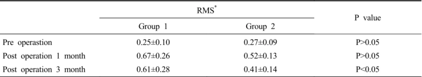
: 양군에서 LASEK전과 비교하여 수술 1개월 후 RMS는 통계적으로 유의하게 증가하였으며 수술 3개월 후에는 수술 1개월 후보다는 감소하는 경향을 보였다. 수술 3개월 후, larger and blend zone 방법으로 조사한 군에서 일반적 인 방법으로 조사한 군보다 RMS의 증가가 통계적으로 유의하게 적었다.
: LASEK 후 술전에 비해 술후 3개월까지 고차광학수차는 증가하였고 larger and blend zone의 방법으로 조사하 는 것이 일반적인 방법으로 조사한 것보다 고차광학수차의 증가를 적게 유발하여 시력을 질적으로 향상 시킬 수 있는 방법으로 생각된다.
557
Table 1. Pre operative spherical equivalent and pre operative RMS* between conventional ablation group and larger zone blend zone ablation group
Grup 1 Grup 2 P value
Spherical equivalent Pre operative RMS 4.46±1.67 0.25±0.10 4.49±1.75 0.27±0.09 P>0.05 P>0.05
*RMS: root mean square of Belle aberration maps (mean±standard deviation: micron).
Group 1 : conventional ablation group.
Group 2 : larger and blend zone ablation group.
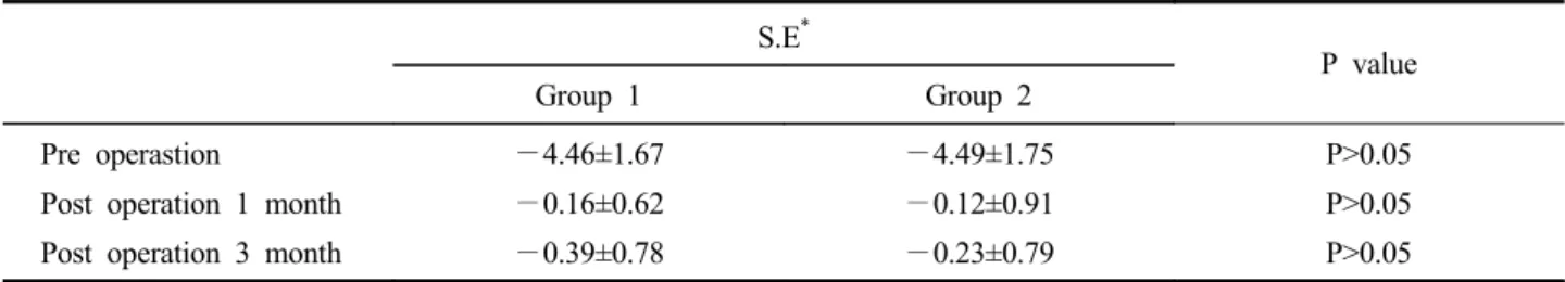
Table 2. Mean refractive data at pre operation, post operation 1 month and post operation 3 months
S.E*
P value Group 1 Group 2
Pre operastion
Post operation 1 month Post operation 3 month
4.46±1.67 0.16±0.62 0.39±0.78 4.49±1.75 0.12±0.91 0.23±0.79 P>0.05 P>0.05 P>0.05 *
S.E.: spherical equivalent (mean±standard deviation: diopter). Group 1 : conventional ablation group.
Group 2 : larger and blend zone ablation group.
Table 3. Visual acuity at pre operation, post operation 1 month and post operation 3 months
V. A*
P value Group 1 Group 2
Pre operastion (corrected)
Post operation 1 month (uncorrected) Post operation 3 month (uncorrected)
0.99±0.03 0.97±0.05 0.96±0.06 0.98±0.03 0.96±0.12 0.97±0.06 P>0.05 P>0.05 P>0.05
*V.A.: visual acuity (mean±standard deviation).
Group 1.: conventional ablation group.
Table 4. Optical aberration between pre operation , post operation 1month in conventional ablation group and in larger and
blend zone ablation group
Pre operation Pre operation 1 month P value
RMS* Gruop 1 Gruop 2 0.25±0.10 0.27±0.09 0.67±0.26 0.52±0.13 P<0.05 P<0.05
*RMS: root mean square of Belle aberration maps (mean±standard deviation: micron).
Group 1 : conventional ablation group.
Group 2 : larger and blend zone ablation group.
Table 5. Optical aberration between post operation 1month and post operation 3month in conventional ablation group and in
larger and blend zone ablation group
Pre operation 3 month Pre operation 1 month P value
RMS* Gruop 1 Gruop 2 0.67±0.26 0.52±0.13 0.61±0.28 0.41±0.14 P>0.05 P<0.05
*RMS: root mean square of Belle aberration maps (mean±standard deviation : micron).
Group 1 : conventional ablation group.
Group 2 : larger and blend zone ablation group.
Table 6. Optical aberration between conventional ablation and larger zone blend zone ablation
RMS*
P value Group 1 Group 2
Pre operastion
Post operation 1 month Post operation 3 month
0.25±0.10 0.67±0.26 0.61±0.28 0.27±0.09 0.52±0.13 0.41±0.14 P>0.05 P>0.05 P<0.05 *
RMS: root mean square of Belle aberration maps (mean±standard deviation: micron). Group 1 : conventional ablation group.
559
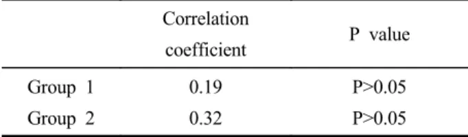
Table 7. Correlation coefficient between spherical equivalent
and optical aberration at post operation 3month Correlation coefficient P value Group 1 Group 2 0.19 0.32 P>0.05 P>0.05 Group 1 : conventional ablation group.
myopic astigmatism. Arch Ophthalmol 2000;118:17-21. 5) Martinez CE, Applegate RA, Klyce SD, et al. Effect of
papillary dilation on corneal optical aberrations after refrective keratectomy. Arch Ophthalmol 1998;116: 1053-62. 6) Oliver KM, Hemenger R, corbett MC, et al. Corneal optical
aberration induced by photorefrective keratectomy. J Refrract Surg 1997;13:246-54.
7) Applegate RA, Howland HC, Sharp RP, et al. Corneal aberrations, visual performance and refractive keratectomy. J Refract surg 1998;14:397-407.
8) Michael J, Carlos E, Martinez, Stephen D, et al. Effect of larger ablation zone and transition zone on corneal optical aberrations after photorefractive keratectomy. Arch Ophthalmol 2001;119:1159-64.
9) Hersh PS, Shah SI, Geiger D, Holladay JT. Summit PRK topography study group. Corneal optical irregularity after excimer laser photorefractive keratectomy. J Cataract Refract Surg 1996;22:197-204.
10) O’Brart DPS, Lohmann CP, Fitzke FW. Disturbances in night vision excimer laser photorefractive keratectomy. Eye 1994;8: 46-51.
11) Naoyuki M. Wavefront technology in ophthalmology. Curr Opin Ophthalmol 2001;12:294-9.
12) Schwiegerling J, Snyder RW. Custom photorefractive keratec-tomy ablations for the correction of spherical and cylindrical refractive error and higher-order aberration. J Opt Soc Am 1998;A15:2572-9.
13) Seiler T, Mrochen M, Kaemmerer M. Operative correction of ocular aberrations to improve visual acuity. J Refract Surg 2000;16:S619-22.
14) Mrochen M, Kaemmerer M, Seiler T. Wavefront - guided laser in situ keratomileusis: early results in three eyes. J Refract Surg 2000;16:116-21.
15) Stulting RD, Carr JD, Thompson KP, et al. Complications of laser in situ keratomileusis for the correction of myopia. Ophthalmol 1999;106:87-94.
561
The Effect of Larger and Blend Zone Ablation on Optical Aberrations
in Laser Epithelial Keratomileusis (LASEK)
Hyun Seok Kwon, M.D., Il Hwan Koh, M.D.,
Kyoung Yul Seo, M.D., Eung Kwon Kim, M.D.
The Institute of Vision Research
Department of Ophthalmology, Yonsei University, College of Medicine, Seoul Korea