■ 접 수 일: 2010년 2월 16일 ■ 심사통과일: 2010년 6월 3일
■ 책 임 저 자: 이 도 형
경기도 고양시 일산서구 대화동 2240 인제대학교 일산백병원 안과
Tel: 031-910-7240, Fax: 031-911-7241 E-mail: [email protected]
* 본 논문은 인제대학교 학술연구조성비 보조에 의한 것임.
pISSN: 0378-6471 eISSN: 2092-9374
DOI : 10.3341/jkos.2010.51.9.1210
= 증례보고 =
안내후방콘택트렌즈 삽입술 후 고위수차의 변화
박세훈1⋅염정훈1⋅최석규1⋅이종현1⋅김진형1⋅이도형1⋅김태진2
인제대학교 의과대학 일산백병원 안과학교실1, 인제대학교 의과대학 서울백병원 안과학교실2
목적: 유수정체 안내후방콘택트렌즈 삽입 후 고위수차의 변화를 알아보았다.
대상과 방법: 2008년 2월부터 2009년 10월까지 삽입술을 시행한 26안을 대상으로 술 전, 술 후 1주, 1개월, 3개월에 Hartmann-Shack 방식의 수차계(KR9000PW)로 눈 전체의 고위수차를 측정하였다. 술 전후에 각막중심 4 mm에서 총고위수차, 총구면수차, 총코마수차, 총트레포일수차 그리고 3차항, 4차항의 RMS값의 변화를 분석하였다.
결과: 평균나이는 25.4세였고, 현성굴절검사상 환자의 평균 구면대응치는 -6.40D였다. 술 후 1주, 1개월, 3개월에 평균 구면대응치는 각각 0.00 ± 0.13D, 0.03 ± 0.21D, -0.013 ± 0.12D이였다. 술 후 1주, 1개월, 3개월에 눈 전체의 총고위수차, 총구면수차, 총코마수차, 총트레포일수차, 3차, 4차항의 고위수차 RMS값 간에는 통계적으로 유의한 차이가 없었으며, 술 전과의 비교도 통계적으로 유의한 변화는 보이지 않았다(Wilcoxon-signed-ranks test, p<0.05).
결론: 유수정체 안내후방콘택트렌즈 삽입술 후 눈 전체의 각막중심 4 mm에서 고위수차는 변화가 없는 것으로 사료된다.
<대한안과학회지 2010;51(9):1210-1216>
안구의 굴절오차를 교정하여 시력을 개선하려는 인류의 노력은 계속되어 왔다. 굴절오차를 해결하는 방법에는 안경 이나 콘택트렌즈를 이용하는 방법에서 엑시머레이저를 이 용한 각막굴절교정수술, 투명수정체적출술 그리고 유수정 체안내렌즈삽입술 등이 있다. 안경과 콘택트렌즈는 비침습 적이고 간단한 방법이지만 미용상의 문제나 착용의 불편함 으로 굴절교정수술을 원하는 사람들이 많다. 엑시머레이저 로 각막굴절교정수술을 하는 방법이 보편화되어 있으나 각 막혼탁과 술 후 각막확장증이1-3발생할 수 있어 대상에 제 한이 있고 특히 술 후에 야간 눈부심이나 빛 번짐을 호소하 는 경우가 많다. 이는 각막굴절교정수술 후 각막곡률의 변 화와 고위수차가 증가하는 것이 그 원인으로 알려져 있 다.4-11또 다른 굴절교정 수술인 유수정체 안내콘택트렌즈 삽입술은 전방렌즈와 후방렌즈가 있으며, 전방렌즈삽입술 에는 전방각지지렌즈와 홍채고정렌즈가 있다.12 안내콘택 트렌즈는 술 후 시력회복이 빠르고 각막혼탁이나 퇴행현상 이 없고 렌즈제거가 용이하며, 조절력이 보전되는 장점과 야간 빛번짐과 눈부심이 적은 굴절교정수술로 술 후 예측
성과 안정성이 높은 수술방법으로 보고되고 있다.13-16 몇 연구에 의하면17,18고도근시 환자에서 전방홍채고정렌즈삽 입술 시행 후에 안구의 고위수차 변화가 적었으며 이것은 술 후 각막의 변화가 적어 술 전 각막곡률을 유지하기 때문 이라 하였다. 안내후방콘택트렌즈 삽입술 후 고위수차의 변 화에 대한 보고는 저자들이 아는 바 아직 없다. 본 연구에 서는 안내후방콘택트렌즈 삽입술을 받은 환자에서 술 전후 에 고위수차의 변화를 알아보고자 하였다.
대상과 방법
2008년 2월부터 2009년 10월까지 안내후방콘택트렌즈 삽입술이 계획된 근시 환자 13명 26안을 대상으로 하였다.
8명 16안 환자에는 ICL (Implantable contact lens, Staar Surgical AG, Switzerland)을 5명 10안에서는 평균 -2.50D의 난시를 보여 toric ICL (toric Implantable con- tact lens, Staar Surgical AG, Switzerland)을, 삽입하였다.
모든 환자에서 술 전에 나안시력 및 최대교정시력, 안압검 사, 각막곡률반경검사, 세극등현미경검사, 각막내피세포검 사, 시야검사, 현성 및 조절마비굴절검사, 안저검사, 각막지 형도검사, 중심부 및 주변부 각막두께검사, 각막직경검사, 초음파검사를 실시하였다. 술 전에 나이가 만 21세 미만이 거나, 각막혼탁, 녹내장, 포도막염 등의 안질환이 있는 경 우, 약시가 의심되는 경우, 당뇨 등의 전신질환이 동반된 경 우, 전방 깊이가 3 mm 이내인 경우, 각막내피세포의 수가
Table 2. Preoperative and postoperative spherical equivalent, uncorrected visual acuity and best-corrected visual acuity in patients
implanted with implantable contact lensMR* Preop. Postop. 1 wk Postop. 1 month Postop. 3 months
Spherical equivalent (Diopters) -6.40 ± 1.99D 0.00 ± 0.13D 0.03 ± 0.21D -0.013 ± 0.12D
UCVA† (logMAR) 1.11 ± 0.18 -0.09 ± 0.03 -0.01 ± 0.03 -0.01 ± 0.04
BCVA‡ (logMAR) 0.00 ± 0.00 -0.16 ± 0.07 -0.17 ± 0.08 -0.16 ± 0.08
* MR = manifest refraction; †UCVA = uncorrected visual acuity; ‡BCVA = best corrected visual acuity.
Table1. Patient characteristics and preoperative data
Variable Myopic patients
Age (mean) 25.4 (19 ~ 30)
Sex (M/F) 5/8
Number of eyes 26
ACD* (mean ± SD, mm) 3.28 ± 0.282 Axial length (mean ± SD, mm) 25.76 ± 0.92 Implanted IOL† power (D) -11.03 ± 1.245
*ACD = anterior chamber depth; †IOL = intraocular lens.
2,000 cells/mm2 미만인 경우는 수술 대상에서 제외하였 다. 안내후방콘택트렌즈 삽입술이결정된 환자는 술 후 발 생할 수 있는 동공폐쇄녹내장을 예방하기 위해서 최소한 수 술 2주전에 Nd-YAG레이저(neodymium YAG laser)를 이 용한 주변부홍채절개술을 10시와 2시 방향에 각각 시행하 였다. 수술 후 목표굴절력은 정시로 잡았으며 삽입할 렌즈 의 도수계산은 현성굴절검사상의 굴절이상을 기준으로 제조 회사(STAAR Surgical AG)의 공식을 따랐다.
수술 3일 전부터 0.3% levofloxacin을 하루에 4회씩 점안 하였고, 술 전에 Mydrin-PⓇ(tropicamaide/phenylephrine HCL)를 10분 간격으로 4회 점안하여 충분히 산동시킨 후 0.5% proparacaine으로 점안마취 하였다. 수술용 현미경하에 ICL 표면의 방향표시점을 확인한 후 제조사의 삽입장치 (STAAR ICL injector system)에 장착하였다. Microsurgery knife (Kai industries Co. Ltd., Japan)를 이용하여 3.2 mm 이측투명각막절개를 만들고 Diamond knife (S) (ME297, Meyco., Switzerland)를 이용하여 주변부 각막 6시, 12시 방향에 전방천자를 시행하였다. 점탄물질(Sodium hyalur- onate)을 주입하여 전방을 채운 후 삽입장치에 준비된 렌 즈를 전방으로 천천히 주입하고, 전방천자된 부위로 ma- nipulator (AE 2531, ASTCO, USA)를 넣어 ICL의 지지부 (footplate)를부드럽게 누르면서 홍채 뒤쪽으로 밀어넣었 다. 렌즈 삽입 후에 주사기에 평형염액(BSS, Alcon labo- ratories, Corp., USA)을 넣어 전방에 천천히 주입하여 점 탄물질(Sodium hyaluronate)을 수동으로 제거하였고, 각 막절개창은 봉합하지 않고 수술을 마쳤다. 수술 중 각막내 피, 수정체 전낭, 홍채에 기계적 손상을 주지 않도록 모든 술기에 세심한 주위를 기울였다. 모든 수술은 1인의 술자에 의해 시행되었으며 술 중 합병증은 발생하지 않았다. 수술
후 1일째부터 0.5% levofloxacin과 0.1% Fluorometholone 점안액을 1일 4회씩 점안하였으며, 경과관찰하면서 사용 횟수를 줄이도록 하였다. 경과 관찰 중 합병증은 관찰되지 않았다.
술 전 및 술 후 1주, 1개월, 3개월에 Hartmann-shack 방식 의 수차계인 KR9000PW (Topcon Corp., Tokyo, Japan)19,20 를이용하여 암실에서 눈 전체의 수차를 측정하였다. 술 전 평균 동공 크기는 6.73 ± 1.52 mm이였고, 암순응 상태에 서 측정된 동공 6 mm에서 눈 전체의 고위수차를 분석하였 다. 또한 삽입된ICL의 optic이 4.65 mm이므로 동공 4 mm 에서 고위수차를 분석하였으며, 동공 4 mm 총고위수차, 총 구면수차, 총코마수차, 총트레포일수차, 3차항(coma-like aberration) 그리고 4차항(spherical-like aberration)의 RMS값들을 눈 전체(ocular), 각막(cornea), 각막전면을 제 외한 안구 내(internal)로 각각 분석하여 비교하였다.
통계학적 처리는 SPSS 12.0 for Window (SPSS Inc.)를 이용하였고, Wilcoxon 부호순위 검정을 시행하였으며, p값 이 0.05 미만일 경우 통계적으로 유의하다고 판단하였다.
결 과
13명 26안은 남자 5명 10안, 여자 8명 16안이었으며, 나 이는 평균 25.4세(19~30)이였으며, 술 전 평균 전방깊이 및 안장축은 3.28 ±0.282 mm, 25.76 ±0.92 mm였다(Table 1).
술 전 구면렌즈 대응치의 평균은 -6.40D (-1.0~-9.75) 였으며, 수술 후 평균구면렌즈대응치는 1주, 1개월, 3개월 에 각각 0.00 ± 0.13D, 0.03 ± 0.21D, -0.013 ± 0.12D이 였다. 수술 전 평균 최대교정시력(logMAR)은 0.00 ± 0.00 이었으며, 술 후 평균나안시력(logMAR)은 1주, 1개월, 3개월에 -0.09 ± 0.03, -0.01 ± 0.03, -0.01 ± 0.04였으 며, 술 후 최대교정시력(logMAR)은 1주, 1개월, 3개월에 -0.16 ± 0.07, -0.17 ± 0.08, -0.16 ± 0.08이었다(Table 2).
대상 가운데 10안에서는 술 전 평균 -2.50 ± 0.87D의 난 시가 있어 Toric ICL을 삽입하였고, 술 후 1주, 1개월, 3개월 에평균 난시는 -0.27 ± 0.52D, -0.27 ± 0.45D, -0.22 ± 0.31D를 보여 술 전에 비해 술 후에 초기 난시양의 90%
이상이 교정되었다. 경과관찰 중 포도막염, 망막박리 등의
Table 3. Changes in mean IOP, specular microscopy and pachymetry during the follow-up period
Variable Preop. Postop.1 wk Postop.1 month Postop.3 months
IOP* (mmHg) 15.54 ± 1.63 17.19 ± 2.59 16.46 ± 2.12 15.85 ± 2.68
Specular (cells/mm2) Pachymetry (μm)
2,656.69 ± 397.58 490.81 ± 40.63
2,584.69 ± 320.19 510.25 ± 25.92
2,657.79 ± 196.55 500.55 ± 34.75
2,561.00 ± 279.84 502.78 ± 34.59
*IOP=intraocular pressure.
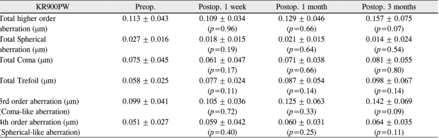
Table 4. The ocular higher order aberrations RMS (root-mean-square) including spherical, coma and trefoil aberration (vector
Zernike analysis up to the 4th order for a 4-mm pupil diameter) preoperatively and postsurgery 1week, 1 month and 3 monthsKR900PW Preop. Postop. 1 week Postop. 1 month Postop. 3 months
Total higher order aberration (μm)
0.113 ± 0.043 0.109 ± 0.034 (p=0.96)
0.129 ± 0.046 (p=0.66)
0.157 ± 0.075 (p=0.07) Total Spherical
aberration (μm)
0.027 ± 0.016 0.018 ± 0.015 (p=0.19)
0.021 ± 0.015 (p=0.64)
0.014 ± 0.024 (p=0.54)
Total Coma (μm) 0.075 ± 0.045 0.061 ± 0.047
(p=0.17)
0.071 ± 0.038 (p=0.66)
0.081 ± 0.055 (p=0.80)
Total Trefoil (μm) 0.058 ± 0.025 0.077 ± 0.024
(p=0.11)
0.087 ± 0.054 (p=0.14)
0.098 ± 0.067 (p=0.14) 3rd order aberration (μm)
(Coma-like aberration)
0.099 ± 0.041 0.105 ± 0.036 (p=0.72)
0.125 ± 0.063 (p=0.33)
0.142 ± 0.069 (p=0.09) 4th order aberration (μm)
(Spherical-like aberration)
0.051 ± 0.027 0.059 ± 0.042 (p=0.40)
0.060 ± 0.031 (p=0.25)
0.064 ± 0.035 (p=0.11)
*Wilcoxon-signed-ranks test.
합병증은 발생하지 않았다.
평균 안압은 술 전 15.54 ± 1.63 mmHg에서 술 후 1주째 17.19 ± 2.59 mmHg로 상승하였으나, 술 후 1개월에는 16.46 ± 2.12 mmHg, 3개월에는 15.85 ± 2.68 mmHg으로 측정되었으며 통계적으로 유의한 차이를 보이지 않았으며, 안압 상승의 합병증은 경과관찰 동안 발생하지 않았다 (p<0.05, Table 3). 또한 각막두께 및 각막내피세포수를 술 전과 술 후 1주, 1개월, 3개월에 측정한 결과 통계적으 로 유의한 차이를 보이지 않았다(p<0.05, Table 3).
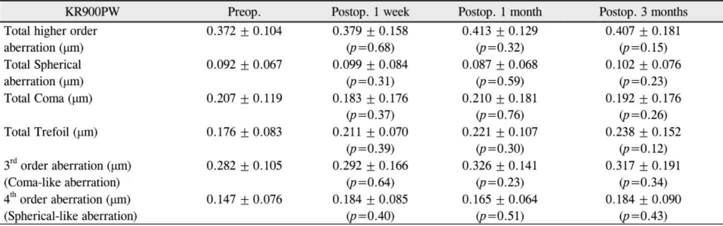
한편, 동공 4 mm에서 눈 전체의 고위수차는 술 전 및 술 후 1주, 1개월, 3개월에 총고위수차가 각각 0.113 ± 0.043 μm, 0.109 ± 0.034 μm, 0.129 ± 0.046 μm, 0.157 ± 0.075 μm로 측정되었으며, 총구면수차는 0.027 ± 0.016 μm, 0.018 ± 0.015 μm, 0.021 ± 0.015 μm, 0.014 ± 0.024 μm, 총코마수차는 0.075 ± 0.045 μm, 0.061 ± 0.047 μm, 0.071 ± 0.038 μm, 0.081 ± 0.055 μm, 총트레포일수차는 0.058 ± 0.025 μm, 0.077 ± 0.024 μm, 0.087 ± 0.054 μm, 0.098 ± 0.067 μm, 3차항의 RMS값은 0.099 ± 0.041 μm, 0.105 ± 0.036 μm, 0.125 ± 0.063 μm, 0.142 ± 0.069 μm, 그리고 4차항의 RMS값은 0.051 ± 0.027 μm, 0.059 ± 0.042 μm, 0.060 ± 0.031 μm, 0.064 ± 0.035 μm로 술전과 비교해 통계적으로 유의한 변화를 보이지 않았다(p<0.05, Table 4). 또한 동공 6 mm에서의 눈 전체의 고위수차는 술 후 1주, 1개월, 3개월에 총고위수차, 총구면수차, 총코마수
차, 총트레포일수차, 3차, 4차항의 고위수차의 RMS값 모두 술 전과 비교해 통계적으로 유의한 차이가 없었다(p<0.05, Table 5).
동공 4 mm에서 각막의 고위수차는 총구면수차와 4차항 의 수차가 각각 술 전에 0.016 ± 0.011 μm, 0.025 ± 0.010 μm였으며, 술 후 1주째 0.031 ± 0.017 μm, 0.041 ± 0.014 μm (p=0.011, p=0.015), 1개월에 0.028 ± 0.016 μm, 0.041 ± 0.014 μm (p=0.012, p=0.015)로 술 전과 비교해 통계적으로 유의한 차이가 있었으나, 술 후 3개월에 0.020
± 0.013 μm, 0.030 ± 0.011 μm로 측정되어 술 전과 비교 해 통계적으로 유의한 차이가 없었고 술 전으로 감소하는 경향을 보였다(Table 6).
동공 4 mm에서 안구내 고위수차는 총구면수차와 4차항 의 수차가 각각 술전에 0.021 ± 0.015 μm, 0.049 ± 0.022 μm였으며, 술 후 1주째 0.072 ± 0.102 μm, 0.077 ± 0.041 μm (p=0.013, p=0.017)로 술 전과 비교해 통계적으로 유 의한 차이가 있었으나 1개월, 3개월에는 술 전과 비교해 통 계적으로 유의한 차이가 없었고 다시 술 전으로 감소하는 경향을 보였다(Table 7).
고 찰
굴절오차를 가진 환자 중에는 안경이나 콘택트렌즈를 사 용해야 하는 불편함과 미용상의 이유등으로 굴절교정수술
Table 5. The ocular higher order aberrations RMS (root-mean-square) including spherical, coma and trefoil aberration (vector
Zernike analysis up to the 4th order for a 6-mm pupil diameter) preoperatively and postsurgery 1week, 1 month and 3 monthsKR900PW Preop. Postop. 1 week Postop. 1 month Postop. 3 months
Total higher order aberration (μm)
0.372 ± 0.104 0.379 ± 0.158 (p=0.68)
0.413 ± 0.129 (p=0.32)
0.407 ± 0.181 (p=0.15) Total Spherical
aberration (μm)
0.092 ± 0.067 0.099 ± 0.084 (p=0.31)
0.087 ± 0.068 (p=0.59)
0.102 ± 0.076 (p=0.23)
Total Coma (μm) 0.207 ± 0.119 0.183 ± 0.176
(p=0.37)
0.210 ± 0.181 (p=0.76)
0.192 ± 0.176 (p=0.26)
Total Trefoil (μm) 0.176 ± 0.083 0.211 ± 0.070
(p=0.39)
0.221 ± 0.107 (p=0.30)
0.238 ± 0.152 (p=0.12) 3rd order aberration (μm)
(Coma-like aberration)
0.282 ± 0.105 0.292 ± 0.166 (p=0.64)
0.326 ± 0.141 (p=0.23)
0.317 ± 0.191 (p=0.34) 4th order aberration (μm)
(Spherical-like aberration)
0.147 ± 0.076 0.184 ± 0.085 (p=0.40)
0.165 ± 0.064 (p=0.51)
0.184 ± 0.090 (p=0.43)
*Wilcoxon-signed-ranks test.
Table 6. The corneal higher order aberrations RMS (root-mean-square) including spherical, coma and trefoil aberration (vector
Zernike analysis up to the 4th order for a 4-mm pupil diameter) preoperatively and postsurgery 1week, 1 month and 3 monthsKR900PW Preop. Postop. 1 week Postop. 1 month Postop. 3 months
Total higher order aberration (μm)
0.113 ± 0.056 0.116 ± 0.043 (p=0.65)
0.110 ± 0.038 (p=0.54)
0.127 ± 0.032 (p=0.14) Total Spherical
aberration (μm)
0.016 ± 0.011 0.031 ± 0.017* (*p=0.01)
0.028 ± 0.016* (*p=0.01)
0.020 ± 0.013 (p=0.25)
Total Coma (μm) 0.104 ± 0.059 0.089 ± 0.049
(p=0.06)
0.099 ± 0.051 (p=0.36)
0.101 ± 0.046 (p=0.65)
Total Trefoil (μm) 0.041 ± 0.029 0.052 ± 0.024
(p=0.45)
0.048 ± 0.024 (p=0.24)
0.052 ± 0.050 (p=0.14) 3rd order aberration (μm)
(Coma-like aberration)
0.110 ± 0.056 0.107 ± 0.046 (p=0.96)
0.095 ± 0.056 (p=0.76)
0.109 ± 0.046 (p=0.58) 4th order aberration (μm)
(Spherical-like aberration)
0.025 ± 0.010 0.041 ± 0.014* (*p=0.02)
0.041 ± 0.014* (*p=0.02)
0.030 ± 0.011 (p=0.13)
*Wilcoxon-signed-ranks test, p<0.05.
Table 7. The internal higher order aberrations RMS (root-mean-square) including spherical, coma and trefoil aberration (vector
Zernike analysis up to the 4th order for a 4-mm pupil diameter) preoperatively and postsurgery 1week, 1 month and 3 monthsKR900PW Preop. Postop. 1 week Postop. 1 month Postop. 3 months
Total higher order aberration (μm)
0.118 ± 0.055 0.127 ± 0.055 (p=0.22)
0.136 ± 0.067 (p=0.12)
0.159 ± 0.094 (p=0.07) Total Spherical
aberration (μm)
0.021 ± 0.015 0.072 ± 0.102* (*p=0.01)
0.017 ± 0.019 (p=0.61)
0.020 ± 0.020 (p=0.32)
Total Coma (μm) 0.082 ± 0.060 0.083 ± 0.045
(p=0.61)
0.093 ± 0.060 (p=0.35)
0.107 ± 0.069 (p=0.11)
Total Trefoil (μm) 0.053 ± 0.029 0.057 ± 0.033
(p=0.77)
0.077 ± 0.051 (p=0.26)
0.087 ± 0.062 (p=0.06) 3rd order aberration (μm)
(Coma-like aberration)
0.106 ± 0.055 0.099 ± 0.050 (p=0.45)
0.099 ± 0.070 (p=0.55)
0.142 ± 0.086 (p=0.11) 4th order aberration (μm)
(Spherical-like aberration)
0.049 ± 0.022 0.077 ± 0.041* (*p=0.02)
0.059 ± 0.044 (p=0.14)
0.055 ± 0.034 (p=0.42)
*Wilcoxon-signed-ranks test, p<0.05.
을 원하는 사람이 많고 굴절교정수술에는 각막굴절교정수 술, 투명수정체적출술, 유수정체 안내콘택트렌즈 삽입술 등 의 방법이 있다. 라식과 같은 각막굴절교정수술은 보편화된 방법으로 안정성과 효용성에 효과적인 수술이지만, 각막의
비가역적인 변화로 몇몇 문제점이 보고 되었다. 술 후 퇴행 가능성, 고도근시 교정시의 안정성과 효용성의 감소, 각막 혼탁, 야간눈부심, 빛번짐 그리고 의인성각막확장증이 발생 할 수 있어서 각막이 얇거나 각막지형도에 이상이 있는 환
자에서는 제한점이 있다.1-11 투명수정체적출술은 초고도 근시 환자에게 시행되었으며 술 후 교정시력의 향상에 효 과적이다는 보고가 있으나 인공수정체 돗수 예측이 어렵고 술 후 조절능력을 상실하게 되고 낭포성황반부종, 망막박리 의 위험성이 있어 수술환자 선별이 중요한 것으로 알려져 있다.21-23
또 다른 굴절교정수술로 유수정체 안내콘택트렌즈 삽입 술이 있다.12 유수정체 안콘택트렌즈에는 전방렌즈와 후방 렌즈가 있으며 현재까지 소개된 전방렌즈에는 Phakic6, Nuvita 렌즈, Vivarte 렌즈 등의 같은 전방각지지렌즈와 Artisan, Artiflex 등의 홍채고정렌즈가 있고, 후방렌즈에는 ICL (Implantable contact lens, Staar Surgical AG, Switzerland) 이 있다. 유수정체 안내콘택트렌즈 삽입술은 각막의 비가역 적인 변화를 주지 않고 조절력을 보전하고 교정가능범위가 크며, 합병증이 발생한 경우 삽입되었던 렌즈제거가 용이한 장점이 있고, 단점으로는 각막내피세포의 손상, 수정체의 전낭혼탁, 인공수정체 중심부 이탈, 녹내장의 발생이 지적
되었다.24-28 본 연구에 사용된 안내후방콘택트렌즈(ICL,
Implantable contact lens, Staar Surgical AG, Switzerland) 는 1997년 국내에 최초로 소개되어 활발히 시행되고 있으 며 중장기임상결과상 예측성, 효용성 그리고 안정성이 높은 렌즈로 보고되고 있다.13-16
굴절교정수술 후 고위수차의 변화에 대한 연구들에서는 라식 등의 각막굴절교정수술 후 고위수차의 증가가 알려져 있으며 고위수차의 증가는 각막연마량(corneal ablation)이 많을수록 더 증가하고 환자의 시력의 질을 나쁘게 하는 원 인으로 보고되었다.24,29,30홍채고정렌즈삽입술 후의 고위수 차 변화에 대한 연구에 의하면 Tehrani and Dick17은 Artiflex 렌즈는 삽입술 후 고위수차의 통계적 유의한 변화 가 없다고 하였다. 반면, Tahzib et al18은 Artisan 렌즈와 Artifex 렌즈를 삽입한 군에서 술 후 구면수차와 트레포일 수차의 변화가 있었으며, 이는 각막절개와 광학부 디자인에 의한 것으로 보고하였다. 그러나 아직 유수정체안내후방렌 즈(ICL) 삽입술 후의 고위수차의 변화에 대한 보고는 저자 들이 아는 바 없다.
이에 본 연구에서는 Hartmann-shack 방식의 수차계이 면서 각막지형도를 분석할 수 있는 KR9000PW (Topcon Corp., Tokyo, Japan)를 이용하여 눈 전체의 고위수차의 측정과 함께 각막의 고위수차변화 및 각막을 제외한 안구 내 고위수차를 각각 분석하였다. 유수정체안내후방렌즈 (ICL)는 광학부가 4.65~5.5 mm이므로154 mm 동공지름 에서 고위수차를 분석하였는데, 술 후 1주, 1개월, 3개월에 눈 전체의 총고위수차, 총구면수차, 총코마수차, 총트레포 일수차, 3차, 4차항의 고위수차의 RMS값 간에는 통계적으
로 유의한 차이가 없었으며, 술 전과의 비교도 통계적으로 유의한 변화는 보이지 않았다(p<0.05). 또한 동공지름 6 mm에서 눈 전체의 고위수차도 분석해 보았는데 술 후 1주, 1개월, 3개월에 총고위수차, 총구면수차, 총코마수차, 총트 레포일수차, 3차, 4차항의 고위수차의 RMS 값간에는 통계 적으로 유의한 차이가 없었으며, 술 전과의 비교도 통계적 으로 유의한 변화는 보이지 않았다(p<0.05, Table 5). 이 는 Tehrani and Dick17이 보고한 홍채고정렌즈삽입술 후 1 주, 3개월, 6개월, 12개월에 3차항(coma-like aberrations : Z31 & Z3-1)의 고위수차가 술 전에 비해 감소하였으나통 계적으로 유의한 차이가 없었으며, 4차항(spherical-like aberration : Z40
)의 고위수차가 술 전에 비해 술 후 1주에 통계적으로 유의하게 절대값의 감소가 있었으나 이후 술 후 3개월, 6개월, 12개월에는 유의한 변화가 없어 홍채고정 렌즈삽입술 후 3차, 4차항의 고위수차는 증가하지 않았다 는 것과 본 연구의 결과가 유사함을 알 수 있다.
한편, 각막의 고위수차 중에는 술 후 1주, 1개월에 총구 면수차 및 4차항의 수차의 증가가 있었으나 술 후 3개월에 술 전과 비교해 통계적으로 유의한 차이 없이 술 전으로 감 소하는 경향을 보이고, 눈 전체의 총고위수차에 변화가 없 는 것으로 볼 때 그 변화는 적은 것으로 판단된다. 각막전면 을 제외한 안구내 고위수차에서는 총구면수차가 술 후 1주 째 증가하였으나 술 1개월 후에는 술 전으로 회복하였다.
최근 백내장수술 후 각막의 고위수차의 변화에 대한 연 구에서 술 후 각막의 고위수차중 트레포일수차와 구면수차 의 증가가 있으며 수술 시 각막절개의 크기에 영향이 있는 것으로 보고되고 있다.31-34또한 Tahzib et al18는 Artisan 렌즈를 삽입한 군에서는 술전 구면수차(Z(4,0)) 및 트레포 일수차(Z(3,-3))가 0.19 ± 0.20 μm, -0.04 ± 0.20 μm에 서 술 후 0.60 ± 0.34 μm, 0.09 ± 0.24 μm로 통계적으로 유의하게 증가하였으며, Artifex 렌즈를 삽입한 군에서는 술 전 구면수차(Z(4,0)) 및 트레포일수차(Z(3,-3))가 0.29 ± 0.18 μm, -0.05 ± 0.15 μm에서 술 후 0.03 ± 0.16 μm, -0.13 ± 0.19 μm로 통계적으로 유의하게 구면수차의 감소 가 있었고, 트레포일수차의 증가가 있었다고 보고하였다.
이는 Artisan 렌즈와 Artiflex 렌즈 삽입 시 각각 6.0 mm와 3.4 mm의 각막절개가 고위수차 증가에 기인한 것이며, Artiflex 렌즈는 광학부의 negative spherical aberration이 있어 구면수차의 감소가 있는 것으로 보고하였고, 더하여 두 렌즈 사이의 구면수차의 차이가 각막절개의 차이와 이 에 따른 상처치유과정의 차이에 기인한 것으로 보고하였다.
본 연구에서도 3.2 mm의 각막절개가 술 후 1주, 1개월에 각막 고위수차의 증가에 영향을 주었을 것으로 생각된다.
반면, 각막전면을 제외한 안구 내의 1주째의 구면수차의 증
가는 수술 시 점탄물질의 제거가 완전하지 못하여 1주째 점 탄물질이 완전 흡수되지 않았거나 수술 시의 안내 정상 구 조의 변화가 있었을 것으로 생각되지만 수술 후 1개월 후에 는 술 전의 상태로 회복될 정도로 가역적이고 미세한 변화 인 것으로 보인다. 또한 본 연구의 대상 가운데 10안에서는 Toric ICL을 삽입하였는데 술 후 3개월까지 초기 난시양의 90% 이상 교정된 상태가 지속되고 있어 Toric ICL의 축정 렬 정도가 고위수차에 미치는 영향을 적을 것으로 사료되 며, 저위수차(defocus and astigmatism)가 고위수차의 변 화에 미치는 영향이 적다는 보고도 있어 저위수차에 의한 영향은 적을 것으로 판단된다.17
안구의 광학적 진단과 치료의 발달과 환자들의 보다 나 은 시력에 대한 욕구의 증가로 고위수차에 대한 관심이 높 아지고 있으며, 수차계의 발달로 눈 전체의 고위수차를 분 석하여 환자의 시력의 질을 평가하고 이해할 수 있게 되었 다. 굴절교정수술 후 환자들의 만족도와 시력의 질을 높이 고자 하는 안과학의 노력은 앞으로도 계속될 것으로 예상 되며, 유수정체 안내후방렌즈(ICL)삽입술은 각막굴절교정 수술 후 고위수차의 증가가 예상되거나 적응증이 되지 않 는 환자에서 대안이 될 수 있을 것으로 생각된다. 본 연구 에서는 26안을 대상으로 안구내후방렌즈삽입술 후 눈 전체 의 고위수차의 변화가 없음을 확인할 수 있었다. 향후 ICL 의 광학적 특성에 대한 실험적 연구와 큰 집단을 대상으로 한 장기적인 추적관찰에 대한 연구가 필요할 것으로 사료 된다.
참고문헌
1) Kim TG, Joo CK. 2 cases of corneal ectasia dected after LASIK. J Korean Ophthalmol Soc 1999;40:846-9.
2) Amoils SP, Deist MB, Gous P, Amoils PM. Iarogenic keratectasia after laser in situ keratomileusis for less than -4.0 to -7.0 diopters of myopia. J Cataract Refract Surg 2000;26:967-77.
3) Geggel HS, Talley AR. Delayed onset keratectasia following laser in situ keratomileusis and photorefractive keratectomy. Ophthalmology 2000;107:640-52.
4) Bailey MD, Mitchell GL, Dhaliwal DK, et al. Patient satisfaction and visual symptoms after laser in situ keratomileusis. Ophthalmology 2003;110:1371-78.
5) Buzzonetti L, Iarossi G, Valente P, et al. Comparison of wavefront aberration changes in the anterior corneal surface after laser-as- sisted subepithelial keratectomy and laser in situ keratomileusis:
preliminary study. J Cataract Refract Surg 2004;30:1929-33.
6) Buzzonetti L, Petrocelli G, Valente P, et al. Comparison of corneal aberration changes after laser in situ keratomileusis performed with mechanical microkeratome and IntraLase femtosecond laser:
1-year follow-up. Cornea 2008;27:174-9.
7) Chalita MR, Chavala S, Xu M, Krueger RR. Wavefront analysis in post-LASIK eyes and its correlation with visual symptoms, re- fraction, and topography. Ophthalmology 2004;111:447-53.
8) Chalita MR, Xu M, Krueger RR. Correlation of aberrations with visual symptoms using wavefront analysis in eyes after laser in situ keratomileusis. J Refract Surg 2003;19:682-6.
9) Yamane N, Miyata K, Samejima T, et al. Ocular higher-order aber- rations and contrast sensitivity after conventional laser in situ keratomileusis. Invest Ophthalmol Vis Sci 2004;45:3986-90.
10) Pesudovs K. Wavefront aberration outcomes of LASIK for high myopia and high hyperopia. J Refract Surg 2005;21:508-12.
11) Sharma M, Boxer Wachler BS, Chan CC. Higher order aberrations and relative risk of symptoms after LASIK. J Refract Surg 2007;
23:252-6.
12) Budo C, Hessloehl JC, Izak M, et al. Multicenter study of the Artisan phakic intraocular lens. J Cataract Refract Surg 2000;
26:1163-71.
13) Jimenez AI, Gomez TG, Bueno JL, Puy P. Contrast sensitivity after posterior chamber phakic intraocular lens implantation for high myopia. J Refract Surg 2001;17:641-5.
14) Lee SY, Cheon HJ, Baek TM, Lee KH. Implantable contact lens to correct high myopia. J Korean Ophthalmol Soc 2000;41:1515-22.
15) Han SY, Lee KH. Long Term Effect of ICL Implantation to Treat High Myopia. J Korean Ophthalmol Soc 2007;48:465-72.
16) Chun YS, Lee JH, Lee JM, et al. Outcomes after Implantable Contact Lens for Moderate to High Myopia. J Korean Ophthalmol Soc 2004;45:480-9.
17) Tehrani M, Dick HB. Changes in higher-order aberrations after im- plantation of a foldable iris-claw lens in myopic phakic eyes. J Cataract Refract Surg 2006;32:250-4.
18) Tahzib NG, MacRae SM, Yoon G, et al. Higher-order aberrations after implantation of iris-fixated rigid or foldable phakic intra- ocular lenses. J Cataract Refract Surg 2008;34:1913-20.
19) Kuroda T, Fujikado T, Maeda N, et al. Wavefront analysis of higher order aberrations in patients with cataracts. J Cataract Refract Surg 2002;28:438-44.
20) Oshika T, Klyce SD, Applegate RA, et al. Comparison of corneal wavefront aberrations after photorefractive keratectomy and laser in situ keratomileusis. Am J Ophthalmol 1999;127:1-7.
21) Fernández-Vega L, Alfonso JF, Villacampa T. Clear lens extraction for the correction of high myopia. Ophthalmology 2003;110:2349- 54.
22) Huber C. Effectiveness of intraouclar lens calculation in high- ametropia. J Cataract Refract Surg 1989;15:667-72.
23) Colin J, Robinet A, Cochener B. Retinal detachment after clear lens extraction for high myopia; seven-year follow-up. Ophthalmology 1999;106:2281-4.
24) AlióJ, Galal A, Montálban R, Pin˝ero D. Corneal wavefront- guided LASIK retreatments for correction of highly aberrated corneas fol- lowing refractive surgery. J Refract Surg 2007;23:760-73.
25) Menezo JL, Peris-Martinez C, Cisneros AL, et al. Phakic intra- ocular lenses to correct high myopia: Adatomed, Staar, and Artisan. J Cataract Refract Surg 2004;30:33-44.
26) Sarikkola AU, Sen HN, Uusitalo RJ, et al. Traumatic cataract and other adverse events with the implantable contact lens. J Cataract Refract Surg 2005;31:511-24.
27) Perez-Santonja JJ, Alio JL, Jimenez-Alfaro I, et al. Surgical correc- tion of severe myopia with an angle-supported phakic intraocular lens. J Cataract Refract Surg 2000;26:1288-302.
28) Baikoff G, Arne JL, Bokobza Y, et al. Angle-fixated anterior cham- ber phakic intraocular lens for myopia of -7 to -19 diopters. J
=ABSTRACT=
Changes in Higher Order Aberration After Implantable Contact Lens Implantation
Se Hoon Park, MD1, Jung Hoon Yum, MD1, Suk Kyue Choi, MD1, Jong Hyun Lee, MD1, Jin Hyoung Kim, MD1, Do Hyung Lee, MD, PhD1, Tae Jin Kim, MD2
Department of Ophthalmology, Ilsan Paik Hospital, Inje University College of Medicine1, Goyang, Korea Department of Ophthalmology, Seoul Paik Hospital, Inje University College of Medicine2, Seoul, Korea
Purpose: To evaluate the changes in higher-order aberrations (HOAs) after implantation of an ICL (implantable contact lens).
Methods: Twenty-six eyes that had undergone ICL implantation between February 2008 and October 2009 were included.
Ocular higher-order aberrations of all the eyes were measured using the Hartmann-Shack aberrometer (KR9000PW).
Examinations were performed preoperatively, as well as one week, one month, and three months after ICL implantation.
Changes in ocular total higher-order aberrations, total spherical aberration, coma aberration, trefoil aberration, and S3 and S4 calculated in the central 4-mm zone and expressed as root mean square (RMS) values were analyzed.
Results: The mean patient age was 25.4 years, and the mean preoperative spherical equivalent was -6.40 diopters(D).
The mean postoperative spherical equivalents were 0.00 ± 0.13D, 0.03 ± 0.21D, -0.013 ± 0.12D at one week, one month, and three months, respectively. There were no statistically significant differences in ocular higher-order aberration RMS, including spherical, coma, or trefoil aberration, at one week, one month, or three months postoperative or between pre- operative and postoperative measurements.
Conclusions: There is no significant change of ocular higher order aberration in the central 4-mm zone after ICL implantation.
J Korean Ophthalmol Soc 2010;51(9):1210-1216
Key Words: Higher order aberrations, Implantable contact lens
Address reprint requests to Do Hyung Lee, MD, PhD Department of Ophthalmology, Ilsan Paik Hospital
# 2240 Daehwa-dong, Ilsanseo-gu, Goyang 411-760, Korea
Tel: 82-31-910-7240, Fax: 82-31-911-7241, E-mail: [email protected] Refract Surg 1998;14:282-93.
29) Oshika T, Miyata K, Tokunaga T, et al. Higher order wavefront aberrations of cornea and magnitude of refractive correction in la- ser in situ keratomileusis. Ophthalmology 2002;109:1154-8.
30) Kohnen T, Mahmoud K, Bühren J. Comparison of corneal high- er-order aberrations induced by myopic and hyperopic LASIK.
Ophthalmology 2005;112:1692-8.
31) Marcos S, Rosales P, Llorente L, et al. Change in corneal aberra- tions after cataract surgery with 2 types of aspherical intraocular lenses. J Cataract Refract Surg 2007;33:217-26.
32) Iseli HP, Jankov M, Bueeler M, et al. Corneal and total wavefront aberrations in phakic and pseudophakic eyes after implantation of monofocal foldable intraocular lenses. J Cataract Refract Surg 2006;32:762-71.
33) Pesudovs K, Dietze H, Stewart OG, et al. Effect of cataract surgery incision location and intraocular lens type on ocular aberrations. J Cataract Refract Surg 2005;31:725-34.
34) Tong N, He JC, Lu F, et al. Changes in corneal wavefront aberra- tions in microincision and small-incision cataract surgery. J Cataract Refract Surg 2008;34:2085-90.