280 PISSN 0304-128X, EISSN 2233-9558
Constitutive Expression of Lipase on the Cell Surface of Escherichia coli using OmpC Anchoring Motif
Seung Hwan Lee*,† and Sang Yup Lee**
*Department of Biotechnology & Bioengineering, Chonnam National University, 77 Yongbong-ro, Buk-gu, Gwangju, 61186, Korea
**Metabolic and Biomolecular Engineering National Research Laboratory, Department of Chemical & Biomolecular Engineering (BK21 Program), Institute of BioCentury, Korea Advanced Institute of Science and Technology, 291, Daehak-ro, Yeseong-gu, Daejeon, 34141, Korea
(Received 17 January 2020; Received in revised form 20 Jaunary 2020; accepted 21 January 2020)
Abstract − We have developed a constitutive display system of the Pseudomonas fluorescens SIK W1 TliA lipase on the cell surface of Escherichia coli using E. coli outer membrane protein C (OmpC) as an anchoring motif, which is an economical compared to induced system. For the constitutive expression of truncated OmpC-TliA fusion proteins, gntT104 promoter was employed. Cell growth was not affected by over expression of fusion protein during entire culture time, suggesting cell lysis was not a problem. The localization of truncated OmpC-TliA fusion protein on the cell surface was confirmed by immunofluorescence microscopy and measuring whole cell lipase activity. Constitutively displayed lipase was very stable, retaining activity enantioselectivity throughout the five repeated reactions. These results suggest that OmpC from E. coli be a useful anchoring motif for displaying enzymes on the cell surface without any inducers, and this stable surface display system can be employed for a broad range of biotechnological applications.
Key words: Cell surface display, Lipase, Constitutive expression, Outer membrane protein, Escherichia coli
1. Introduction
Lipases (triacylglycerol ester hydrolases, EC 3. 1. 1. 3) are ubiquitous enzymes found in various living organisms, including bacteria, plants and animals. Originally, this enzyme catalyzes the hydrolysis of oils, triglycerides and fatty acid esters under aqueous condition and also mediates the transesterification of esters under the non- aqueous solvents. It is one of the most commonly used enzymes in industry, because of its excellent enantioselectivity, commercial availability and broad substrate specificities to natural and unnatural esters of different structures [1-5]. However, there are some drawbacks for the practical usage of soluble enzyme, including difficulty of separation from solution and decrease or elimination of its activity under the non-conventional conditions such as organic solvents, extreme pH or temperature. These problems can be solved by immobilization of enzyme on the solid supports by physical methods such as adsorption on a water-insoluble matrix, gel entrapment, microencapsulation with a solid membrane and formation of enzymatic Langmuir- Blodgett film, and chemical methods including covalent attachment, crosslinking and co-crosslinking with other substances [6-10]. However, these methods also present disadvantages. For example, the interaction between enzymes and supports can be weak, which decreases total activity of immobilized enzyme. In the case of chemical methods, immobilization is rather expensive and covalent bonding often causes
structural change of the enzyme [11].
Cell surface display, which is a technique to display peptides or proteins on the surface of gram-negative and gram-positive bacteria, fungi, or even mammalian cells by appropriately fusing them to surface anchoring motifs, can be used a non-conventional method for immobilization of enzyme [12-15]. Several researchers have been reported successful expression of lipase on the cell surface of bacteria such as E. coli, Psedomonas sp., and yeast strains by employing various anchoring techniques and cell surface displaying enzyme shows excellent enzymatic characteristics [16-19]. However, the expression of fusion proteins was induced by adding expensive isopropyl-β-D- thiogalactopyranoside (IPTG), which results in the increase of total cost. Finally, the industrial application is hampered for this reason.
Therefore, a constitutive expression should be considered for further process development for economic consideration.
In this paper, we developed the constitutive display of P. fluorescens SIK W1 TliA lipase on the cell surface of E. coli using OmpC as an anchoring motif and further investigated stability of surface displayed lipase by repeated usage of genetically immobilized enzyme.
2. Materials and Methods
2-1. Bacterial Strains and plasmids
E. coli XL10-Gold Tetr ∆(mcrA)183 ∆(mcrCB-hsdSMR-mrr)173 endA1 supE44 thi-1 recA1 gyrA96 relA1 lac Hte [F' proAB lacIqZ∆M15 Tn10 (Tetr) Amy Camr] was used as a host strain for general cloning works and protein expression. Plasmids used in this study are listed in Table 1. All the DNA manipulations, including restriction digestion, ligation and agarose gel electrophoresis, were carried out by standard
†To whom correspondence should be addressed.
E-mail: [email protected]
This is an Open-Access article distributed under the terms of the Creative Com- mons Attribution Non-Commercial License (http://creativecommons.org/licenses/by- nc/3.0) which permits unrestricted non-commercial use, distribution, and reproduc- tion in any medium, provided the original work is properly cited.
procedures. Recombinant E. coli was cultivated at 37 °C and 250 rpm in a 250 mL flask containing 100 mL of Luria-Bertani (LB) medium (10 g/liter bacto-tryptone, 5 g/liter bacto-yeast extract and 5 g/liter NaCl) supplemented with 50 mg/mL of ampicillin.
2-2. DNA manipulation
Polymerase chain reaction (PCR) was performed with the PCR Thermal Cycler MP (Takara Shuzo Co., Shiga, Japan) using Expandä High Fidelity PCR System (Roche Molecular Biochemicals, Mannheim, Germany). DNA sequencing was carried out using the Bigdye terminator cycle sequencing kit (Perkin-Elmer Co., Boston, MA), Taq polymerase, and ABI PrismTM 377 DNA sequencer (Perkin-Elmer Co.). All DNA manipulations including restriction digestion, ligation, and agarose gel electrophoresis were carried out by standard procedures [20].
Primers used in this study are listed in 2. E. coli ompC were amplified from the genomic DNA of E. coli W3110 using the primers designed based on the reported genome sequences [21].
2-3. Immunofluorescence microscopy
Cells were harvested by centrifugation at 3,500 × g for 5 min at 4 °C, washed with phosphate buffered saline (PBS) solution and resuspended in PBS solution supplemented with 3% (wt/vol) of bovine serum albumin (BSA; Sigma Co., St. Louis, MO). Cells were incubated with mouse anti-6His antibodies diluted (1:1000) in PBS solution containing 3% (wt/vol) BSA at 4 °C for 4 h. After washing five times with PBS solution, the cell-antibody complex was incubated overnight at 4 °C with rabbit anti-mouse IgG conjugated with fluorescein isothiocyanate (FITC) (Sigma) at a dilution of 1:3000. Prior to microscopic observation, cells were washed five times with PBS solution to remove unbound anti-mouse IgG conjugated with FITC.
For the immunofluorescence microscopy, cells were mounted on poly-L-lysine coated microscopic slide glasses and examined by confocal microscopy (Carl Zeiss, Jena, Germany). Photographs were taken with a Carl Zeiss LSM 410.
2-4. Measurement of lipase activities
Cells were cultivated in a 250 mL flask containing 100 mL LB medium at 37°C and 250 rpm. Cells were harvested by centrifugation for 5 min at 5,590 g and 4 °C, washed with distilled water, and were lyophilized with freeze dryer (TFD5505, Ilshin Lab., Gyeonggi-do, Korea) for 48 h. Lipase activity was assayed by spectrophotometric
method using p-nitrophenyl decanoate as a substrate. The p-nitrophenyl decanoate was dissolved in acetonitrile at a concentration of 10 mM.
Ethanol and 50 mM Tris-HCl (pH 8.0) were subsequently added to make a substrate solution having a volume ratio of 1:4:95 (10 mM p-nitrophenyl decanoate in acetonitrile: ethanol: Tris-HCl). Lyophilized cells (0.15 mg), culture aliquot (200 uL) or culture supernatant (500 µL) was added to 3 mL substrate solution for the determination of lipase activity. After incubating the reaction mixture at 37 °C for 10 min, the activity was assayed by detecting the product, p-nitrophenol, spectrophotometrically at 405 nm. The reaction was terminated by adding 2 uL of 0.5 M ethylene diamine tetraacetic acid (EDTA).
For the examination of stability of cell surface displayed lipase, 10 mg of lyophilized cell was resuspended in 10 mL Tris-HCl (pH 8.0) and incubated at 37 °C for 2 weeks. The 0.1 mL aliquots were taken, and added to 1 mL substrate solution for the measurement of residual activity at 37 °C for 10 min.
2-5. Preparation of enantiomerically pure compound For enantioselective hydrolysis, 300 mg of lyophilized cells was resuspended in 30 mL of 50 mM Tris-HCl (pH 8.0), into which 100 mg of racemic methyl mandelate (Aldrich, St. Louis, MO) was added.
The reaction mixture was incubated at 37 °C and 250 rpm. Small aliquots of reaction mixture were removed at 6, 12, 24, 36 and 48h of reaction, and the products were analyzed by high performance liquid chromatography (HPLC; 1100 HPLC system, Agilent, Palo Alto, CA).
2-6. Reusability of the cell surface displayed lipase
300 mg of lyophilized cells was resuspended in 30 mL of 50 mM Tris-HCl (pH 8.0), into which 100 mg of racemic methyl mandelate was added. The reaction mixture was incubated at 37 °C and 250 rpm.
The reaction was terminated by centrifugation for 5 min at 559 × g and 4 °C, and the supernatant was sequentially extracted with 30 mL each of ethyl acetate and ethyl ether twice at pH 3.0. Harvested cells were resuspended in 30 mL of 50 mM Tris-HCl (pH 8.0) and reused for the next reaction up to five times.
2-7. Analytical methods
Cell growth was monitored by measuring the optical density at 600 nm (OD600) using a spectrophotometer (BECKMAN DU650, Fullerton, CA). For the analysis of product ((S)-mandelic acid), Chiralcel Table 1. Plasmids used in this study
Plasmid Relevant Characteristics Reference
p10499A Apr; gntT104 promoter Park et al., 2002
p104OC1 p10499A derivative; containing 339-bp fragment of ompC of E. coli This study p104OC2 p10499A derivative; containing 927-bp fragment of ompC of E. coli This study p104OC3 p10499A derivative; containing 1056-bp fragment of ompC of E. coli This study
p104OC1PL p104OC1 derivative; P. fluorescens SIK WI lipase gene This study
p104OC2PL p104OC2 derivative; P. fluorescens SIK WI lipase gene This study
p104OC3PL p104OC3 derivative; P. fluorescens SIK WI lipase gene This study
OD-H column (Daicel) was employed using a mixture of hexane, isopropanol and trifluoroacetic acid having a volume ratio of 80:20:1 as a mobile phase at a flow rate of 0.5 mL/min. Reaction products and substrates were detected by measuring the absorbance at 210 nm using a diode array detector (1100 HPLC DAD, Agilent).
3. Results
3-1. Development of constitutive display system
For the display of P. fluorescens SIK W1 TliA lipase on the surface of E. coli, we first searched for the possible anchoring motif. Previously, OmpC of E. coli had been used for the anchoring motif of surface display; we selected this as an anchoring motif [22]. For the constitutive expression of lipase on the cell surface, the truncated ompC (ompCt) genes encoding the 92, 288 and 331 amino acids from the N-terminus were amplified by using primer sets shown in Table 2, and were cloned into the SacI and XbaI sites of p10499A [23], which has the constitutive gntT104 promoter, to make p104OC1, p104OC2 and p104OC3, respectively. Serine and arginine were additionally inserted at the C-terminus by introducing the XbaI site at the 3' end of the ompCt genes. The P. fluorescens SIK W1 lipase gene from pTacOprF188PL [24] was inserted into the XbaI and HindIII sites of p104OC1, p104OC2 and p104OC3 to make p104OC1PL, p104OC2PL and p104OC3PL, respectively. Recombinant XL-10 Gold cells harboring all six plasmids were cultivated at 37oC. Growth defects were not severe for all of the recombinant cells throughout the culture time. Therefore, recombinant XL-10 Gold harboring p104OC1PL, p104OC2PL and p104OC3PL were used in further studies.
3-2. Confirmation of cell surface display
To examine the surface display of lipase, lipase on the E. coli cell surface was analyzed by immunofluorescence microscopy. As shown in Fig. 1B, E. coli XL-10 Gold (p104OC1PL) became fluorescent due to the binding of anti-His antibody followed by binding of FITC- conjugated secondary antibody. On the other hand, E. coli XL-10 Gold cells harboring p10499A was not fluorescent at all (Fig. 1A).
These results suggest that lipase was successfully displayed in an active form using the OmpCt as an anchoring motif on the surface of E. coli without significant cell lysis.
After confirmation of successful display of lipase on the cell surface, whole cell lipase activity was measured during the cultivation.
Fig. 2(A) shows the relative activity of cell surface displayed lipase after 24 h culture. The highest whole cell lipase activity was obtained in the case of p104OC2PL. Fig. 2(B) shows the time profiles of specific
lipase activity of XL10-Gold harboring p104OC1PL, p104OC2PL and p104OC3PL during cultivation at 37 °C. The whole cell lipase Fig. 1. Immunofluorescence micrographs of recombinant E. coli XL- 10 Gold harboring p10499A (A) and p104OC1PL (B). Cells were incubated with rabbit anti-lipase probe antibody followed by probing with goat anti-rabbit IgG-FITC conjugate.
Table 2. Primers used in this study
Primer Primer sequencea Gene to be amplified Template DNA used
Primer 1 5'-cgagctcatgaaagttaaagtactgtcc
truncated ompC E. coli W3110 chromosome Primer 2 5'-ggctctagaacgaccgttgttagttacgcc
Primer 3 5'-ggctctagagccacgacccaggtttttacc Primer 4 5'-ggctctagagccagcgtcacgagtgaactg
aRestriction enzyme sites are shown in bold.
Fig. 2. Relative activity (A) and time profiles of the lipase activity (B) of recombinant E. coli XL-10 Gold harboring p104OC1PL (●), p104OC2PL (■) and p104OC3PL (○) continuously dis- playing lipase at 37oC in LB medium.
activity was detected from the early exponential phase to stationary phase in all recombinant E. coli. However, little lipase activity was detected in the supernatant. These results suggest that lipase was constitutively expressed on the cell surface with active form under the control of gntT104 promoter without significant cell lysis during the entire culture period.
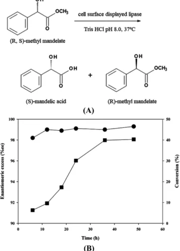
3-3. Biocatalytic applications of constitutive cell surface dis- played lipase
For the possible application as a biocatalyst, we investigated the enantioselective resolution of racemic methyl mandelate. The scheme of this reaction is shown in Fig. 3(A). Time profiles of reaction during enantioselective resolution are shown in Fig. 3(B). The conversion of reaction and enantiomeric excess of the product, (S)-mandelic acid obtained in 48 h were over 40% and 99%, respectively.
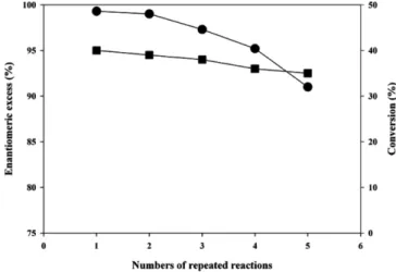
Next, the stability of cell surface displayed lipase was investigated by carrying out repeated cell recycle reactions with methyl mandelate as a model substrate. The conversion and enantiomeric excesses of
product obtained after the repeated reactions are shown in Fig. 4. The catalytic activity of cell surface displayed lipase was maintained for five repeated reactions (120 h) with slight decrease of enantiomeric excess of mandelic acid (99% to 91.0%). These results suggest that the lipase displayed on the E. coli cell surface using the OmpC seemed to be maintained stably active during the repeated usage.
4. Discussion
Microbial cell surface display has been employed in a wide range of applications including live vaccine, adsorbents, biosensors, and whole cell biocatalyst along the enzyme displayed [12,13]. Among the various surface displayed enzymes, lipase expressed on the cell surface showed high stability and activity under harsh condition and could be used for the many biotechnological applications [11,16-19].
Expression of protein on the cell surface sometimes causes membrane instability or growth inhibition. This problem can be solved by optimization of fusion point, tightly regulation of expression [17,24].
In this paper, we developed a stable expression of lipase on the cell surface of E. coli by combination of constitutive expression and optimization of fusion point using outer membrane protein OmpC from E. coli as an anchoring motif. Previously, we reported that E. coli OmpC can be used as a successful fusion point for the display of poly-histidine peptides on the surface of E. coli by sandwich fusion strategy [22]. However, this strategy is not available for the display of larger protein. There have been several papers that show successful display of larger proteins by applying C-terminal deletion-fusion [18,24]. Therefore, we decided to employ the C-terminal deletion- fusion as a fusion strategy. After searching the possible anchoring motif and fusion strategy, we found the possible fusion point of anchoring motif for the C-terminal deletion. Based on the predicted secondary structure and information found in the literature, we chose Arg92, Gly288 and Gly331 as potential fusion sites for displaying P.
fluorescens SIK W1 lipase [22,25]. As shown in Fig. 2, all these sites Fig. 3. Reaction scheme (A) and time profiles (B) of the enantiose-
lective resolution of methyl mandelate using lipase displayed on the surface of E. coli XL-10 Gold (p104OC2PL). Time profiles of conversion (■) and enantiomeric excess (●) of reaction products are shown.
Fig. 4. Time profiles of conversion (■) and enantiomeric excess (●) during the repeated resolution of racemic methyl mande- late using XL-10 Gold (p104OC2PL).
were suitable for the surface display of protein without induction, and membrane instability or growth inhibition was not shown in all cases.
For industrial applications, stability is one of the most important aspects. The constitutive display using OmpC anchor shows good stability during five repeated reactions. Whole cell lipase activity of surface displayed lipase and the enantiomeric excess of product maintained over 90% during the reactions (Fig. 4). The stability of cell surface displaying lipase has been reported, and the enzymatic characteristics were enough to apply to industry [18,24,26,27] However, all the reported systems were based on the inductive expression system.
Therefore, addition of expensive inducer such as IPTG is needed before use, which results in the increase of total production cost. On the contrary, the constitutive display system, reported in this paper, does not need any inducer to express enough proteins on the cell surface suitable for the production of enantiomerically pure compounds without induction. These findings suggest that constitutive surface display system can be used as a new, cost-effective immobilized enzyme for the enantioselective biocatalysis.
In conclusion, we developed the constitutive and stable display of lipase on the cell surface using E. coli OmpC as an anchoring motif via C-terminal deletion-fusion strategy. The expression of enzyme can be maintained throughout the cultivation without expensive inducer such as IPTG. We also demonstrated the repeated usage of constitutive displayed lipase as an enantioselective biocatalyst of racemic methyl mandelate, suggesting that it can be used for a cost-effective biocatalyst for the production of enantiomerically pure compounds.
Acknowledgment
This study was financially supported by Chonnam National University (Grant number: 2015-3022).
References
1. Jaeger, K.-E., Dijkstra, B. W. and Reetz, M. T., “Bacterial Bio- catalysts: Molecular Biology, Three-dimensional Structures, and Biotechnological Applications of Lipase,” Annu. Rev. Microbiol., 53, 315-351(1999).
2. Jaeger, K.-E. and Eggert, T., “Lipases for Biotechnology,” Curr.
Opin. Biotechnol., 13(4), 390-397(2002).
3. Reetz, M. T., “Lipases as Practical Biocatalysts,” Curr. Opin.
Chem. Biol., 6(2), 145-150(2002).
4. Stergiou, Y. S., Foukis, A., Filippou, M., Koukouritaki, M., Para- pouli, M., Theodorou, L. G., Hatziloukas, E., Afendra, A., Pandey, A. and Papamichael, E. M., “Advances in Lipase-catalyzed Esteri- fication Reactions,” Biotechnol. Advan., 31(8), 1846-1859(2013).
5. Gotor-Fernandez, V., Brieva, R. and Gotor, V., “Lipases: Useful Biocatalysts for the Preparation of Pharmaceuticals,” J. Mol.
Catal. B, 40(3-4), 111-120(2006).
6. Rodrigues, R. C., Virgen-Ortiz, J. J., dos Santos, J. C. S., Alca- ntar, A. R., Barbosa, O., Ortiz, C. and Fernandez-Lafuente, R.,
“Immobilization of Lipases on Hydrophobic Supports: Immobi-
lization Mechanism, Advantages, Problems, and Solutions,” Bio- technol. Adv., 37(5), 746-770(2019).
7. Van Beilen, J. B. and Li, Z., “Enzyme Technology: an Over- view,” Curr. Opin. Biotechnol., 13(4), 338-344(2002).
8. Rodrigues, R. C., Ortiz, C. Berenguer-Murcia, A., Torres, R., and Fernández-Lafuente, R., “Modifying Enzyme Activity and Selectivity by Immobilization, Chem. Soc. Rev., 42(15), 6290-6307 (2013).
9. Linqiu, C., Carrier-bound immobilized enzymes: principles, applica- tion and design, WILEY-VCH, Weinheim, Germany (2005).
10. Krajewsk, B., “Application of Chitin- and Chitosan-based Mate- rials for Enzyme Immobilizations: a Review,” Enzyme Microbial.
Technol., 35(2-3), 126-139(2004).
11. Shiraga, S., Kawakami, M., Ishiguro, M. and Ueda, M., “Enhanced Reactivity of Rhizopus oryzae Lipase Displayed on Yeast Cell Surfaces in Organic Solvents: Potential as a Whole-cell Biocat- alyst in Organic Solvents,” Appl. Environ. Microbiol., 71(8), 4335- 4338(2005).
12. Georgiou, G., Stathopoulos, C., Daugherty, P. S., Nayak, A. R., Iverson, B. L. and Curtiss, R. I., “Display of Heterologous Proteins on the Surface of Microorganisms: from the Screening of Com- binatorial Libraries to Live Recombinant Vaccines,” Nat. Biotech- nol., 15, 29-34(1997).
13. Lee, S. Y., Choi, J. H. and Xu, J., “Microbial Cell Surface Dis- play,” Trends Biotechnol., 21, 45-52(2003).
14. Smith, M. R., Khera, E. and Wen, F., “Engineering Novel and Improved Biocatalysts by Cell Surface Display,” Ing. Eng. Chem.
Res., 54(16), 4021-4031(2015).
15. Liu, Z., Ho, S-H., Hasunuma, T., Chang, J.-S., Ren, N.-Q. and Kondo, A., “Recent Advances in Yeast Cell-surface Display Tech- nologies for Waste Biorefineries,” Bioresour. Technol., 215, 324- 333(2016).
16. Matsumoto, T., Fukuda, H., Ueda, M., Tanaka, A. and Kondo, A., “Construction of Yeast Strains with High Cell Surface Lipase Activity by Using Novel Display Systems Based on the Flo1p Flocculation Functional Domain,” Appl. Environ. Microbiol., 68(9), 4517-4522(2002).
17. Shimazu, M., Nuyen A., Mulchandani, A. and Chen., W., “Cell Surface Display of Organophosphorus Hydrolase in Pseudomonas putida Using Ice Nucleation Protein Anchor,” Biotechnol. Prog., 19, 1612-1614(2003).
18. Lee, S. H., Choi, J-I., Park, S. J., Lee, S. Y. and Park, B. C., “Display of Bacterial Lipase on the Escherichia coli Cell Surface by Using FadL as An Anchoring Motif and Its Use in Enantioselec- tive Biocatalysis,” Appl. Environ. Microbiol., 70, 5074-5080(2004).
19. Han, Z., Han, S., Zheng, S. and Lin, Y., “Enhancing Thermosta- bility of a Rhizomucor miehei Lipase by Engineering a Disulfide Bond and Displaying on the Yeast Cell Surface,” Appl. Micro- biol. Biotechnol., 85, 117-126(2009).
20. Sambrook, J., and Russell, D. W., Molecular cloning: a labora- tory manual, 3rd ed., Cold Spring Harbor Laboratory Press, Cold Spring Harbor, NY (2001).
21. Blattner, F. R., Plunkett, G., Bloch, C. A., Perna, N. T., Burland, V., Riley, M., Collado-Vides, J., Glasner, J. D., Rode, K., May- hew, G. F., Gregor, J., Davis, N. W., Kirkpatrick, H. A., Goeden, M. A., Rose, D. J., Mau, B. and Shao, Y., “The Complete Genome Sequence of Escherichia coli K-12,” Science, 277, 1453-1462(1997).
22. Xu, Z. and Lee, S. Y., “Display of Polyhistidine Peptides on the Escherichia coli Cell Surface by Using Outer Membrane Protein C as An Anchoring Motif,” Appl. Environ. Microbiol., 65, 5142-5147 (1999).
23. Park, S. J., Park, J. P. and Lee, S. Y., “Metabolic Engineering of Escherichia coli for the Production of Medium-chain-length Polyhydroxyalkanoates Rich in Specific Monomers,” FEMS Micro- biol. Lett., 214, 217-222(2002).
24. Lee, S. H., Choi, J., Han, M-J., Choi, J. H. and Lee., S. Y., Display of Lipase on the Cell Surface of Escherichia coli Using OprF as An Anchor and Its Application to Enantioselective Resolution in Organic Solvent,” Biotechnol. Bioeng., 90, 223-230(2005).
25. Jeanteur, D., Lakey, J. H. and Pattus, F., “The Bacterial Porin Superfamily: Sequence Alignment and Structure Prediction,” Mol.
Microbiol., 5, 2153-2164(1991).
26. Matsumoto, T., Ito, M., Fukuda, H. and Kondo, A., Enantioselec- tive Transesterification Using Lipase-displaying Yeast Whole-cell Biocatalyst,” Appl. Microbiol. Biotechnol., 64, 481-485(2004).
27. Lee, H., Park, S. J., Han, M.-J., Eom, G. T., Choi, M.-J., Kim, S. H., Oh, Y. H., Song, B. K. and Lee, S. H., “Expression of a Lipase on the Cell-surface of Escherichia coli Using the OmpW Anchoring Motif and Its Application to Enantioselective Reactions,” Biotech- nol. Lett., 35(10), 1677-1683(2013).