AN EXPERIMENTAL STUDY OF NEWLY DESIGNED IMPLANT WITH RBM SURFACE IN THE RABBIT TIBIA : RESONANCE FREQUENCY ANALYSIS
AND REMOVAL TORQUE STUDY
Mi-Kyoung Won, D.D.S., Chan-Jin Park, D.D.S., M.S.D., Ph.D.,
Kyoung-Soo Chang, D.D.S., M.S.D., Ph.D., Chang-Whe Kim, D.D.S., M.S.D., Ph.D., Yung-Soo Kim, D.D.S., M.S.D., Ph.D., Zakiahbt Mohd Isa, D.D.S., M.S.D., Ph.D.*, Yusnidar Tajul Ariffin, D.D.S., M.S.D., Ph.D.*
Department of Prosthodontics, College of Dentistry, Seoul National University, Univ. of Malaya* Statement of problem. The importance of fixture design and surface treatment.
Purpose. The clinical success of dental implants is affected by many factors such like as degree of osseointegration, the effective load dispersion for the prostheses, and a lot of attempts have been made to overcome the difficulties. In this study, efforts were made to find the possibility of clinical acceptance of the dental implants of newly designed surface and resorbable blast media surcace.
Materials and methods.In this study, two groups of custom-made, screw-shaped implants were prepared. Tthe first with the consisting of Bra。nemark clone design and the other with the new design. These implants were divided into four groups according to the kinds of surface treat- ment. Four implants(AVANA�, Osstem, Busan, Korea)of each group were installed in twenty rab- bits. Group A was consisted of Bra。nemark clone implant left as machined, Group B with Bra。nemark clone implants with RBM(Resorbable blast media) surface, Group C with newly designed implants left as machined and Group D with newly designed implants with RBM surface. One of the twenty rabbits died from inflammation and the observation was made for six weeks. Specimens from four groups were observed using scanning electron microscopy with 40, 100, 1000 magni- fication power and microsurface structures were measured by white-light scanning interferometry for three dimensional surface roughness measurements(Accura 2000�, Intek-Plus, Korea.).
Removal torque was measured in 17 rabbits using digital torque gauge(MGT 12R, Mark-10 corp., NY, U.S.A.) immediately after the sacrifice and two rabbits were used for the histologic prepa- ration(EXAKT 310�, Heraeus Kulzer, Wehrheim, Germany) of specimens and observed under light microscope. Resonance frequency measurement(Osstell�) was taken with the 19 rabbits at the begin- ning of the implant fixation and immediately after the sacrifice.
Results. Following results were taken from the experiment.
1. The surface of the RBM implants as seen with SEM had rough and irregular pattern with retic- ular formation compared to that of turned specimens showing different surface topographies.
2. The newly designed implant with RBM surface had high removal torque value among four groups with no statistical significance. The average removal torque was 49.95±6.70Ncm in Group A, 51.15±4.40Ncm in Group B, 50.78±9.37Ncm in Group C, 51.09±4.69Ncm in Group D.
3. The RFA values were 70.8±4.3Hz in Group A, 71.8±3.1Hz in Group B, 70.9±2.5Hz, 72.7
±2.5Hz in Group D. Higher values were noted in the groups which had surface treatment compared to the untreated groups with no statistical significance.
4. The results from the histomorphometric evaluation showed a mean percentage of bone-to- implant contact of 45±0.5% in Group A, 55±3% in Group B, 49.5±0.5% in Group C, and 55±3% in Group D. Quite amount of newly formed bone were observed at the surface RBM-treated implants in bone marrow space.
Key Words
Dental implant, Resonance frequency analysis, Removal torque, Digital torque gauge, Resorbable J Korean Acad Prosthodont : Volume 41, Number 6, 2003
F
or the past 30 years the use of osseointegrated implants has become a scientifically accepted and well-documented treatment modality for the reha- bilitation of completely and partially edentulous patients. Osseointegration is a treatment concept based on stability1and the rigid fixation seems to be a pre- requisite for a favorable long-term clinical out- come.2 The term osseointegration has been used to define a direct structural and functional con- nection between ordered living bone and the surface of a load carrying implant, and it has been mechan- ically defined as continuity between implant and adja- cent hard tissue.3-5Successful long-term stability of osseointegrated dental implants has been report- ed in a large number of clinical studies using com- mercially pure titanium implants.6-8Although the implants have high survival rates,6,9 other clinical studies,9-11however, reported increased failure rates in area with poor bone quality such as low bone density or insufficient bone volume and height, mainly in the posterior maxilla, especially for screw-type implants with a turned surface. Failures related to these situations are caused by bone loss as a result of lack of primary and secondary implant sta- bility.
The density and quantity of the bone, the surgical technique, and the design of implant determine primary stability. Secondary stability is the stabili- ty of implant after primary healing and can be in- creased by bone formation and remodeling at the im- plant-bone interface.
Successful osseointegration of endosseous im- plants results from a favorable interaction between the implant geometry, surface texture and the tissues at the bone site.12Implant material, the macro-design and surface structure are among the crucial fac- tors influencing the clinical outcome of an im- plant.13Many attempts have been made over the past decade to improve bone anchorage of dental implants.
Especially, surface macrostructure and microstructure have developed to establish a stable fixation be-
tween the implant and the bony tissue and to improve load transfer and to evoke favorable bone and cell response.
The current paper will focus on the in vivo in- vestigations, especially those that were evaluated with biomechanical tests, i.e. resonance frequency analy- sis(RFA) and removal torque. From a mechanical standpoint, the removal torque technique mea- sures the strength of the bone-implant interface in terms of shear, while the RFA is considered to measure the stability during bending.14
The purpose of the present study was to evaluate bone tissue reaction around a newly design thread- ed implants (AVANA�, Osstem, Busan, Korea) with new surface treatment called resorbable blast media (RBM) placed in the rabbit tibia using main- ly resonance frequency analysis and removal torque measurement. The resonance frequency measurement and anchorage of newly design implants with RBM surface was compared with those of conventional Bra�- nemark clone with turned surface, Bra�nemark clone with RBM surface, and the new design with turned surface screws.
MATERIAL AND METHODS 1. Animals and Surgical technique
Twenty adult New Zealand white rabbits of both sexes weighing 3.5 to 4 kg were included in this study.
Prior to surgery, animals were acclimated to the vi- varium for a period of observation to insure that they were healthy and stable. The animals were anes- thetized using intramuscular injections of keta (8.8mg/kg) for surgical procedure. Prior to surgery, the shaved skin was carefully washed with a mix- ture of iodine and 70% ethanol. 1.8mL of lidocaine 2% were injected locally into the surgical sites. The tibial metaphysis was exposed by incisions through the skin, fascia, and periosteum. By intermittent drilling using low rotary speed (not exceeding 2000r.p.m) with copious saline irrigation, 2 holes were
drilled 7mm apart in the central portion of each tib- ia and sequentially enlarged to 3mm. After tap- ping and slightly countersinking the sites, the implants were gently screwed into place, until the implant shoulder was leveling with the bone surface. All im- plants penetrated the first cortical layer only, nev- er engaging the opposite cortical side. Each rabbit re- ceived four implants, from each of the different groups (group A, B, C, D) which were randomly as- signed to their implantation sites. Then, the skin and fascia layers were closed separately using resorbable sutures. After the operation, the animals received Cefazolin IM injection per day for 7 days. The ani- mals were kept in separate cages and immediately after surgery they were allowed to bear full weight.
The follow-up time was six weeks. One week after surgery, one animal died due to an unknown in- flammation. The other nineteen animals were sac- rificed using an overdose of carbon dioxide.
2. Implant preparation: design and surface treat- ment
A total of 80 custom-made, screw-shaped im- plants were used. 40 implants were Bra�nemark clone designed, and other 40 implants had a new de-
sign(AVANA�, Osstem, Busan, Korea). These im- plants were divided into 4 groups according to surface treatment.) Group A and B Bra�nemark clone implants have 0.6mm pitch, and group C, D newly design implants have 0.8mm pitch.
�Group A: 20 Bra�nemark clone implants left as machined.
�Group B: 20 Bra�nemark clone implants with RBM surface.
�Group C: 20 newly designed implants left as machined.
�Group D: 20 newly designed implants with RBM surface.
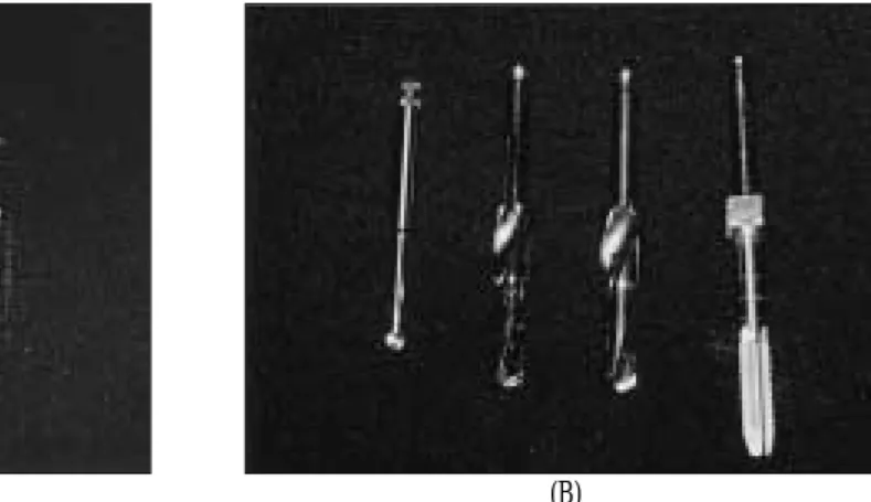
(A) (B)
Fig. 1.A : drills for Bra�nemark clone implants. B : drills for new-design implants.
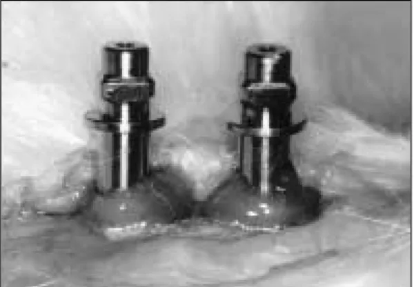
Fig. 2.Implants from each group. Group A, B, C, D
3. Scanning electron microscopy and scanning in- terferometry
The electron microscopy used to observe the characteristics of the microstructure of the RBM implants and to compare it with the surface of the machined implants was performed on a scanning elec- tron microscopy. (×40, 100, 1000, JSM-840A, JEOL, Japan)
Microsurface structure(of 2 implants of each ma- terial from each group) was measured by white-light scanning interferometry for three dimensional sur- face roughness measurements: Acurra 2000(IN- TER PLUS, Korea). In 2 samples from each of the 4 groups, 3 threads were selected randomly and scanned along their circumference in 4 different area, yielding 12 measurements for each surface topography. Three height parameters, Ra, Rq, and Rz, were used for quantitative characterization of the surface roughness. Radescribes the arithmetic mean of departures of the roughness profile from the midline., Rqis the root mean square parameter cor- responding to Ra, and Rz measures the average height difference between the 5 highest peaks and the 5 lowest valleys.
4. Resonance frequency measurement
Resonance frequency analysis (RFA), a novel technique for the clinical measurement of implant stability and osseointegration, was presented by Meredith et al.15This method is non-invasive tech- nique that measures the implant stability in terms of interfacial stiffness (Hz). The frequency response of the system was measured by attaching the transducer, i.e. an L-shaped cantilever beam, to a screw im- plant. The excitation signal was a sine wave varying in frequency from 5kHz to 15kHz with peak am- plitude of 1 volt. The first flexural (bending) resonance frequency of the resulting system was measured.16,17 The resonance frequency was measured immediately
after implant placement (20 rabbits) and at sacrifice six weeks later (19 rabbits).
5. Removal torque
To test the implant stability, specifically the strict- ly interfacial shear strength, removal torque was mea- sured at the time of animal sacrifice, i.e. six weeks after implantation. According to Roberts et al, in rab- bits it takes 6 weeks for the woven bone to be replaced by lamellar bone with adequate strength for load bear- ing.18
The small diameter hex top was 0.7mm and too short to measure. Using resin cement (Panavia 21, Kuralay, Japan), small diameter fixture mount was attached (Fig. 3) and the force needed to unscrew the implants (n=72, 18 rabbits) was measured using a dig- ital torque gauge (Model MGT12, Mark-10 Corp., 458 West John Street Hicksville, NY 11801 USA) It has the measuring torque range of 135Ncm and the accuracy of 0.5% of full scale of 1 digit, i.e. 0.3ozin (about 0.2Ncm) The implants were subjected to slowly increasing torque until loosening was detected, and the peak torque value was measured.
6. Histologic preparation of specimens
The remaining two animals were sacrificed with- out subjecting the implants to removal torque, and the specimens (including implants; n=4) and sur- rounding tissues were washed in saline solution and fixed in 4% paraformaldehyde and 0.1% glu- taraldehyde in 0.15mol/L cacodylate buffer at 4℃
and pH 7.4. The specimens were further dehy- drated in ascending concentrations of alcohol rins- es and infiltrated with glycolmethacrylate resin (Technovit 7200 VLC, Kulzer & Co, Wehrheim, Germany). After polymerization, the specimens were sectioned longitudinally at about 100μm and ground to a final thickenss of about 25μm (EXAKT 310, GMBH & Co, Germany) as described by donath19
One section was obtained for each implant and stained with hematoxylin and eosin. The histo- morphometric analysis was performed using a light microscope connected to a personal comput- er. The percentage of bone to implant contact around threads, which engaged only in cortical plate was observed with 40 magnification power of light microscope. No measurement was taken from the cancellous bone area in the rabbits, because not much of significance was there to measure the percentage of contact in hollow marrow space.
7. Statistical analysis
Mean value of resonance frequency analysis were calculated and subjected to a repeated measure ANOVA to test for significant differenced between 4 investigated groups. Scheffe's test between all groups was perfomed for removal torque evaluation.
Statistical testing was carried out at the 5% signifi- cance level.
RESULTS
1. Topographic Evaluation
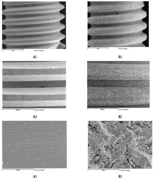
The scanning electron microscopy analysis and sur- face roughness measurements demonstrate the different surface topographies between the surface of the RBM implants and the turned implant. The sur- face of the RBM implants as seen with SEM had a rough and irregular pattern, while the turned spec- imens showed relatively smooth surface. (Fig. 6) At higher magnification, the surface appears reticulated, with undermining deformation of the metal re- maining after impaction of the resorbable hydrox- yapatite material blasted under pressure on the
Fig. 4.Small diameter fixture mount was attached to the implant. Without this procedure, the hex top of implant would be worn-out.
Fig. 5.Digital torque gauge for removal torque mea- surement.
Fig. 3.Transducer attached to implant which placed into rabbit tibia.
surface of the implant (Fig. 6, B3, ×1000)
The Bra�nemark clone implant with turned surface showed an average surface roughness 0.26μm; cor-
responding values for newly designed implant with RBM surface were 1.12μm.
A1 B1
A2 B2
A3 B3
Fig. 6.Scanning electron microscopy of custom-made implants. (A1) Implant with machined surface ×40 mag. (A2) Grooved pattern on machined surface ×100 mag. (A3) smooth turned surface ×1000 mag. (B1) Implant with RBM surface. It has coarse roughness ×40 mag. (B2) ×100 mag. (B3) At higher magnification, the metal presents a uni- form reticulation (×1000)
2. Resonance frequency analysis (RFA)
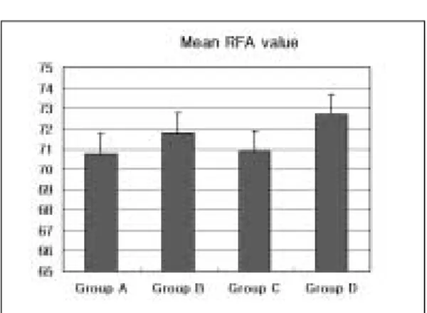
After initial implants installation, all implants of 4 groups were measured by connecting transducer to implant and orienting it parallel to long axis of im- plant. The mean RFA values were 68.9 for group A implants, 66.2 for group B implants, 70.2 for group C implants, 65.9 for group D implants. The mean RFA values six weeks after implant insertion were 70.8±
4.3 for group A implants, 71.8±3.1 for group B implants, 70.9±2.5 for group C implants, and 72.7
±2.5 for group D implants. There were no statisti- cal significant differences between groups.
3. Removal torque measurement
Six weeks after implant placement, the average re- moval torque was 49.99±6.70Ncm for group A implants, 51.15±4.40Ncm for group B implants, 50.78±9.37Ncm for group C implants, and 51.09±
4.69Ncm for group D implants. The removal torque results are summarized in Table II. The torque
Table I. Mean surface roughness(in μm)
Group Ra(SD) Rq(SD) RZ(SD)
Group A (Bra�nemark clone, turned) 0.26 0.31 1.13
Group B (Bra�nemark clone, RBM) 1.02 1.18 4.53
Group C (New design, turned) 0.29 0.34 1.24
Group D (New design, RBM) 1.12 1.26 4.36
Fig. 7. Mean values of the resonance frequency mea- surements (Hz) after six weeks of healing time. (n=19) There were no statistically significant differences between the groups.
Table III. Torque values after 6 weeks (n=17) Group Group A Group B Group C Group D Mean RT 49.99 51.15 50.78 52.09
SD 6.70 4.40 9.37 4.69
Fig. 8. Torque values for implants inserted in the tibia of rabbits 6 weeks (n=17)
Table II. Mean values of RFA after six weeks of healing time.
Group Group A Group B Group C Group D
Mean RFA 70.8 71.8 70.9 72.7
SD 4.3 3.1 2.5 2.5
measurements yielded no statistical significance. The highest removal torque corresponded to the newly designed implant with RBM surface, while the lowest was demonstrated by the turned, Bra�ne- mark clone implants. Higher torque was needed to unscrew RBM surface implants compared to the torque needed for the turned, Bra�nemark clone implants.
4. Histomorphometric evaluation
A typical cross-section showed the triangular- shaped tibia and a central bone marrow cavity with the implant inserted through the superior cortex.
On the microscope, all 8 implants were well inte-
grated into bone and a mature cortical bone sur- rounded the implants. The implants were in con- tact with cortical bone layer exclusively along the upper threads in the cortical region, while the threads in the bone marrow were in contact with much of mar-
A B
C D
Fig. 9.25micron ground sections of 4 implants from each group. All implants were well integrated into bone. (A) Turned Bra�nemark clone implant as Group A, (B) Bra�nemark clone implant with RBM surface as Group B, (C) Newly designed implant with turned surface as Group C, (D) Newly designed implant with RBM surface as Group D. Note newly formed bone in the marrow space. Note the bone formed at the implants body.
Table IV. Percentage of bone-to-implant con- tact 6 weeks after Implant Placement
Rabbit No. Group A Group B Group C Group D
1 45 52 49 55
2 46 58 50 60
Mean 45.5 55 49.5 57.5
SD 0.5 3 0.5 2.5
row tissue. Some new bone formation was observed only newly designed with RBM surface in contact with marrow space. Qualitative histologic differences among the remaining 3 groups were not seen.
Results showed a mean percentage of bone-to-im- plant contact was 45±0.5% for the turned Bra�nemark clone implants, 55±3% for the Bra�nemark clone im- plants with RBM surface, 49.5±0.5% for the turned newly designed implants, and 57.5±2.5% for the newly designed implants with RBM surface. The his- tomorphometric analyses were summarized in Table IV.
DISCUSSION
The quality of the implant surface is one of the six factors described by Albrektsson13et al which in- fluence the healing at the implantation site and subsequently affect the osseointegration. The rough- ness of the implant surfaces favors distribution of stress, retention of implants in the bone, and bone response with bone trabeculae growing in a per- pendicular direction to implant surface.20, 21Studies have reported that adequate growth of bone in the interior of the pores or cavities left by the surface treat- ment requires that these must be approximately 100μm in size. The growth of bone tissue into cav- ities of this size allows a mechanical interlocking of the implants with bone.22Wong et al.20have re- ported that bone matrix may be deposited in pore sizes of only 1-2μm, which resulted in an increase in push-out force. Analysis of RBM implants using scan- ning electron microscopy revealed rough surface and pore size of (2.5-4 μm). This allowed bone growth in- to these pores so that mechanical interlocking of the implant with bone was possible and resulted in an improved bone -implant interface as described by other articles.23-27
Implants with RBM surface had higher value than control on RFA measurement and histomor- phometric evaluation. These implants also achieved higher degree of bone-to-implant contact compared
to turned implants. And newly designed implant with RBM surface had new bone formation around bone marrow space contrary to implants of other groups.
This result is thought to arise from the novel design and the RBM surface. In vitro studies have also shown that the superficial roughness of the materials can influence cell function, matrix deposition, and mineralization.28-30In a series of studies, Wennerberg et al, systematically investigated the effect of surface roughness of implants and the response in rabbit bone.31-34And the authors concoluded that implants with a surface roughness of Ra 1 to 1.5μm seemed to be at an optimal roughness with regard to retention in bone as well as bone-to-implant contact as mea- sured by histomorphometry. And this obervation is also in accordance with observations by von Recum
& van Kooten35that reported excellent tissue at- tachment without signs of inflammation when im- planting filter membranes with pore size of 1-3μm.
Based on these results, it seems that the benefit of in- creasing roughness on a micrometer scalereaches a maximum level between 1.0 to 1.5μm. The Ra value of RBM treated implants was within this limits seemed to have resulted in favorable bone response.
Irrespective of whether resonance frequency analysis (RFA) or removal torque (RT) was used, the biomechanical data showed the similar results.
Newly designed implant with RBM surface im- plants had the highest value on both experiments but not much statistically significant difference. Both new design and surface characteristics of the implant en- abled it. The newly designed of dental implant with half-rounded shape at the apex, flatness at the top of the threaded surface, and 45 degrees of re- verse bevel at the bottom surface set the stress to the apex of the dental implant during lateral or oblique load. Forcing the stress to the top part rather than the apex part of the dental implant is not highly rec- ommended, because there is much accumulation of stress at the top part of the dental implant. The cortical bone which integrate with the top part of the dental implant is unable to disperse the attained stress
causing the concentration of stresses at the referred area. The dental implants, which took part in the ex- periment, are much capable of dispersing the stress especially to the direction of apex compared with gen- eral threaded types or other models of dental im- plant.30
RT and RFA are well-documented biomechanical techniques that evaluate the stability and stiffness of the osseointegrated implant interface. Removal torque evaluated the biomechanical bond between implant and the bone. Several studies have report- ed that RT techniques offer reliable test-to-control com- parisons.12,36RFA is a recently introduced non-invasive test method and this technique has been utilized in a number of experimental tests to record a change in implant stiffness.17,31,37-40RT and RFA are quantitative method for assessment of implant stability and os- seointegration and also enable us to compare different implant system, such as changes in implant geom- etry or surface modifications.
CONCLUSION
1. The surface of RBM implants as seen with SEM had a rough, irregular pattern than that of the turned implants that seemed to have a smoother surface. At higher magnification, the RBM surface showed reticular pattern.
2. Ra values for machined implants were 0.2-0.3μm;
corresponding values for newly designed implants with RBM surface were 1.0-1.2μm.
3. The average removal torque was 44.99±6.70Ncm for group A implants, 51.15±4.69Ncm for group B implants, 50.78±9.37Ncm for group C im- plants, 51.15±4.40Ncm for group D implants.
There was no statistically significant difference.
4. The mean RFA values were 70.8 for group A implants, 71.8 for group B implants, 70.9 for group C implants, 72.7 for group D implants. There was no statistically significant difference.
5. A mean percentage of bone-to-implant contact was 45±0.5% for group A implants, 55±3% for
group B implants, 49.5±0.5% for group C im- plants, 57.5±2.5% for group D implants. The light microscopic picture demonstrated implants with RBM surface had higher values with respect to bone-to-implant contact and group D im- plants showed new bone formation around im- plant body.
Within the limits of this 6-week experimental study, it may be stated that the overall pattern of new- ly designed implants with RBM surface resulted in significantly higher percentage of bone-to-implant contact and comparable removal torque and reso- nance frequency measurement when compared to machined, Bra�nemark clone implants. And the pre- sent investigation revealed the fact that RBM surface treatment has some advantage over turned sur- face in bone-to-implant contact, removal torque and resonance frequency measurement.
REFERENCES
1. Zarb GA, Albrektsson T. Osseointegration : a re- quiem for the periodontal ligament? Int J Periodontics Restorative Dent 1991;11:88-91.
2. Albrektsson T, Sennerby L. State of the art in oral implants. Journal of Clinical Periodontologi 1990;18, 474-481.
3. Bra�nemark PI. Osseointegration and its experimental background. J Prosthet Dent 1983;50: 399-410.
4. Albrektsson T, Hansson HA, Ivarsson B. Interface analysis of titanium and zirconium bone implants.
Biomaterials 1985;6:97-101.
5. Bra�nemark P.I, Hansson BO, Adell R . Osseoint- egrated implants in the treatment of the edentulous Jaw. Experience from a 10 year period. Scand J Plast Reconstr Surg 1997;16.
6. Adell R, Lekholm U, Rockler B, Bra�nemark PI.
A 15-year study of osseointegrated implants in the treatment of the edentulous jaw. J Oral Surg 1981;10:387-416.
7. Adell R, Eriksson B, Lekholm U, Bra�nemark PI, Jemt T. A long-term follow-up study of osseoin- tegrated implants in the treatment of totally eden- tulous jaws. Int J Oral Maxillofac Implants 1990;5:347-359.
8. Lekholm U, van Steenberghe D, Herrmann I, Bolender C, Folmer T, Gunne J, et al. Osseoint- egrated implants in the treatment of partially edentulous jaws: A prospective 5-year multicen- ter study. Int J Oral Maxillofac Implants 1994;9:627- 635.
9. Adell R. Clinical results of osseointegrated im- plants supporting fixed prosthesis in edentulous jaw. Jprosthet Dent 1983; 50:251-4.
10. Adell R, Lekholm U, Rockler B, Bra�nemark PI, Linddhe J, Eriksson B, et al. Marginal tissue reac- tions at osseoitegrated titanium fixtures I. A three- year longitudinal prospective study. Int J Oral Surg 1986;15:39-52.
11. Albrektsson B, Zarb G,Worthington P,Eriksson A. The long-term efficacy of currently used dental implants. A review and proposed criteria for suc- cess. Int J Oral Maxillofac Implants 1986;1:11-25.
12. Carlsson L, Rostlund T, Albrektsson B, Albrektsson T. Removal torque for polished and rough titani- um implants. Int J Oral Maxillofac Implants 1988;3:21-24.
13. Albrektsson T, Bra�nemark P.I, Hansson HA, Lindstrom J. Osseointegrated titanium implants.
Acta Orthop Scand 1981;52:155-170.
14. Rasmusson L, Meredith N, Cho I.H, Sennerby L.
The influence of simultaneous versus delayed placement on the stability of titanium implants in onlay bone grafts. Int J of Oral Maxillofac Surg 1999;28:224-231.
15. Meredith N, Alleyene D, Cawley P. Qualitative de- termination of the stability of the implant-tissue in- terface using resonance frequency analysis. Clin Oral Implants Res 1996; 7:261-269.
16. Meredith N. On the clinical measurement of implant stability and osseointegration. Go¨teborg, Sweden:
Department of Biomaterials/Handicap Research, University of Go¨teborg. Ph D Thesis 1997.
17. Meredith N, Shagaldi F, Alleyne D, Sennerby L, Cawley P. The application of resonance frequen- cy measurements to study the stability of titanium implants during healing in the rabbit tibia. Clin Oral Imp Res 1997;8:234-243.
18. Robert RW, Smith RK, Zilberman Y, Mozsary PG, Smith R: Osseous adaptation to continuous load- ing of rigid endosseous implants. Am J Orthod 1984;86:95-111.
19. Donath K. Die Trenn-Du¨nnschliff-Technik zur Herstellung histologischer Pra¨paraten von nicht schneidbaren Geweben und Materialien. Der Pra
¨parator 1988;34:197-206.
20. Buser D, Schenk R, Steinemann S. Influence of surface characteristics on bone integration of ti- tanium implants: A histomorphogenic study in miniature pigs. J Biomed Mater Res. 1991;25:889- 902.
21. Wong M, Eulenberger J, Schenk R. Effect of surface topology on the osseointegration of implant ma- terialws in trabecular bone. J Biomed Mater Res.
1995;29:1567-1575
20. Johansson C.B, Albrektsson T. Integration of screw implants in the rabbit. A 1-year follow-up of removal torque of Ti implants. Int J oral Maxillofac Implants 1986;2:69-75.
21. Carlsson L, Ro¨stlund T, Albrektsson B, Albrektsson T. Removal torques for polished and rough titanium
implants. Int J oral Maxillofac Implants 1988;3:21- 24.
22. Bobyn J, Pilliar R, Cameron H. The optimum pore size for the fixation of porous-surface metal implants by the ingrowth of bone. Clin Orthop. 1980;150:263- 270.
23. Ericsson I, Johansson CB, Bystedt H et al. A histo- morphometric evaluation of implant contact on ma- chined-prepared and roughened titanium den- tal implants. Clinical Oral Implant Res. 1994;5:202- 206.
24. Gotfredesen K, Nimb L, Ho¨rting-Hansen E.
Histomorphometric and removal torque analy- sis for TiO2-blasted titanium implants. An exper- imental study in dog. Clinc Oral Implants Res.
1992;3:77-84.
25. Martin JY, Schwarttz, Hummer TW. Effect of ti- tanium surface roughness on proliferation, dif- ferentiation and protein synthesis of human os- teoblast-like cells(MG63). J Biomater Res. 1995;29:389- 401.
26. Breme J, Wadewitz V, Fu¨rbacher B. Production and mechanical properties of porous sintered specimens of the implant alloy TiAl5Fe2. Advances in Biomaterials. 1990;9:63-68.
27. Martin J, Schwartz Z, Hummert T. Effect of titanium surface roughness on proliferation, differentia- tion and protein synthesis of human osteoblast-like cells. J Biomed Mater Res. 1995;29:389-401.
28. Brunette D. The effects of implant surface topog- raphy on the behavior of cells. Int J Oral Maxillofac Implants. 1988;3:231-246.
29. Boyan BD, Hummert T, Kieswetter K. Role of material surfaces in regulating bone and carti- lage cell respone. Biomaterials. 1996;17:137-146.
30. Kim Y.S. 3-dimensional finite element study on sur- face characteristics and stress of implants. SNUDH Dept of Prosthodontics. Unpublished.
31. Wennerberg A, Albrektsson T, Andersson B, Krol JJ. A histomorphometrifc and removal torque study of screw-shaped titanium implants with three different surface topographies. Clin Oral Implants Res 1995;6:24-30.
32. Wennerberg A, Albrektsson T, Lausmaa J. Torque and histomorphometric evaluation of c.p.titanium screws blasted with 25-and 75-micron-sized par- ticles of Al2O3. J Biomed Mater Res 1996;30:251-260.
33. Wennerberg A, Albrektsson T, Anderssen B. Bone tissue response to commercially pure titanium implants blasted with fine and coarse particles of aluminum oxide. Int J Oral Maxillofac Implants 1996;11:38-45.
34. Wennerberg A, Albrektsson T, Johansson C, Andersson B. Experimental study of turned and grit- blasted screw-shaped implants with special em- phasis on effects of blasting materials and sur- face topography. Biomaterials 1996;17:15-22.
35. Von Recum AF, van Kooten TG. The influence of microtopography on cellular response and the implications for silicone implants. J Biomater Sci
polymer Edition 1995;7:181-198.
36. Rasmusson L, Meredith N, Sennerby L.
Measurements of stability changes of titanium implants with exposed threads subjected to barrier membrane induced bone augmentation. A study in the rabbit tibia. Clin oral Imp Res 1997;8:103-116.
37. Rasmusson L, Meredith N, Kahnberg K.E, Sennerby L. Stability assessments and histology of titanium implants placed simultaneously with autogenous onlay bone grafts in the rabbit tibia. Int J Oral Maxillofac Surg 1998;27:229-235.
38. Park C. A study on the measurement of the implant stability using resonance frequency analysis. PhD thesis, 2000, Dankook University, Korea.
39. Park CJ. In vitro comparative study between ISQ
and Periostest� values on the implant stability measurements according to the increased effective implant length. J Korean Academy of Prosthodontics 2001;39:625-635.
40. Park CJ. A study on the change of implant stabil- ity using resonance frequency analysis. Ph D the- sis(Manuscript). 2000, Seoul National University, Korea.
Reprint request to:
DR.YUNG-SOOKIM, D.D.S., M.S.D., Ph.D.
DEPT. OFPROSTHODONTICS,COLLEGE OF DENTISITY,
SEOUL NATIONAL UNIV.
28-1 YEONGUN-DONG, CHONGNO-GU, 110-749, SEOUL KOREA