I. INTRODUCTION
Staphylococcus aureus is the major pathogen of the genus of Staphylococcus and an important nosocomial pathogen causing a variety of clinical infections including septicemia, pneumonia, wound sepsis, septic arthritis, osteomyelitis, post-surgical toxic shock syndrome and food-borne disease (Vincent et al., 1995; Boyce, 1997).
Many S. aureus accessory genes encode virulence factors such as enterotoxins A through I (sea to sei, respectively), toxic shock syndrome toxin 1 (tst), exfoliative toxins A and B (eta, etb) (Projan and Novick, 1997; Munson et al., 1998). These genes are often carried
Corresponding Author : Seok-Jae Jeon, 730-717, Indong Community Health Substation, Gumi-si, Gyeongsangbuk-do, Korea.
Tel : 054-473-2287, 017-538-9669.
E-mail : [email protected]
on mobile genetic elements, such as phages and pathogenicity islands (SaPIs), which transfer horizontally between strains (sea, tst, eta) (Betley and Mekalanos, 1985; Lindsay et al., 1997; Yamaguchi et al., 2000;
Yoshizawa et al., 2000) or which are suspected to do so (etb, seb) (Jackson and Iandolo, 1986; John et al., 1998).
Transfer of tst can occur at an extremely high frequency (Lindsay et al., 1997) in vitro, although it is not known if this occurs in the clinical setting. There is also evidence that mobile accessory virulence genes are not distributed uniformly among strains. For example, seb and tst are never seen in the same strain (Bohach at al, 1990).
The purpose of the present study was to investigate the distribution of genomic differences in clinical strains of S.
aureus, particularly those that might have an effect on pathogenicity. We studied 259 epidemiologically unrelated strains collected from 5 hospitals located in Daegu and
Virulence Genes of Staphylococcus aureus Isolated in Daegu and Gyeongsangbuk-do Areas
Seok-Jae Jeon1 and Hee-Moo Lee2
Indong Community Health Substation, Gumi 730-717, Korea1
Department of Biology Science, Andong National University, Andong 760-749, Korea2
Nine types of staphylococcal enterotoxin (SE) genes (sea~see, seg~sej), 3 types of virulence genes (eta, etb, tst), mecA and 16S rRNA as internal positive control were detected from 187 clinical MRSA (methicillin resistance Staphylococcus aureus) strains isolated from a variety hospitalized patients in Daegu and Gyeongsangbuk-do areas using the multiplex PCR. The frequency of the S. aureus strains harboring recently reported SE genes (seg~sej) were found to be very high (65.9%) and greater than that of the strains harboring classical SE (sea~see) genes (47.8%) as previously established. Taking into account that the newly described pairs form SE genes (i.e., sec+seg+sei, seg+sei) were many, in the other hand, single form SE genes (i.e., seg, seh, sei and sej) were rarely detected. The S. aureus with pairs form enterotoxigenic genes become more potentially toxigenic strains. Furthermore, this work indicated a systematic association between the seg and sei genes and their high incidence among the S, aureus strains, which suggests that these two SE’s could be an important phylogenetic link among the staphylococcal enterotoxins.
Key Words : Staphylococcus aureus, Virulence genes, Enterotoxin, PCR ISSN 1738-3544
Gyeongsangbuk-do in Korea.
II. MATERIALS AND METHODS
We characterized 187 strains of MRSA. The strains were isolated from 5 hospitals which are located at Daegu and Gyeongsangbuk-do areas in Korea. No isolates were known to be epidemiologically related, and the isolates comprised strains were isolated from different patients on different wards and taken at different times in 2005.
The reference S. aureus strains used for this study were ATCC13565 for sea and sed genes, ATCC14458 for seb, ATCC19095 for sec, ATCC23235 for sed, ATCC27664 for see and previously analyzed clinical isolates for seg, seh, sei, sej, eta, etb, mecA, tst and 16S rRNA.
1. Selection of genes and primer design
Primers for the sea, seb, sec, sed, see, seg, seh, sei, sej, eta, etb, tst, mecA and 16S rRNA gene were designed to be specific for each gene and to have similar melting temperatures for multiplex PCR from published sequences.
The absence of homologous regions in any other known DNA sequence available on the GenBank database was ensured by using Blast software (Altschul et al., 1990).
2. DNA extraction
Chromosomal DNA was isolated as described by Hookey (Hookey et al, 1998) with some modification. All S. aureus isolates were incubated in brain heart infusion broth (Becton & Dickinson Company, Sparks, MD., USA) for 15~18h at 37℃. One mL of each cultured broth was pelleted by centrifugation at 12,000× g for 5 min, and washed two times with 700 μL of TE buffer (Bioneer Corporation, Deajeon, Korea). The bacterial pellet was resuspended in 565 μL of TE buffer, 30 μL of 20%
sodium dodecyl sulfate (Sigma Chemical Company, St
Louis, Mo., USA), 50 μL of 200 μg/mL lysostaphin (Sigma Chemical Company, St Louis, Mo., USA), 5 μL of 20 mg/mL proteinase K (Bioneer Corporation, Deajeon, Korea) and then incubated for 90 min at 37℃. Addition of 5 M NaCl 100 μL and 80 μL of CTAB/NaCl (Sigma Chemical Company, St Louis, Mo., USA) was followed and incubated for 20 min. at 65℃, and then centrifuged at 12,000× g for 15 min at 4℃. The lystate was extracted with equal volume of phenol- chloroform-isoamylalcohol (Bioneer Corporation, Deajeon, Korea) and centrifuged at 12,000× g for 15 min at 4℃. The supernatent was mixed with two or three times more absolute ethanol by volume and then fixed for over night at -20℃ to precipitate DNA, centrifuged at 12,000× g for 15 min at 4℃. The pellet was washed with 70% ethanol and dried in vacuum condition. The DNA pellet was resuspended in 30 μL of TE buffer and stored at -20℃ until PCR amplification.
3. Mutiplex PCR
Three sets of multiplex PCR were performed to inve- stigation of the endotoxin genes. The first primer set contained sea, seb, sec, sed, and see primers. The second set contained seg, seh, sei, and sej primers. The third set contained eta, etb, tst, mecA and 16S rRNA primers. Each PCR content was made up of 2 μL of templete DNA, 2 μ L of primer pools (for 1st primer set, 0.2 μL each; 2nd primer set, 0.25 μL each; 3rd primer set, 0.25 μL each), 10 μL of PCR PreMIX (Bioneer Corporation, Deajeon, Korea) contained 1 U of Taq DNA polymerase, 250 μM dNTP, 10 mM Tris-HCl, 40 mM KCl, 1.5 mM MgCl2, stabilizer and tracking dye, and DW upto 20 μL. Each mixture was heated at 95℃ for 2 min before 28 cycles of amplification (denaturation at 95℃ for 1 min, annealing at 53℃ for 1 min, extension at 72℃ for 2 min) and final extension was performed at 72℃ for 5 min in a Mygene 96 Thermal Block (Bioneer Corporation, Deajeon, Korea).
4. Electrophoresis
The amplified DNA products (7 μL) were electro- phoretically separated in 1.5% agarose gel (Bioneer Cor-
poration, Deajeon, Korea) in 1× TAE buffer (Bioneer Corporation, Deajeon, Korea) by Mupid-eX (Advance electronics Inc. Tokyo, Japan), stained with ethidium bromide, visualized on a UV transilluminator, and photo- graphed with Polaroid.
M 1 2 3 4 5 M 6 7 8 9 10 M
500 bp → 100 bp →
Fig. 1. Multiplex PCR amplification products of reference strains. Lane 1~5 are ATCC control strains, lane 7~10 are clinical isolated strains. Lane 1 is ATCC13565 (sea and sed), lane 2 is ATCC14458 (seb), lane 3 is ATCC19095 (sec), lane 4 is ATCC23235 (sed), lane 5 is ATCC27664 (see), lane 6 is negative control, lane 7 is D-hospital strain-4 (seg, sei), lane 8 and 10 are A-hospital strain-1 and D-hospital strain-31 (mecA and, 16S rRNA). Lane 9 is E-hospital strains-7 (mecA, tst and 16S rRNA genes). M is a 100 bp DNA ladder (Bioneer Cor- poration, Deajeon, Korea).
Gene A-hospital*
n=39
B-hospital n=35
C-hospital n=26
D-hospital n=48
E-hospital n=38
H-institute n=1
Total n=187
sea 20 (51.3) 20 (57.1) 5 (19.2) 8 (16.7) 23 (60.5) 0 (0.0) 76 (40.6)
seb 0 ( 0.0) 0 ( 0.0) 0 ( 0.0) 0 ( 0.0) 1 ( 2.6) 0 (0.0) 1 ( 0.5)
sec 11 (28.2) 10 (28.6) 19 (73.1) 40 (83.3) 15 (39.5) 0 (0.0) 95 (50.8)
sed 1 ( 2.6) 0 ( 0.0) 0 ( 0.0) 0 ( 0.0) 0 ( 0.0) 0 (0.0) 1 ( 0.5)
see 0 ( 0.0) 0 ( 0.0) 0 ( 0.0) 0 ( 0.0) 0 ( 0.0) 0 (0.0) 0 ( 0.0)
seg 19 (48.7) 16 (45.7) 24 (92.3) 40 (83.3) 27 (71.1) 1 (100.0) 127 (67.9)
seh 0 ( 0.0) 0 ( 0.0) 0 ( 0.0) 0 ( 0.0) 0 ( 0.0) 0 (0.0) 0 ( 0.0)
sei 18 (46.2) 12 (34.3) 22 (84.6) 40 (83.3) 21 (55.3) 0 (0.0) 113 (60.4)
sej 0 ( 0.0) 0 ( 0.0) 0 ( 0.0) 0 ( 0.0) 0 ( 0.0) 0 (0.0) 0 ( 0.0)
eta 0 ( 0.0) 0 ( 0.0) 0 ( 0.0) 0 ( 0.0) 0 ( 0.0) 0 (0.0) 0 ( 0.0)
etb 1 ( 2.6) 0 ( 0.0) 0 ( 0.0) 0 ( 0.0) 0 ( 0.0) 0 (0.0) 1 ( 0.5)
tst 11 (28.2) 16 (45.7) 20 (76.9) 39 (81.3) 17 (44.7) 0 (0.0) 103 (55.1)
mecA 36 (92.3) 34 (97.1) 26 (100.0) 48 (100.0) 37 (97.4) 0 (0.0) 181 (96.8)
16S rRNA 39 (100.0) 35 (100.0) 26 (100.0) 48 (100.0) 38 (100.0) 1 (100.0) 187 (100.0)
* The percentile of investigated isolates was presented in ( ).
Table 1. The distribution of S. aureus strains harboring the genes coding for the virulence factors collected by 5 hospitals and 1 public research institution
127 bp271 bp →→
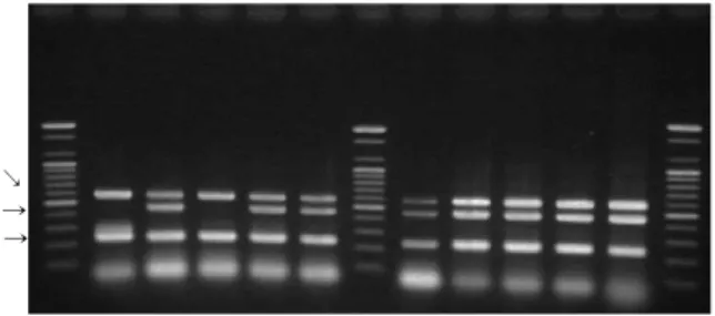
M 1 2 3 4 5 M 6 7 8 9 10 M
Fig. 2. Results of multiplex PCR with the 1st primer set. Amp- lificaton products for sea presented at 127 bp size are lane 1, 3, 5, 7 and 9 (obtained from A-hospital strain-12, B-hospital strain-7, C-hospital strain-20, E-hospital strain-12, respectively).
Amplification products for sec presented at 271 bp size are lane 2, 4, 6, 8 and 10 (obtained from A-hospital strain-37, B-hospital strain-30, C-hospital strain-35, D-hospital strain-18, E-hospital strain-35, respectively). Lane M is a 100 bp DNA ladder (Bioneer Corporation, Deajeon, Korea).
III. RESULTS
Of all 187 clinically identified MRSA isolated in Daegu and Gyeongsangbuk-do areas, 181 strains were mecA positive (Table 1). All S. aureus strains carried mecA presented at least one enterotoxin gene of classical SE or recently described SE genes (Table 1). Frequencies of those genes in MRSA were presented in the order of seg, sei, sec, sea at 67.9%, 60.4%, 50.8%, 40.6%, respectively (Table 1). There were no S. aureus carring see or seh or
sej gene. The seb and sed genes were rarely presented in only one strain respectively.
Among other virulence factor encoding gene, toxic shock syndrome toxin 1 gene (tst) was expressed at 55.1%, but exfoliative toxins A and B genes (eta, etb) were not or rarely presented at 0% and 0.5% respectively (Table 1).
Of the total 181 MRSA strains, 125 strains carried two or more enterotoxin genes as described as Table 2. There were 12 kinds of multiple enterotoxin gene carrying type in this study. The most frequent multiple enterotoxin genes carrying type were in the order of [sec+seg+sei] type and
Table 2. Distribution of enterotoxin gene in specimens
Specimens None
sea sec seg sea
sec sea seg
sec seg
seg sei
sea seg sei
sec seg sei
sea sec seg sei
sea sed seg sei
seb sec seg sei
Total
Urine 0 1 0 0 0 0 2 4 0 2 1 0 0 10
Blood 2 3 0 0 0 1 0 1 2 5 0 1 0 15
Wound 2 16 1 1 1 1 0 2 2 4 0 1 0 31
Pus 0 5 3 2 0 1 1 1 5 7 0 0 0 25
Sputum 1 9 6 1 1 1 2 5 10 38 4 0 0 78
Other 1 3 5 0 1 0 0 1 6 10 0 0 1 28
Total 6 37 15 4 3 4 5 14 25 66 5 2 1 187
M 1 2 3 4 5 M 6 7 8 9 10 M
228 bp → 445 bp → 575 bp ↘
Fig. 4. Multiplex PCR results with the 3rd primer set are presented.
Lane 2, 4, 5, 6, 7, 8, 9 and 10 (obtained from A-hospital strain- 27, B-hospital strain-12, C-hospital strain-14, C-hospital strain-15, D-hospital strain-6, D-hospital strain-7, E-hospital strain-7, E-hospital strain-11, respectively) are PCR positive against mecA, tst and 16S rRNA genes. Lane 1 (A-hospital strain-16) and lane 3 (B-hospital strain-11) are amplified mecA and 16S rRNA genes.
M is a 100 bp DNA ladder (Bioneer Corporation, Deajeon, Korea).
465 bp →
M 1 2 3 4 5 M 6 7 8 9 10 M
327 bp →
Fig. 3. Multiplex PCR results with the 2nd primer set are presented. Amplification products for seg (327 bp) and sei (465 bp) are expressed together on lane 1, 2, 3, 4, 5, 6 and 7 (obtained from A-hospital strain-6, A-hospital strain-17, B-hospital strain-9, B-hospital strain-26, C-hospital strain-13, C-hospital strain-36, D-hospital strain-3, respectively). Lane 9 is E-hospital strain-6, presented the only seg product. On lane 8 (D-hospital strain-18) and 10 (E-hospital strain-33), there are no products of any genes.
M is a 100 bp DNA Ladder (Bioneer Corporation, Deajeon, Korea).
[sea+seg+sei] type, which respectively accounted for 35.3%, 13.4%. Especially, seg and sei genes were concurrently presented in 113 strains (62.4%) of the total 181 MRSA (Table 2, Fig. 3). It is full of significance for closely relation as enterotoxin gene rearrangement or propagation between S. aureus strains.
In distribution analysis of enterotoxin gene according to specimens, there were no significant differences between enterotoxin gene types or multiple enterotoxin gene carrying types and the isolated group from the specimens (Table 2).
IV. DISCUSSION
Many articles used the PCR method to deteet staphylococcal enterotoxin genes (Tornadijo et al., 1996;
Holeckova et al., 2002). All of them found a high variability (75~80%) in the presence of enterotoxin genes.
Our experiments confirmed this fact as well. We detected the genes for nine types of enterotoxin (sea~see, seg~sej), 3 types of virulence genes (eta, ,etb, tst), mecA and 16S rRNA as the internal positive control in our set of 187 S.
aureus strains. The majority of enterotoxin positive staphylococci (97.3%) carry genes for enterotoxin. Some data published by other authors show that the staphylococcal enterotoxin type [seg+sei] was the most frequent type (Rosec et al., 1998) but our most frequent type was [sec+seg+sei], without the production any of the sea, seb, sec, sed or see, which harbored one or more of the seg, seh, sei, or sej genes (Mac Launchlin et al., 2000). Thus, it is likely that the corresponding SE's should be the causative agent of these outbreaks. A similar observation was made with clinical strains impli- cated in staphylococcal toxic shock syndrome and scarlet fever without producing classical SE's or toxic shock syndrome toxin 1, leading to the suggestion that seg or sei caused these diseases (Jarraud et al., 1999)
When taking into account the seg, seh, sei and sej, the
incidence of potentially enterotoxogenic S. aureus considerably increases among the strains, which were isolated from the clinical environment. The seg gene was systematically detected in association with sei, as was sed with sej. The latter association has been described (Zhang et al., 1998) and the former was also observed in clinical strains (Jarraud et at., 1999) where seg and sei were in tandem orientation and separated by 1.9 kb length intergenic DNA. Nevertheless, this systematic seg and sei association is surprising since these two genes were initially cloned from two different strains (Munson et al., 1998) and Mac Lauchlin et al. (2000) found 8 strains which harbored seg alone, and over 109 ones which were seg and/or sei.
Furthermore, seg and sei were the sole SE gene which could be detected in association with each other (Table 2).
The apparent systematic association between seg and sei and the horizontal spread of these two genes among our S. aureus strains could suggest that they are either a reservoir or a part of a reservoir for an enterotoxin gene rearrangement in S. aureus, as has been hypothesized for the region where sei was identified and sequenced (Munson et al., 1998)
The result conclude that the incidence of the recently described SE genes (seg~sej), and particularly seg and sei, among the clinical S. aureus isolated in Korea is very high. Thus, the role of seg, seh, sei and sej in the staphylococcal disease is probably underestimated, unless their corresponding genes are rarely or weakly expressed.
Given the results of this work, there is a need to improve or develop detection methods for these SE's.
Further, this work indicates a systematic association between seg and sei, and a wide distribution of these two genes among the S. aureus strains. Thus, extensive studies about the seg and sei genes could be of great interest to investigate the existence of regions for the enterotoxin gene rearrangement in S. aureus and the phylogenetic aspects of the staphylococcal enterotoxins.
REFERENCES
1. Altschul SF, Warren G, Maller W, Eugene W, Myers W, David JL. Basic local alignment search tool. J Mol Biol 215: 403-410, 1990.
2. Betley MJ, Mekalanos JJ. Staphylococcal enterotoxin A is encoded by phage. Science 229:185-187, 1985.
3. Bohach, GA, Fast DJ, Nelson RD, Schlievert PM.
Staphyloccocal and streptococcal pyrogenic toxins involved in toxic shock syndrome and related illnesses. Crit Rev Microbiol 17:251-272, 1990.
4. Boyce JM. Epidemiology and prevention of nosocomial infections, In Crossley KB, Archer GL(ed.), The staphylococci in human disease. p309- 329, Churchill Livingstone Publishing New York, 1997.
5. Holeckova B, Holoda, AE, Fotta M, Kalinacova V, Gondol J, Grolmus J. Occurrence of Enterotoxigenic Staphylococcus aureus in Food. Ann Agric Environ Med 9:179-182, 2002.
6. Hookey JV, Richardson JF, Cookson BD. Molecular typing of Staphylococcus aureus based on PCR restriction fragment length polymorphism and DNA sequence analysis of coagulase gene. J Clin Microbiol 36:1083 1089, 1998.–
7. Jackson MP, Iandolo JJ. Cloning and expression of the exfoliative toxin B gene from Staphylococcus aureus. J Bacteriology 166:574-580, 1986.
8. Jarraud S. Cozon G, Vandenesch F, Bes M, Etienne J, Lina G. Involvement of enterotoxins G and I in staphylococcal toxic shock syndrome and staphy- lococcal scarlet fever. J Clin Microbiol 3:2446-2449, 1999.
9. John VH, Judith FR, Barry DC. Molecular typing of Staphylococcus based on PCR restriction fragment length ploymophism and DNA sequence analysis of the coagulase gene. J Clin Microbiol 36(4): 1083-1089, 1998.
10. Lindsay JA, Kurepina N, Novick RP. Clinical isolates
of Staphylococcus aureus encode TSST-1 on genetic elements related to S. aureus pathogenicity, island-1 (SaPI1). European Conference on Toxic Shock Syndrome, Royal Society of Medicine, London, UK.
1997.
11. Mac Lauchlin J, Narayanan GL, Mithani V, O'Neill G. The detection of enterotoxins and toxic shock syn- drome toxin genes in Staphylococcus aureus by polymerase chain reaction. J Food Port 63:479-488, 2000.
12. Munson SH, Tremaine MT, Betley MJ, Welch TA.
Identification and characterization of staphylococcal enterotoxin types G and I from Staphylococcus aureus. Infect Immun 66:3337-3348, 1998.
13. Projan, SJ, Novick RP. The molecular basis of pathogenicity. In Crossley KB, Archer GL. (ed.), The staphylococci in human disease. p5-81 Churchill Livingstone, Publishing, New York, 1997.
14. Rosec JP, Dalet C, Paul O, Guiraud JP. Staphy- lococcal isolates de produits alimentaires : detection des souches enterotoxinogenes par PCR. Ann Falsif Expert Chim 943:157-173, 1998.
15. Tornadijo ME, Fresno JM, Carballo J, Sarimiento MR. Population levels, species and characteristics of Micrococcaceae during the manufacturing and pre- paring of Armada-sobado goat's milk cheese. J Food Prot 59:1200, 1996.
16. Vincent JL, Bihari DJ, Suter PM, Bruining H, White AJ, Nicolas MH, Chanoin M, Wolff R, Spencer C, Hemmer M. The prevalence of nosocomial infection in intensive care units in Europe. Result of the european prevalence of infection in intensive care (EPIC) study. EPIC International Advisory Com- mittee. JAMA 274:639-644, 1995.
17. Yamaguchi TT, Hayashi H, Takami K, Nakasone M, Ohnishi K, Nakayama S, Yamada H, Komatsuzawa, M Sugai. Phage conversion of exfoliative toxin A production in Staphylococcus aureus. Mol Microbiol 38:694-705, 2000.
18. Yoshizawa Y, Sakurada J, Sakurai S, Machida K, Kondo I, Masuda S. An exfoliative toxin A-conv- erting phage isolated from Staphylococcus aureus strain ZM. Microbiol Immunol 44:189-191, 2000.
19. Zhang S, Iandolo JJ, Stewart GC. The enterotoxin D plasmid of Staphylococcus aureus encodes a second enterotoxin determinant (sej). FEMS Microbiol Lett 168:227-233, 1998.
대구광역시와 경상북도 지역에서 분리한 Staphylococcus aureus 병독소 유전자의 분자적 연구
을 이용하여 대구와 경상북도 내의 다양한 입원 환자들로부터 분리된 주의 균주를
Multiplex-PCR 187 MRSA
재료로 가지 종류의 내독소9 (sea see, seg sej~ ~ ), 3종류의 병독소(eta, etb, tst) 그리고 내부 양성 지표로써 16S rRNA와 MecA 유전자를 검출하였다. S. aureus 균주에서 새로운 형태의 내독소 유전자(seg~sej 의 빈도가)
로 매우 높게 잠복하고 있었으며 고전적 형태의 내독소 유전자
65.9% , (sea~see 도) 47.8%로 선행 연구에서 검출된
것만큼 높게 잠복하고 있었다 새로운 형태의 내독소 중 쌍을 이룬 형태 즉. ( , sec+seg+sei, seg+sei 는 많이 검출된) 반면 단일 형태의 내독소 즉( , seg, seh, sei, sej 는 거의 검출되지 않았거나 없었으며 쌍을 이룬 내독소 유전자를) , 가진 S. aureus는 잠재적으로 보다 더 독성균주가 될 것으로 판단된다 더 나아가. S. aureus 균주들 사이의 높은 보유율을 보이는 seg와 sei 사이의 체계적인 관련성은staphylococcal enterotoxin들 사이에 중요한 계통발생적 연 계가 있을 수 있다는 것은 암시한다.