INTRODUCTION
Chronic hemo- and peritoneal dialysis have been estab- lished as live-saving and live-sustaining modalities for the treatment of patients with end-stage renal disease (ESRD).
Hundreds of thousands of persons are alive today as a re- sult of receiving dialysis. Despite the widespread availabil- ity and advancements in dialysis, the expected remaining lifetime for children with ESRD remains low, between 35%
Corresponding Author : Kim, Eun-Sook Department of Clinical Laboratory Science, Wonkwang Health Science University, Iksan 570-750, Korea
Telephone: 82-63-840-1214, Fax: 82-63-840-1219, E-mail: [email protected]
Received : 6 August 2012
Return for modification : 14 September 2012 Accepted : 15 September 2012
Korean J Clin Lab Sci. 2012, 44(3) : 128 - 135 ISSN 1738-3544
and 47% of that of an age- and race-matched US popula- tion (Mitsnefes, 2002).
The kidney and cochlea have similar physiological characteristics, specifically the active transport of fluid and electrolytes by the stria vascularis and the glomeru- lus, accounting for similar effects of aminoglycosides and some immunological factors on the two organs (Ozturan and Lam, 1998). Development of both the inner ear and the kidney is affected by similar genetic factors in certain hereditary diseases as reported in Alport’s syndrome and branchio-oto-renal syndrome (Thodi et al., 2006). It has been reported in a number of variable cases that hearing impairment is associated with renal failures, including use of ototoxic medications, electrolyte imbalance, alterations in blood urea nitrogen and lipids(Johnson and Mathog, 1976), hypertension (Gartland et al., 1991), and haemodi- alysis treatment itself (Bazzi et al. 1995; Serbetcioglu et al.
Association of ND4L gene 10609 mutation and hearing loss in a Korean with ESRD patients
Eun Sook Kim
Department of Clinical Laboratory science, Wonkwang Health Science University, 344-2 Shinyong-dong, Iksan, Jeonbuk 570-750, Korea
The kidney and cochlea have similar physiological characteristics, specifically the active transport of fluid and electrolytes, similar effects of aminoglycosides and some immunological factors. Several mitochon- drial DNA (mtDNA) defects have been identified to be associated with hearing impairment either in syn- dromic or nonsyndromic forms. Dialysis patients had more oxidative stress than healthy subjects and this elevated oxidative stress leads to alterations of the mtDNA. To generate a more comprehensive analysis of the relationship between mitochondrial variation and hearing loss, two SNPs of 10609, 14668 position showed nominal levels of association with hearing loss. In our result, the mean PTA values in the ESRD patients were 28±13.9 (mean±SD) dB and 51.0±23.2 dB in low and high frequencies, which were sig- nificantly higher than those in the normal controls. 10609T>C and 14668C>T were significantly associated with hearing loss in the ESRD patients. In summary, our results suggest that the polymorphisms of the ND4L subunit gene might be association with ESRD patients and hearing loss.
Key words : ESRD; hearing loss; mitochondrial DNA; ND4L 10609 gene
Materials and methods
Audiological evaluation
After informed consent, 114 ESRD patients and 58 nor- mal individuals were subjected to otological examination, such as otoscopy and tympanometry to check if there was any fluid or drainage in the ears. All of the patients and normal controls showed normal external and middle ear conditions. Further, the patients did not present any abnor- mal neurological conditions that could be associated with hearing loss. For hearing loss test, the patient and control groups underwent a basic audiological evaluation (pure- tone, high frequency and speech audiometry), using a clin- ical audiometer (Madsen Electornics). The degree of hear- ing loss was defined according to the pure-tone averages (PTA), which were based on the average dB values at low (0.25, 0.5, 1.0, and 2.0 kHz) or high (4.0 and 8.0 kHz).
The mean ages of the ESRD patients and normal controls were 63 years and 60years, respectively.
Six SNP markers
we selected six SNP markers that showed relatively high- er variation frequency (18 - 44 %) in the test with 50 ESRD patients. The six SNP markers were 6962G>A, 10609T>C, 14668C>T, 14783T>C, 15043G>A, and 15301G>A. All the other SNPs showed allele variation frequencies of less than 12%. All of the selected markers are present in the coding regions of the mitochondrial respiratory chain.
PCR amplification
Genomic DNA that includes the mitochondrial genome was isolated from leucocytes using the differential de- proteinization method as previously reported (Lahiri and Nurberger, 1981). The entire mitochondrial genome was PCR amplified with 13 sets of primers. The sequences of oligonucleotide primers are available upon request. Am- plification reactions were performed in a final volume of 2001). The incidence of sensorineural hearing loss among
patients with chronic renal failure (CRF) is considerably higher than in the general population (Bazzi et al.,1995;
Ozturan and Lam, 1998; Johnson et al., 1976; Antonelli et al., 1990)
Cochlear function requires a high rate of ATP produc- tion that is produced in mitochondria, and the human mitochondrial genome encodes for 13 subunits of the mi- tochondrial respiratory chain, 22 tRNAs, and two rRNAs.
Genetic damages more often occur to mtDNA than to nuclear DNA, apparently due to lack of efficient DNA re- pair mechanisms. Further, the lack of protective histone proteins in mitochondria makes mtDNA more susceptible to DNA damaging agents (Nagy et al., 2003). Several mi- tochondrial DNA (mtDNA) defects have been identified to be associated with hearing impairment either in syndromic or nonsyndromic forms, including A1555G in the 12S rRNA gene and an insertion of C at position 7472 and T7445C in the tRNASer gene (Estivill et al., 1998; Hutchin et al., 1993;
Tiranti et al., 1995; Reid et al., 1994). Also, it is reported that mtDNA at position 4977 seems to predict survival in ESRD, but a reduced mitochondrial copy number seems to predict a poor outcome (Rao et al., 2009), that there may be an association between mtDNA4977 del and SNHL(Bai and Seidman. 2001). However, It is not yet established whether haemodialysis treatment itself can cause negative or positive effects on hearing in patients with CRF.
Therefore, this study analyzed the mitochondrial DNA mutation for finding relation of hearing loss in patients with ESRD. Our data sequenced that mtDNA coding region was compared in a group of ESRD patients without hearing loss(n=51) and hearing loss(n=63).
25 l, containing approximately 100 ng of genomic DNA, 10 pmole of each primer, 0.2 mM of each dNTP, and Taq DNA polymerase (Takara). Thermocycling consisted of 30 cycles at 94 C for 1 min, at 57 - 61 C for 1 min, and at 72 C for 1 min, with a predenaturation at 94 C for 5 min and a final extension at 72 C for 7 min. Amplification was carried out in a GeneAmp PCR system 9700 thermocycler (Applied Biosystem). The amplification product was verified in 1.5%
agarose gel.
Sequencing analysis of mtDNA
Association of polymorphic markers with hearing loss was performed by DNA sequencing analysis of the PCR fragments amplified from the total mtDNA. DNA sequenc- ing was carried out with the primers used for the PCR amplifications, using the BigDye Terminator v3.1 cycle Sequencing Kit (Applied Biosystems). Thermocycling for DNA sequencing consisted of 25 cycles at 96 C for 10 sec, 55 C for 5 sec, and 60 C for 4 min. Electrophoresis of the extension products was carried out in the ABI Prism 3100 Genetic Analyzer.
Statistic analysis
Fisher’s exact probability test was utilized to assess p and X2 values. p -values less than 0.05 were considered to in- dicate statistical significance.
Results
PTA analysis in ESRD patients
The prevalence of hearing loss in ESRD patients and normal controls was measured by pure tone audiometry.
The degree of hearing loss was defined according to the pure-tone average (PTA) value at both low (0.25, 0.5, 1.0, and 2.0 kHz) and high (4.0, and 8.0 kHz) frequencies. The PTA values less than 25 dB was defined as clinically nor-
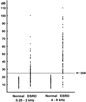
mal, and the values equal to or more than 25 dB was de- fined as hearing loss as previously described (Hutchinson and Klodd, 1982). Out of 114 ESRD patients, 55% (63/114) and 87% (99/114) of the patients showed PTA values high- er than 25 dB in low and high frequency thresholds, re- spectively (Fig. 1, Table 1). None of the normal controls showed PTA values higher than 25 in both low and high frequencies (Fig. 1, Table 1). The mean PTA values in the ESRD patients were 28 ± 13.9 (mean ± SD) dB and 51.0 ± 23.2 dB in low and high frequencies, respectively, which were significantly higher than those in the normal controls (14.0 ± 4.13 dB and 14.5 ± 4.3 dB in low and high frequencies, respectively: Fig. 1).
Fig. 1. Analysis of Pure-Tone Average(PTA) in normal people and ESRD patients. 58 of normal people without ESRD and 114 of ESRD patients were analyzed PTA. The mean age of normal people is approximately 60 years, mean age of patients was 63years. It was analyzed low(0.25-2 KHz) and high(4-8 KHz) frequency of PTA. (normal control average;
low and high frequency, 14 dB and 15 dB ESRD patients average; low and hight frequency, 28 dB and 51dB)
Table 1. Analysis of Pure- Tone Averages in the ESRD(114 patients)
Freq(kHz) Normal Hearing loss
Right ear Left ear Right ear Left ear
low frequency* 51‡ 51 63 63
high frequency† 15 18 99 96
* : 0.25 – 2 KHz, †: 4 – 8 KHz, ‡: number of patients
Association of 10609T>C and 14668C>T markers with hearing loss.
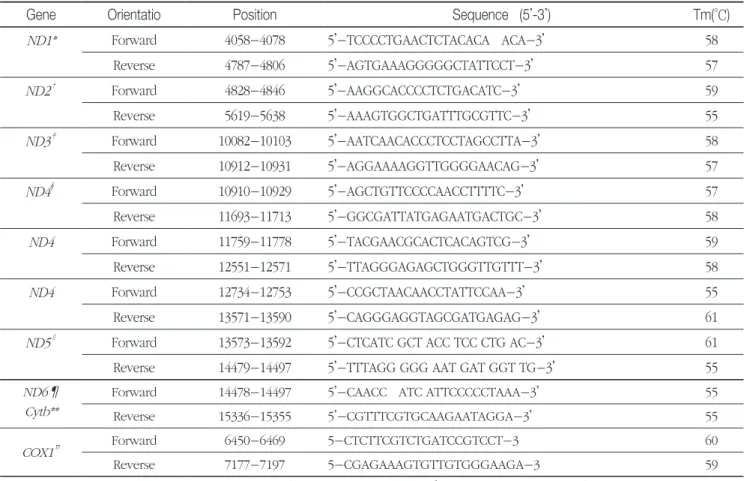
For screening of mitochondrial mutations in patients with ESRD, we screened for single nucleotide polymorphic (SNP) markers in the mitochondrial genome. Using 9 sets of oligonucleotide primers that cover the entire mitochon- drial coding genome, we amplified the mtDNA in 50 ESRD patients and then sequenced the PCR amplicons (Table 2).
We found 104 single nucleotide polymorphisms (SNPs) in 50 ESRD patients. Among the identified SNPs, 75 SNPs were reported ones in the Mitomap database (http://www.
mitomap.org), and 29 SNPs were novel ones (Table 3).
Majority (83.6 %) of the 104 SNPs were transition substitu- tion, whereas 16.3 % of the SNPs were transversion substi- tutions.
To the relevance of hearing loss and mitochondrial mu- tations in 114 ESRD patients, we divided the ESRD patients into two groups based on hearing loss and compared al- lele frequency of the six SNP markers between the pa- tient groups. Allele frequency of A6962G>A, 14783T>C, 15043G>A, and 15301G>A were not significantly different between the groups with or without hearing loss. In con- trast, 10609T>C and 14668C>T were significantly associated with hearting loss in the ESRD patients, showing a p-value of 0.007 and 0.045, respectively (Fig. 2, Table 4).
Discussion
Compared with normal controls, ESRD patients with he-
Table 2. Oligonucleotide primers used for PCR amplifications
Gene Orientatio Position Sequence (5’-3’) Tm(°C)
ND1* Forward 4058-4078 5’-TCCCCTGAACTCTACACA ACA-3’ 58
Reverse 4787-4806 5’-AGTGAAAGGGGGCTATTCCT-3’ 57
ND2† Forward 4828-4846 5’-AAGGCACCCCTCTGACATC-3’ 59
Reverse 5619-5638 5’-AAAGTGGCTGATTTGCGTTC-3’ 55
ND3‡ Forward 10082-10103 5’-AATCAACACCCTCCTAGCCTTA-3’ 58
Reverse 10912-10931 5’-AGGAAAAGGTTGGGGAACAG-3’ 57
ND4§ Forward 10910-10929 5’-AGCTGTTCCCCAACCTTTTC-3’ 57
Reverse 11693-11713 5’-GGCGATTATGAGAATGACTGC-3’ 58
ND4 Forward 11759-11778 5’-TACGAACGCACTCACAGTCG-3’ 59
Reverse 12551-12571 5’-TTAGGGAGAGCTGGGTTGTTT-3’ 58
ND4 Forward 12734-12753 5’-CCGCTAACAACCTATTCCAA-3’ 55
Reverse 13571-13590 5’-CAGGGAGGTAGCGATGAGAG-3’ 61
ND5∥ Forward 13573-13592 5’-CTCATC GCT ACC TCC CTG AC-3’ 61
Reverse 14479-14497 5’-TTTAGG GGG AAT GAT GGT TG-3’ 55
ND6¶
Cytb**
Forward 14478-14497 5’-CAACC ATC ATTCCCCCTAAA-3’ 55
Reverse 15336-15355 5’-CGTTTCGTGCAAGAATAGGA-3’ 55
COX1∏ Forward 6450-6469 5-CTCTTCGTCTGATCCGTCCT-3 60
Reverse 7177-7197 5-CGAGAAAGTGTTGTGGGAAGA-3 59
*;NADH dehydrogenase 1, †;NADH dehydrogenase 2, ‡; NADH dehydrogenase 3, §; NADH dehydrogenase 4, ∥; NADH dehydrogenase 5,
¶; NADH dehydrogenase 6, **; Cytochrome b, ∏; Cytochrome c oxidase (COX) subunit 1
modialysis treatment showed a highly significant bilateral sensorineural hearing loss at all frequencies, which was more marked in higher frequencies (Fig. 1). It was report- ed that many factors have been implicated for hearing im- pairment (Gatland et al., 1991; Yassin et al., 1970). There are contrasting results as to whether hemodialysis therapy may be a significant factor for hearing impairment (Rossini et al., 1984; Bazzi.et al., 1995; Hutchinson et al., 1982). Of course, the negative factors related to hemodialysis treat- ment which could lead to hearing impairment are: acute hypotension, reduction in blood osmotic pressure, acute clearance of urea, increased red blood cell mass. But the direct impact of hearing impairment is not.
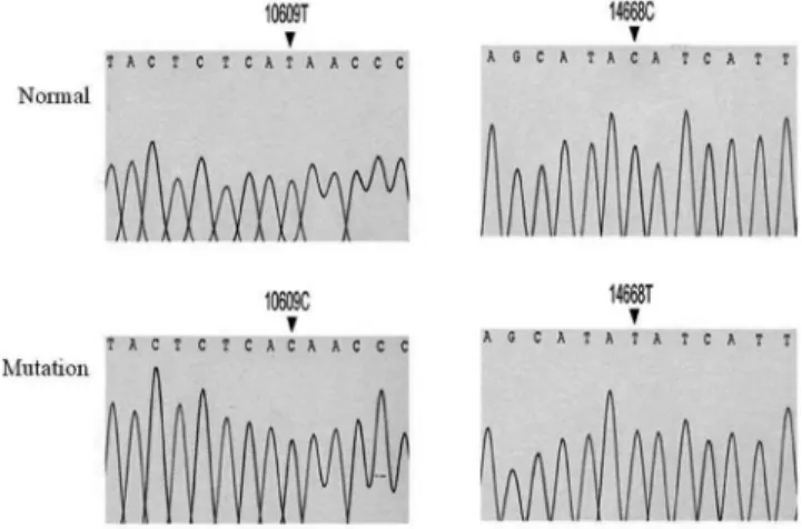
Dialysis patients had more oxidative stress than healthy subjects (Chen et al., 2009), and this elevated oxidative stress leads to alterations of the mtDNA. Since mitochon- dria are a major source of ROS and are highly susceptible to oxidative damage, we evaluated the corelation of mtD- Fig. 2. Results of mtDNA sequencing. hearing health (H)
show a wild-type sequence, whereas hearing loss (L) have a T to C (*) transition at base position 10609 affecting the ND4L gene, base position 14668 in the ND6 gene transition C to T(*)
Table 3. Novel mtDNA SNPs found in ESRD patients
Gene Location
(nt)
Amino acid
change % Gene Location
(nt)
Amino acid
change %
ND1* 3835C>G P177A 4 CO3 9777 G>C G191R 2
3851C>G A182G 8 9786 G>C G194R 2
3871A>C T189P 2 9933 C>G H243D 2
3960C>G P218P* 8 9949 T>G V248G 2
CO1† 6192 A>C M97T 2 9987 T>G S261A 2
6195 A>C N98H 2 ND3§ 10073 A>C L5F 2
6701 A>G E266E* 2 ND4L∥ 10689 G>C G74R 2
6818 C>T F305F* 2 10743 A>C N92H 2
7110 T>C Y403H 2 ND4¶ 10831 A>G W24W* 2
7408 A>C Y502S 8 11511 A>C N1187T 2
CO3‡ 7687 C>T I34I& 6 ND5** 12424 A>C N30H 2
7777 C>A V64V& 8 ND6∏ 14166 T>C N6N* 2
7879 A>G K98K& 8 CYTBⅡ 15036 A>C H97P 2
8015 C>G T144V 8 15037 C>T H97H* 8
9303 A>T M33L 2
*;NADH dehydrogenase 1, †; Cytochrome c oxidase (COX) subunit 1, ‡;Cytochrome c oxidase (COX) subunit 3, §; NADH dehydrogenase 3 ∥; NADH dehydrogenase 4L, ¶; NADH dehydrogenase 4, **; NADH dehydrogenase 5, ∏;NADH dehydrogenase 6, Ⅱ; Cytochrome b, &; synonym
NA mutation in the ESRD patients with hearing loss. Muta- tion of mtDNA in renal failure desease reported in a wide variety of nonpathogenic polymorphisms, but no data in hearing loss. To generate a more comprehensive analysis of the relationship between mitochondrial variation and hearing loss, we have analysed chi-square. We genotyped 104 mtDNA variants in screening test and analysed data from the six SNP markers. Two SNPs of 10609, 14668 posi- tion showed nominal levels of association with hearing loss (Table 3).
The ND6 (NADH dehydrogenase 6) subunit is one of the least conserved mitochondrially encoded proteins in com- plex I and involved in a ubiquinone binding site. Muta- tions in ND6 subunit were found to decrease enzyme affin- ity towards decylubiquinone (DB) (Chinnery et al., 2001).
In our result, a similar substitution, 14668C>T was detected
in the coding region for ND6 subunit in ESRD patiant with hearing loss (Fig. 2). Although it will be need to be verified with using a large number of normal control samples, this result suggests that the 14668C>T polymorphisms of the ND6 gene are associated with these factors in the ESRD pa- tients. Also, the ND4L (NADH dehydrogenase 4L) subunit of mitochondrial NADH:ubiquinone oxidoreductase (com- plex I) is an integral membrane protein. The ND4L subunit that contains two highly conserved glutamates is essential for NADH-1 enzyme activity (Kao et al., 2005; Kervinen et al., 2004). We detected the presence of 10609T>C muta- tion in the ND4L subunit, this position would explain the pattern of differences of amino acid (Met to Thr) (Fig. 2).
The polymorphisms within the conserved site of the glu- tamates region might have influence on the expression level by suppressing the NADH-1 enzyme activity. Further Table 4. Allele frequency of 6 SNP markers in ESRD patients
Position Gene Amino Acid
ESRD
without hearing loss (n=51)
ESRD
without hearing loss (n=63)
p values*
6962G>A COI 353
Leu>Leu
G 86%
(44)
A 14%
(7)
G 84%
(53)
A 16%
(10)
1.000
10609T>C ND4L 47Met>Thr
T 96%
(49)
C 4%
(2)
T 76%
(48)
C 24%
(15)
0.007*
14668C>T ND6 2Met>Met
C 73%
(37)
T 27%
(14)
C 52%
(33)
T 48%
(30)
0.045*
14783T>C CYB 13
Leu>Leu
T 45%
(23)
C 55%
(28)
T 33%
(21)
C 67%
(42)
0.28
15043G>A CYB 99Gly>Gly
G 45%
(23)
A 55%
(28)
G 33%
(21)
A 67%
(42)
0.28
15301G>A CYB 185
Leu>Leu
G 49%
(25)
A 51%
(26)
G 33%
(21)
A 67%
(42)
0.132
*; Value was determined by χ2 test from 2 × 2 contingency table
analysis using the transmitochondrial cell cybrid system will be necessary to establish the functional significance of the 10609T>C mutation in the complex I gene of mtDNA.
In summary, our results suggest that the SNP of the ND4L subunit gene might be association with ESRD pa- tients and hearing loss.
Acknowledgements
This paper (exhibition practice etc) was supported by Wonkwang Health Science University in 2011
References
1. Antonelli AR, Bonfioli F, Garrubba V, Ghisellini M, Lamoretti MP, Nicolai P, et al. Audiological findings in elderly patients with chronic renal failure. Acta Otolaryngol Suppl. 1990, 476:54-68.
2. Bai U, Seidman MD. A specific mitochondrial DNA deletion (mtDNA4977) is identified in a pedigree of a family with hear- ing loss. Hearing res 2001, 154:73-80.
3. Bazzi C, Venturini CT, Pagani C, Arrigo G, D’’Amico GD.
Hearing loss in short and long term haemodialysed patients.
Nephrol Dial Transplant 1995, 10:1865-1868.
4. Chen JB, Lin TK, Liao SC, Lee LC, Liou CW, Wang PW, et al. Lack of association between mutations of gene-encoding mitochondrial D310 (displacement loop) mononucleotide re- peat and oxidative stress in chronic dialysis patients in Taiwan.
Journal of Negative Results in BioMedicine 2009, 8:10-16.
5. Chinnery PF, Brown DT, Andrews RM, Singh-Kler R, Rior- dan-Eva P, Lindley J, et al. The mitochondrial ND6 gene is a hot spot for mutations that cause Leber’s hereditary optic neu- ropathy. Brain 2001, 124:209-218
6. Estivill XN, Govea A, Barcelo E, Perello C, Badenas E, Romero L, et al. Familial progressivesensorineural deafness is mainly due to the mtDNA A1555G mutation and is enhanced by treat- ment with aminoglycosides. Am J Hum Genet 1998, 62:27-35.
7. Gatland D, Tucker B, Chalstrey S, Keene M, Baker L. Hearing loss in chronic renal failure-hearing threshold changes follow- ing hemodialysis. J R Soc Med 1991, 4:587-589.
8. Hutchinson J, Klodd D. Electrophysiological analysis of
auditory,vestibular, and brainstem function in chronic renal failure. Laryngoscope 1982, 92:833-843.
9. Hutchin, TI, Haworth K, Higashi N, Fischel-Ghodsian M, Stoneking N, Saha C,Arnos Cortopassi G. A molecular basis for human hypersensitivity to antibiotics. Nucleic Acids Res 1993, 21:4174-4179.
10. Johnson DW, Mathog RH. Hearing function and chronic re- nal failure. Ann Otol Rhinol Laryngol. 1976, 85: 43-49.
11. Johnson DW, Wathen RL, Mathog RH. Effects of hemodialy- sis on hearing threshold. ORL J Oto Rhino lary 1976. 38:129- 139.
12. Kao MC, Nakamaru-Oqiso E, Matsuno-Yagi A, Yaqi T.
Characterization of the membrane domain subunit NuoK (ND4L) of the NADH-quinone oxidoreductase from Esch- erichia coli. Biochemistry 2005, 12:9545-9554.
13. Kervinen, Patsi J, Finel M, Hassinen IE. A pair of membrane- embedded acidic residues in the NuoK subunit of Escherichia coli NDH-1, a counterpart of the ND4L subunit of the mito- chondrial complex I, are required for high ubiquinone reduc- tase activity. Biochemistry 2004, 43:773-781.
14. Lahiri DK, Nurberger JL. A rapid non-enzymatic method for the preparation of HMW DNA from blood for RFPLs studies.
Nucleic Acid Res 1981, 19:5444.
15. Rao M, Li L, Demello C, Guo D, Jaber BL, Pereira BJ, et al.
HEMO Study Group. Mitochondrial DNA Injury and Mor- tality in Hemodialysis Patients. J Am Soc Nephrol. 2009, 20(1):189-196.
16. Mitsnefes MM. Pediatric end-stage renal disease: Heart as a target. J Pediatr 2002, 141:162-164.
17. Nagy A, Wilhelm M, Kovacs G. Mutations of mtDNA in re- nal cell tumours arising in end-stage renal disease. J Pathol 2003, 199:237-242.
18. Rossini M, Stefano D, Febbo A, Paolo D, Bascini M. Brain- stem auditory responses (BAERs) in patients with chronic re- nal failure. Electroen Clin Neuro 1984, 57:507-514.
19. Ozturan O, Lam S. The effect of hemodialysis on hearing us- ing pure-tone audiometry and distortion-product otoacous- tic emissions. ORL J of Oto Rhino lary 1998, 60:306-313.
20. Rao M, Li L, Demello C, Guo D, Jaber BL, Pereira BJ, et al. HEMO Study Group. Mitochondrial DNA Injury and Mortality in Hemodi- alysis Patients. J Am Soc Nephrol 2009, 20:189-196.
21. Reid FM, Vernham GA, Jacobs HT. A novel mitochondrial pointin a maternal pedigree with sensorineural deafness.
Hum Mutat 1994, 3:243-247.
22. Serbetcioglu B, Erdogan S, Sifil A. Effects of a single session of Hemodialysis on Hearing Abilities. Acta Otolaryngol. 2001, 121:836-838.
23. Thodi C, Thodis E, Danielides V, Pasadakis P, Vargemezis
V. Hearing in renal failure. Nephrol Dial Transplant. 2006, 21:3023-3030.
24. Tiranti VP, Chariot F, Carella A, Toscano P, Soliveri P, Gir- landa F, et al. Maternally hearing loss, ataxia and myoclonus associated with a novel pointin mitochondrial tRNASer (UCN)
gene. Hum Mol Genet 1995, 4:1421-1427.
25. Yassin A, Badry A, Fatthi A. The relationship between elec- trolyte balance and cochlear disturbances in cases of renal failure. J Laryngol Otol 1970, 84:429-435.