Culturing the Human Dental Pulp cells in the Collagen Matrix and on the ground tooth surface
Sang-Hyuk Park
Department of Conservative Dentistry, College of Dentistry, Kyung-Hee University
콜라젠 기질(COLLAGEN MATRIX)과 마모된 치아표면에서의 치수세포 배양에 관한 연구
박 상 혁
경희대학교 치과대학 치과보존학 교실
이 연구의 목적은 원래의 치수조직과 유사한 조직을 재생하기 위한 pulp tissue engineering의 한 방법으로 건전한 조직으로부터 배양된 치수세포와 쥐의 조섬유세포(NIH 3T3 cell)를 Rat tail type I collagen solution에서 3차원적으 로 관찰하기 위한 것으로, 콜라젠 젤의 수축량과 세포의 증식량을 비교하였으며, 또한 마모된 사람치아의 표면과 배양용 기에서 두 세포의 증식량을 비교하여 다음과 같은 결과를 얻었다.
1. 콜라젠 젤에 NIH 3T3 세포를 배양한 경우 그 수축량은 최소였으나, 치수세포를 배양한 경우 그 수축량은 현저하였다.
2. 서로 다른 수의 치수세포를 콜라젠 젤에서 배양시킨 경우 세포 수가 많을수록 수축량이 증가하였으며, 세포가 없는 콜라젠 젤은 수축하지 않았다.
3. 치수세포를 콜라젠 젤에서 18일간 배양시킨 후 세포의 증식은 거의 없는 반면, NIH 3T3 세포는 계속 증식하였다.
4. 마모된 사람 치아 표면과 배양 용기에서 치수세포와 NIH 3T3세포를 배양한 경우 NIH 3T3세포가 치수세포에 비 해 빠르게 증식 하였으며, 특히 사람 치아의 표면에서 NIH 3T3세포가 현저히 빠른 증식을 보였다.
이상의 결과는 치수세포를 type I collagen gel에서 3차원 적으로 배양 후 치수조직의 재생을 유도하는 pulp tissue engineering에 관한 연구에 발판이 될 것으로 사료된다.
K
Keeyy wwoorrddss : 치수세포, NIH 3T3세포, 제 1형 콜라젠 젤, 치아표면, 세포증식, 기질수축
Ⅰ. INTRODUCTION
Tissue engineering science has been in varying stages of development and progress to provide an alternative method for repairing damaged tissues.
The dental pulps have long been considered a vul- nerable tissue to infection and trauma which fre- quently leads to irreversible inflammatory condi- tions. The root canal treatment by removing the diseased pulps has been the most common treat- ment option. While some investigators in the past attempted to regenerate the pulp tissue, no desir- able results were obtained and these efforts have not been actively pursued in the recent decades.
Even the more conservative approach such as vital pulp therapy has not been a popular clinical choice due to lack of predictable results.
Dental pulp is a loose connective tissue, with extensive vascular and nerve supply, containing predominantly fibers and ground substance that make up the extracellular matrix (ECM), and cel- lular component including mainly fibroblasts and undifferentiated mesenchymal cells. The uniquely specialized pulp cells(odontoblasts) are lining against the dentinal wall and their processes extend into the dentinal tubules. The volume of the mature pulp tissue is very small and so it appears relatively less difficult to regenerate this small vol- 국문초록
ume of soft tissue than larger organs or tissues.
However, it is considered rather difficult to regen- erate the entire pulp tissue due to the following reasons: 1) the unique anatomical location of pulp tissue-enclosed in dentin having limited blood sup- ply from one end for the in-growth of new tissue elements; 2) the small size of the pulpal canal space rendering a technically sensitive procedure to implant the regenerated pulpal tissue in vitro into the canal; and 3) the unique microscopic anatomy of pulp tissue, e.g., the odontoblasts and their relationship with the nerve fibers and vessels.
While trying to regenerate the exact pulp tissue to its pristine condition appears difficult to achieve, regenerating a pulp connective tissue with vascular structure and nerve fibers appears not impossible.
The goal of tissue engineering is to regenerate living tissue substitutes in order to replace or enhance lost tissue function or structure. To accomplish this, the cells out of tissue must be organized and behave as if they are part of the original tissue by interacting with the surrounding ECM which acts as a supporting material created by the cells as a scaffolding on which to reside1). The ECM is composed of a variety of macromole- cules which can be grouped into four major classes (collagens, proteoglycans, cell interactive glycopro- teins, elastic fibers), each of which is responsible for specific ECM characteristics. Type I collagen is the most abundant type of collagen isolated from many adult connective tissues (skin, bone, ten- don). Pulp fibroblasts can produce both type I and type III collagens, whereas the majority of collagen molecules produced by odontoblasts are type I2).
Synthetic matrices fabricated from naturally- derived (e.g., collagen) or synthetic materials (e.g., polyglycolic acid or PGA) are often utilized as a delivery vehicle for these cells and to guide the process of tissue formation3). It has been shown by Rutherford’s group (1999) that the fibroblast cul- tured from human adult dental pulps seeded onto PGA formed new tissue similar to that of native pulp after implanted into mouse dermis4). They also found that when pulp cells seeded onto the three different synthetic matrices (PGA fibers, type I col- lagen hydrogel, or alginate), the growth of the cells
was moderate in collagen gel5). Type I collagen gel has been used to measure the contraction of the collagen gel matrix mixed with bovine aortic endothelial cell6)or human lung fibroblast7).
In this study, to characterize the growth behavior of pulp cells in collagen matrix, rat tail type I colla- gen was mixed with pulp cells from human dental pulp tissue, and the growth of pulp cells and con- traction of the collagen gel was estimated. The result of this investigation may help to establish pulp tissue engineering protocol in vitro and in vivo.
Ⅱ. MATERIALS AND METHOD
Sample collection and Cell cultureFreshly extracted, intact, caries-free third molars (n=10) were obtained from healthy individuals (15-25 years of age) in the Department of Oral Surgery at the UCLA School of Dentistry according to a protocol approved by the UCLA Medical Institutional Review Board. Immediately after extraction, teeth were stored in phosphate buffered saline (PBS) and transported to the laboratory.
Teeth were split open and the maximal amount of pulp tissue extracted was divided into several small fragments approximately 2×2 mm in size each. Repeated washing with PBS, pulp fragments were placed in a 60 mm culture dish containing Dulbecco’s Modified Eagle Medium (DMEM; Life Technologies/GIBCO BRL, Gaithersburg, MD) sup- plemented with 10% fetal bovine serum (FBS).
Pulp cells outgrown from pulp fragments exhibited a fibroblast-like phenotype and they were allowed to reach confluence and passed at 1:2 ratio until used for experiments (passages 3-8 were used).
Cell culture media were supplemented with 100 units/ml penicillin-G, 100
μ
g/ml streptomycin, and 0.25μ
g/ml fungizone (Gemini Bio-Products, Inc., Woodland, CA). In this study, primary pulp cells of fibroblast-like phenotype outgrown from human pulp tissue(passage 3-5) were used for experi- ments and NIH 3T3 cells grown in the same cul- ture medium for pulp cells were used as a compari- son.1. Collagen gel preparation and collagen gel contraction assay
Rat tail type I collagen was purchased from BD Biosciences (Bedford, MA). Pulp cells were mixed with collagen gel solution [9 parts type I collagen (3.97 mg/ml), 0.023 part 1N NaOH and 1 part 10
×PBS] and placed in a microcentrifuge tube kept on ice. This neutralized, isotonic collagen solution was used to minimize injury of pulp cells. The mix- ture of pulp cells and collagen gel were placed into 96-well plates (0.1 ml/well) and incubated at 37℃
for 30 min to allow gelation. Culture medium (0.2 ml/well) was then added into each well rendering cell/collagen mixture lifted from the bottom and suspended in the medium. The plates were incu- bated at 37℃ with 5% CO2 and the medium changed every 2-3 days. The area of the gels was measured daily using the grid of the microscope.
2. Cell proliferation on the culture dish and tooth surface
After the pulp tissue from the human 3rd molar were collected, the tooth surface was prepared with diamond bur to fit the 96-well plates and ground with #600 SiC paper, and then sterilized with one hundred times more concentrated penicillin, strep- tomycin, and fungizone than those used for cell cultures, followed by Betadine, 2% chlorohexidine, and 5.25% NaOCl, in sequence. Trypsinized human dental pulp cells and NIH 3T3 cells (120 cells/mm2) were seeded on the tooth surface and 96 well-plates to compare the proliferation of the two cells. The seeded cells on the tooth surface were trypsinized in every two days for 9 days, and those in 96 wells were trypsinized everyday for 10 days.
Detached cells were counted with the Coulter counter.
3. Cell proliferation in the collagen gel matrix
Dental pulp cells and NIH 3T3 cells (3×10⁴ cells/well) were mixed in the collagen gel solution
(0.1 ml) prepared as mentioned above and incu- bated at 37℃, 5% CO2for 30 minutes. 0.2 ml of a 3 mg/ml solution of type I collagenase (Sigma) in PBS was used for 45 minutes to detach the cells from the cast collagen gel and the number of the cells was counted with the Coulter counter at the different time point for 18 days.
Ⅲ. RESULTS
1. Collagen gel preparation and collagen gel contraction assay
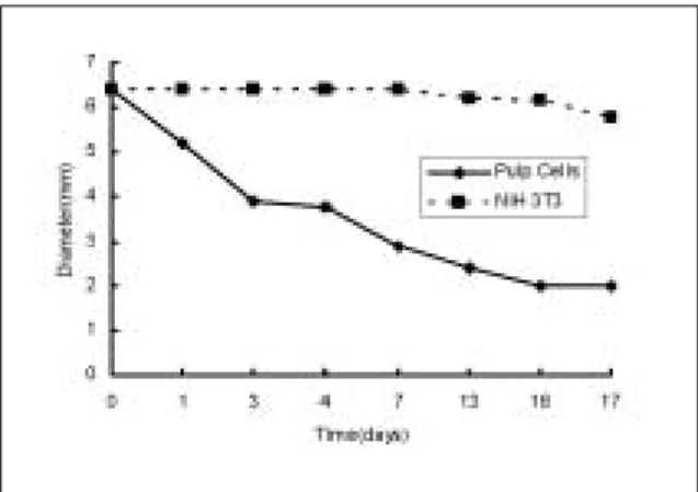
There was no contraction of the collagen gel matrix with NIH 3T3 cells until after day 7. Only minimal contraction (down to 90%) was observed after 17 days. In contrast, the contraction was dra- matic with pulp cells through out the course of observation (down to 45% on day 7 and to 31% on day 17) (Fig. 1).
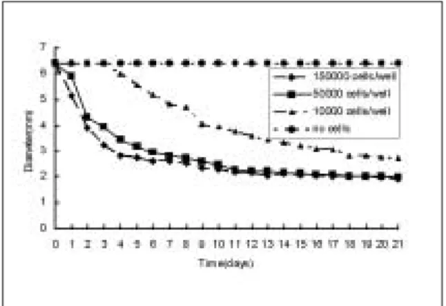
To determine the contraction of the collagen gel matrix affected by the concentration of pulp cells, various numbers of cells were mixed with the colla- gen and cultured in 96-well plates. The results showed that the collagen matrix with 1.5×10⁵ cells/well and 5×10⁴ cells/well, contracted down to 30% after 21 days, the matrix with 104 cells/well to 35%, and the collagen gel without cells had no
Fig. 1. Contraction analysis of collagen gel matrix seeded with pulp cells and NIH 3T3 cells.
contraction (Fig. 2). It took 3, 5, and 15 days for collagen gel matrix with 1.5×10⁵, 5×10⁴, and 104 cells/well to be half size of the original matrix respectively.
2. Cell proliferation on the culture dish and tooth surface
When cells grown in 96-well plates, the dental pulp cells and NIH 3T3 cells had a similar growth rate up to 4 days after seeding, after which NIH 3T3 cells grew at a faster rate than pulp cells till the end of experimental period (10 days) as demonstrated in Fig. 3. NIH 3T3 cells had a 25.4- fold increase of the number of the cells, while pulp cells had 19.7-fold increase compared with original number of the cells plated on the 96 well-plates
after 10 days(Fig. 3).
The pulp cells plated on the ground dentin sur- face proliferated less than the ones grown on the culture dish after 9 days, whereas NIH 3T3 cells proliferated significantly on the tooth surface than on the culture dish. There was a significant increase of NIH 3T3 cells (185-fold) and less increase of pulp cells (7.7-fold) grown on the dentin surface after 9 days (Fig. 4).
3. Cell proliferation in the collagen gel matrix
Pulp cells showed either minimal or no prolifera- tion in collagen gel matrix up to 18 days after plat- ing, whereas NIH 3T3 started to proliferate signifi- cantly after 8 days and continued to grow till 18 days (Fig. 5).
Fig. 2. Contraction test of the collagen gel matrix seeded with different cell number of pulp cells.
Fig. 3.Proliferation test of the cells grown on the 96- well plates.
Fig. 4.Proliferation test of the cells grown on dentin surface.
Fig. 5. Proliferation test of cells grown in collagen gel.
Ⅳ. DISCUSSION
This is the first investigation to compare the con- traction of type I collagen in Vitro by culturing pulp-derived human fibroblasts and mouse skin fibroblast(NIH 3T3 cells) three dimensionally. The use of type I collagen gels did not lead to the devel- opment of new tissues which resembled native pulp, while the cells adhered and proliferated in the collagen gel.
Collagen-Glycosaminoglycan matrix seeded with porcine dental pulp cells in Vitro decreased in size to less than half of their original diameter by 28 days and the level of α-Smooth-muscle action (SMA) increased with passage number, so SMA containing pulp cells had the capability to contract a collagen-glycosaminoglycan analog of extracellu- lar matrix in vitro.8)
Type I collagen has been widely utilized to engi- neer a variety of tissue type due to its ability to promote cell adhesion and allow for cell-based remodeling, type I collagen is the most abundant type of collagen isolated from many adult connec- tive tissues (skin, bone, tendon)1), and out of the collagen molecules occurring in the pulp type I and III represent the bulk of the pulp tissue collagen.
Because type I is the predominant type and may contribute to the establishment of the architecture of the pulp, in this study commercial rat tail type I collagen was purchased and used as the scaffolds to grow the pulp cells and NIH 3T3 cells three dimensionally3).
Human lung fibroblasts were cast into two differ- ent density of type I rat tail collagen(0.75 mg/ml and 2 mg/ml), the size of the lower density gel after 15 days was smaller than the higher density one7). The contraction of type I collagen by bovine aortic endothelial(BAE) cells and human dermal fibroblast was compared in collagen gel contraction assay, the fibroblasts contracted rigid gels more effectively in comparison to BAE cells9).
In this study, human dental pulp cells contracted significantly comparing with NIH 3T3 cells in rat tail type I collagen, nevertheless the proliferation rate of the NIH 3T3 cells cultured in culture dishes and type I collagen gel increased more than human
dental pulp cells. The basis for this reduced cellu- lar proliferation rate on the collagen matrices was not identified but seemed to depend on fibrillar organization and required the native(triple helical) conformation of the collagen molecule10). The more contraction of the collagen gels were made with the increased number of the pulp cells mixed with the same condition of collagen solution, nor contraction without any cells.
When skin fibroblasts are cultured on or in colla- gen gels, there was a significant reduction in their rate of proliferation10). Skin fibroblast cultured in the collagen gel and culture dish with human serum had the more proliferation rate than bovine serum, and platelet-derived growth factor reduced mitotic rate observed with the cells on collagen gels11).
Human dental pulp cells were expected to grow much better on the prepared human tooth surface than NIH 3T3 cells, but it was surprising that the proliferation rate of NIH 3T3 cells was significantly higher than human dental pulp cells. It is not clear if there is some growth factor in the human tooth surface to induce the proliferation of the mouse skin fibroblast.
The pulp tissue engineering may ultimately find clinical application as a novel approach to repair and/or regenerate dental pulp, and they may also provide a useful system to assess the biocompati- bility of chemicals utilized in dental practice which come in contact with native pulp. It is possible that engineered dental pulp tissue will provide a model system in which reparative dentinogenesis can be studied. This work may also provide the first step to engineer an entire tooth, and these cell behav- iors on the collagen matrix may provide informa- tion needed to establish pulp tissue engineering protocols.
Ⅴ. CONCLUSION
The purpose of this study is to reveal if the rat tail type I collagen can be used as scaffold to grow human dental pulp cells and mouse skin fibrob- lasts(NIH 3T3 cells) and to characterize the behaviors of those two cells on the collagen gel
matrix and ground tooth surface. For the purpose the contraction of the collagen matrix and prolifer- ation rate after those cells were mixed with the type I collagen solution three dimensionally and on the ground tooth surface.
According to this study, the results were as fol- lows:
1. The contraction of the collagen mix was minimal with NIH 3T3 cells (down to 90% on 17 days), whereas the contraction was dramatic with pulp cells (down to 45% on 7 days and to 31% on 17 days).
2. When the different number of pulp cells were cultured in collagen gel, the more contraction of the collagen matrix occurred with the more pulp cells, nor contraction of collagen matrix without any cells.
3. The pulp cells showed either minimal or no pro- liferation in Rat tail type I collagen up to 18 days after plating, whereas NIH 3T3 started to proliferate significantly after 3-5 days and con- tinued to grow till 18 days.
4. NIH 3T3 cells proliferated better than pulp cells on the ground tooth surface and culture dishes, proliferation rate of NIH 3T3 cells were signifi- cantly high.
These data indicate that pulp cells contract the collagen fibers extensively, whereas NIH 3T3 cells only minimally contract the collagen matrix. While pulp cells proliferate in culture dishes, they grow minimally on human dentin surface and in collagen matrix. NIH 3T3 cells, in contrast, proliferated sig-
nificantly in all three conditions tested in our experiment, particularly on dentin surface. These cell behaviors may provide information needed to establish pulp tissue engineering protocols.
REFERENCES
1. Mikos AG and Mcintire LV: Frontiers in tissue engi- neering. Pergamon. Houston. TX. 1998.
2. Hargreaves KM and Goodis HE: Seltzer and Bender’s Dental Pulp. Quintessence Publishing Co. Inc. Chicago.
IL. 2002.
3. Putnam AJ and Mooney DJ: Tissue engineering using synthetic extracellular matrices. Nature Med, 2:824- 826,1996.
4. Mooney DJ, Powell C, Piana J and Rutherford B.
Engineering dental pulp-like tissue in Vitro. Biotechnol.
Prog, 12:865-868,1996.
5. Bohl KS, Shon J, Rutherford B and Mooney DJ : Role of synthetic extracellular matrix in development of engi- neered dental pulp. J Biomater Sci. Polymer Edn, 9:
749-764,1998.
6. Bernon RB and Sage EH : Contraction of fibrillar type I collagen by endothelial cells: A study in vitro. J Cell Biochem, 60:185-197,1996.
7. Zhu YK, Umino T, Liu XD, Wang HJ, Romberger DJ, Spurzem JR and Rennard SI : Contraction of fibroblast- containing collagen gels: initial collagen concentration regulates the degree of contraction and cell survival. In vitro Cell. Dev. Biol.-Animal, 37:10-16,2001.
8. Brock DP, Marty-Raix R and Spector M : A-Smooth- muscle actin in and contraction of porcine dental pulp cells. J Dent Res, 81:203-208,2002.
9. Vernon RB and Sage EH : Contraction of fibrillar type I collagen by endothelial cells: A study in vitro. J Cell Biochem, 60:185-197.1996.
10. Schor SL : Cell proliferation and migration on collagen substrata in vitro. J Cell Sci, 41:159-175,1980.
11. Rhudy RW and Mcpherson JM : Influence of the extra- cellular matrix on the proliferative response of human skin fibroblasts to serum and purified platelet-derived growth factor. J Cell Physiol, 137:185-191,1988.
박 상 혁
경희대학교 치과대학 보존과 임상강사
서울시 동대문구 회기동 1번지 경희대학교 치과대학 보존학교실 Tel : 02-958-9330
E-mail : [email protected]