Characterization of Human Dental Pulp Cells from Supernumerary Teeth by Using Flow Cytometry Analysis
Yonsook You
1, Jongbin Kim
2, Jisun Shin
2, June-Haeng Lee
2, Jongsoo Kim
21
Misa Seoul S Dental Clinic,
2Department of Pediatric Dentistry, School of Dentistry, Dankook University
The aim of this study was to analyze cells from human dental pulp tissue of impacted supernumerary teeth as stem cells with flow cytometry. Human dental pulp cells from 15 supernumerary teeth were identified their characteristics as stem cells by expression of mesenchymal stem cell markers through flow cytometry analysis at passage 3 and passage 10. Cluster of differentiation (CD) 73, CD 90, CD 34, CD 45 and STRO-1 cell surface markers were used to figure out characteristics of dental pulp stem cells from supernumerary teeth. At passage 3, the cell population showed positive expression of CD 73, CD90 and STRO-1, lacked expression of CD 34 and CD 45. At passage 10, CD 73, CD 90 and STRO- 1 showed positive expression while CD 34 and CD 45 showed negative expression. This study indicated that dental pulp stem cells of supernumerary teeth had the properties of mesenchymal stem cells at both early and late passage.
Impacted supernumerary teeth could be considered as a noble source of stem cells because of rapid growth and maintaining characteristics of stem cells until late passage.
Key words : Supernumerary tooth, Dental pulp stem cell, Flow cytometry Abstract
Corresponding author : Jongsoo Kim
Department of Pediatric Dentistry, College of Dentistry, Dankook University, 119 Dandae-ro, Dongnam-gu, Cheonan, 31116, Korea Tel: +82-41-550-0222 / E-mail: [email protected]
Received March 27, 2019 / Revised June 28, 2019 / Accepted June 25, 2019
Ⅰ. Introduction
Stem cells in human body have infinite potentials for cure or acceleration of healing in damaged tissue. Nevertheless, sourc- es of stem cells to harvest out from human bodies are limited because of the difficulty of extracting many pure stem cells and the necessity of additional invasive procedure. Recently, it has been trying to find various sources of stem cell[1]. Embry- onic stem cells have been treated as conventional stem cells for the last half century[2]. But the embryonic stem cells have limited supply and ethical issues. To replace the embryonic stem cells, studies on adult stem cells from dental tissues had
been reported on various sources including dental pulp, ex- tracted deciduous tooth, periodontal ligament, dental sac and apical follicle[3-7]. Most of stem cell researches in the dental field were related to the cells from wisdom teeth or deciduous teeth. However, the wisdom teeth have limitation of relatively late extraction time. Deciduous teeth exfoliate in an early age, but most of them has a physiologic root resorption and in- complete pulp tissue remnants. Impacted supernumerary teeth (SNT) can removed without damage of pulp tissue during sur- gery procedure at an early age[8].
The International Society for Cellular Therapy (ISCT) pro-
posed minimal criteria for identifying mesenchymal stem cell
(MSC) in 2006[9]. Previous studies of adult stem cells con- firmed as MSCs by applying these criteria[10-12] while most studies used data obtained from a few or one donor.
The purpose of this study is to analysis characteristics of MSC of dental pulp from 15 impacted SNT.
Ⅱ. Materials and Methods
1. Materials
Impacted supernumerary teeth in the anterior maxilla were extracted from 15 healthy patients who were between 6 and 9 years old. This study was approved by the Dankook university dental college institutional ethics committee (H-1506/006/001).
Supernumerary teeth were kept in α-minimum essential medium (α-MEM, Gibco) with 20% fetal bovine serum (FBS, Gibco), 100 U/mL penicillin, 100 μg/mL streptomycin (Gibco), 2 mM L-glutamine (Gibco) and 10 nM L-ascorbic acid (Sigma) immediately after extraction.
2. Methods
1) Cell preparation and culture
Supernumerary teeth were cut around at cementoenamel junction with dental disc until before exposing dental pulp.
The crown and root were split along the groove to obtain dental pulp tissue with sterile dental file. Dental pulp tissue was chopped to fine slices less than 1 mm. Dental pulp cells were extracted using enzymatic digestion method with 3 mg/
mL type I collagenase (Sigma-Aldrich) and 4 mg/mL Dispase (Sigma-Aldrich) under the shaking incubator at 37°C for 1 hour. Cells were filtered by 70 μm Falcon strainer (Corning) and cultured in α-MEM with 20% FBS, 100 U/mL penicillin, 100 μg/mL streptomycin, 2 mM L-glutamine. Culture solution was changed every 2 - 3 days, and floating particles were rinsed out. The cells were separated for next passage using trypsin- EDTA (Corning). Subculture until passage 10 was lasting for stemness comparison between cells of early and late passages.
2) Flow cytometry analysis
Passage 3 and passage 10 cells were chosen to compare the expression of cell surface markers of early and late pas- sage. Cells were counted to put same number of cells for each cell surface markers. 1 × 10
5of the cells and 1 μg cell surface markers were leaved in refrigerator at 4°C for 1 hour. The sur-
face markers used in this study were cluster of differentiation (CD) 73, CD 90, CD 34, CD 45 (BD Biosciences) and STRO-1 (R&D system). Inactive cell surface markers were removed and the fluorescent antibodies were used for florescence-activated cell sorting (FACS) analysis. The cell-antibody complexes were analyzed with BD FACSCalibur flow cytometry (BD Biosciences).
The cells that were not reacted to any markers were used as control group.
3) Statistical analysis
Data were analyzed using SPSS 23.0 statistical software (SPSS Inc., USA).
Ⅲ. Results
1. The culture of supernumerary dental pulp stem cells
Dental pulp cells were extracted from SNT of 15 patients; SNT 01 - SNT 15. Dental pulp cells were cultured to passage 10.
2. Immunophenotype
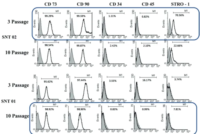
The expression of the cell surface markers in the control group and the experimental group was obtained at passage 3 and passage 10 of human dental pulp cells from SNT 01 and SNT 02 (Fig. 1). CD 73, CD 90, STRO-1 as MSC surface mark- ers were highly expressed in the dental pulp cells from SNT, compared to only a small degree expression of CD 34, CD 45 as hematopoietic and endothelial markers. The expression of STRO-1 at passage 3 of SNT 02 and passage 10 of SNT 01 satisfy the criteria presented by ISCT.
Table 1 and 2 showed the expression of CD markers of the dental pulp cells from SNT at passage 3 and passage 10. CD 73 and CD 90 were positively expressed, while CD 34 and CD 45 were negatively expressed. STRO-1 was expressed. Most of dental pulp cells from SNT were satisfied with the criteria of ISCT.
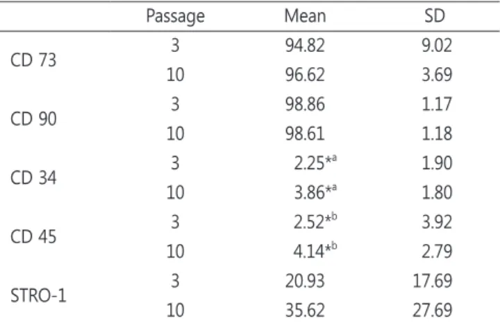
Table 3 showed the mean value of expression of CD markers
at passage 3 and passage 10. At passage 3, dental pulp cells
from SNT showed positive expression of CD 73 (94.82%), CD
90 (98.86%) and STRO-1 (20.93%), negative expression of CD
34 (2.25%) and CD 45 (2.52%). At passage 10, dental pulp cells
from SNT showed positive expression of CD 73 (96.62%), CD
90 (98.61%) and STRO-1 (35.62%), negative expression of CD
34 (3.86%) and CD 45 (4.14%). The expression of CD 73, CD90
Table 1. Flow cytometry analysis of the mean percentile values of the expression of CD marker at passage 3
CD73 CD90 CD34 CD45 STRO-1 SNT01 95.62 97.44 3.55 16.17 3.74 SNT02 99.29 99.59 1.11 0.82 70.56 SNT03 95.83 98.66 0.88 0.91 12.81 SNT04 98.73 99.56 8.34 4.13 6.51 SNT05 99.33 99.41 1.56 1.69 19.77 SNT06 99.26 99.70 1.40 1.07 7.91 SNT07 97.83 99.82 1.54 1.44 13.17 SNT08 99.25 99.48 0.49 0.18 32.76 SNT09 73.21 95.50 2.79 3.02 10.84 SNT10 99.12 99.33 2.19 1.40 16.96 SNT11 99.68 99.76 3.36 2.89 45.40 SNT12 99.59 99.67 1.28 1.11 21.08 SNT13 97.72 98.26 2.33 1.59 29.15 SNT14 95.21 98.26 1.54 1.13 8.55 SNT15 72.63 98.48 1.40 0.25 14.77 CD : Cluster of differentiation, SNT : Supernumerary tooth
Table 2. Flow cytometry analysis of the mean percentile values of the expression of CD marker at passage 10
CD73 CD90 CD34 CD45 STRO-1 SNT01 98.92 98.90 0.83 0.99 7.81 SNT02 98.54 99.87 2.42 2.10 22.66 SNT03 96.20 99.70 4.89 3.86 47.06 SNT04 98.52 98.77 2.52 2.17 14.59 SNT05 92.41 97.58 1.14 1.11 39.01 SNT06 96.64 96.26 3.41 2.90 16.17 SNT07 85.02 98.72 3.41 2.63 15.26 SNT08 95.90 96.10 6.79 6.27 18.00 SNT09 98.54 99.56 3.71 3.23 11.29 SNT10 97.81 98.52 6.37 5.41 84.66 SNT11 99.69 99.35 6.50 5.00 9.36 SNT12 98.60 98.87 3.45 11.61 74.37 SNT13 95.79 99.49 3.57 2.34 59.61 SNT14 98.81 99.57 3.91 7.37 30.62 SNT15 97.90 97.86 5.03 5.08 83.78 CD : Cluster of differentiation, SNT : Supernumerary tooth
Fig. 1. Expression of cell surface markers in flow cytometry analysis of supernumerary tooth 01 and supernumerary tooth 02 at passage 3 and passage 10.
SNT : Supernumerary tooth, CD : Cluster of differentiation
and STRO-1 between two passages was not significantly differ- ent. There was a significant difference between two passage in expression of CD 34 and CD 45.
The average values of G-mean were calculated in flow cy- tometry results (Table 4).
IV. Discussion
The adult stem cells exist as undifferentiated cells in hu- man organs[2,13,14]. Pittenger et al .[15] were classified the adult stem cells into MSCs and hematopoietic stem cells, and proved that the adult stem cells could be differentiated into osteoblasts, chondroblasts, myoblasts and nerve cells by con- trolling culture condition. The other studies reported the adult stem cells could be obtained from various tissues including bone marrow, pancreas, adipose, muscle, blood, hair follicles, skin and dental tissues[16].
In 2006, ISCT presented the minimal 3 criteria of MSCs[9].
First, they have characteristic of adhesion to plastic culture flask. Second, they must show positive expression of CD 105, CD 73 and CD 90, while as well as showing negative expres-
sion of CD 45, CD 34, CD 14 or CD 11b, CD 79alpha or CD 19 and HLA-DR. Thirdly, they should have a possibility of differen- tiation to osteoblasts, adipocytes and chondroblasts in vitro.
The aim of this study was to identify MSCs in 15 SNT with flow cytometry analysis. CD 73, CD 90 and STRO-1 were used as positive CD markers, and CD 34 and CD45 were used as negative CD markers.
STRO-1 is early mesenchymal stem cell marker, which was detected around blood vessels or nerve fascicles while not detected in size-sieved stem cells[12,17,18]. In 2007, Kolf et al .[19] reported STRO-1 was the best CD marker to identify mesenchymal stem cells. Gay et al .[20] reported that STRO-1 showed 27% expression in the cells from periodontal ligament of impacted wisdom teeth, and Park et al .[21] presented that STRO-1 showed 33.4% expression in the cells from periodon- tal ligament of supernumerary teeth. In this study, STRO-1 showed 20.93% expression at passage 3 and 35.62% at pas- sage 10. The expression of the STRO-1 was even slightly in- creased in passage 10 comparing to passage 3. However, there was no significant difference between the two passages.
The expression of positive CD markers was not statistically different between passage 3 and passage 10 in flow cytometry analysis. This meant later passage of dental pulp cells from SNT still contained stemness and could have value of use as a source of stem cells. The expression of negative CD markers was a statistical significance between passage 3 and passage 10. According to ISCT’s recommend, the negative CD marker should be detected lower, further study is required.
The dental pulp stem cells (DPSCs) of SNT from 15 donors showed characteristic of MSCs by flow cytometry using CD markers. The characteristic of MSCs was maintained until pas- sage 10. However, FACS results alone are not enough to verify the stem cell function of cells. The differentiation to hard tis- sues or fat should also be examined. Also additional research- es including real time qPCR, would be needed to confirm gene expression in follow-up studies.
Table 3. The mean percentile values of flow cytometry analysis at passage 3 and passage 10
Passage Mean SD
CD 73 3 94.82 9.02
10 96.62 3.69
CD 90 3 98.86 1.17
10 98.61 1.18
CD 34 3 2.25*
a1.90
10 3.86*
a1.80
CD 45 3 2.52*
b3.92
10 4.14*
b2.79
STRO-1 3 20.93 17.69
10 35.62 27.69
Mann-Whiteny test (* : p < 0.05), MSC : Mesenchymal stem cell, CD : Clus- ter of differentiation, SD : Standard deviation
Table 4. Average G-mean values of flow cytometry analysis at passage 3 and passage 10
CD 73 CD 90 CD 34 CD 45 STRO-1
Passage 3 10 3 10 3 10 3 10 3 10
G-Mean 114.95 144.82 338.40 493.86 0.11 3.22 -0.01 3.29 3.15 9.23
SD 81.76 60.15 214.38 475.05 0.41 0.60 0.37 0.85 3.00 5.88
CD : Cluster of differentiation, SD : Standard deviation
V. Conclusions
The result indicated that DPSCs of impacted SNT had the characteristics of MSCs. Comparing passage 3 and passage 10 in flow cytometry analysis, the cell marker expressions were similar. There was no significantly difference between the posi- tive CD marker values of passage 3 and passage 10. There was a significant difference in the negative CD marker values be- tween the two passages. DPSCs maintained stemness until the late passage. It is confirmed that the possibility of using DPSCs of impacted SNT from young children as a donor of stem cells.
References
1. Kim J : Characterization of differentiation of the supernu- merary dental pulp stem cells toward the odontoblast by application period of additives. J Korean Acad Pediatr Dent , 42:312-318, 2015.
2. Evans MJ, Kaufman MH : Establishment in culture of pluri- potential cells from mouse embryos. Nature , 292:154-156, 1981.
3. Gronthos S, Mankani M, Shi S, et al . : Postnatal human dental pulp stem cells (DPSCs) in vitro and in vivo. Proc Natl Acad Sci USA , 97:13625-13630, 2000.
4. Miura M, Gronthos S, Shi S, et al . : SHED: stem cells from human exfoliated deciduous teeth. Proc Natl Acad Sci USA , 100:5807-5812, 2003.
5. Seo BM, Miura M, Shi S, et al . : Investigation of multipotent postnatal stem cells from human periodontal ligament.
Lancet , 364:149-155, 2004.
6. Morsczeck C, Gotz W, Hoffmann KH, et al . : Isolation of precursor cells (PCs) from human dental follicle of wisdom teeth. Matrix Biol, 24:155-165, 2005.
7. Sonoyama W, Liu Y, Shi S, et al . : Mesenchymal stem cell- mediated functional tooth regeneration in swine. PLoS One , 1:e79, 2006.
8. Kim J : Managing Complications Related to Multiple Super- numerary Teeth. J Korean Acad Pediatr Dent , 41:180-186, 2014.
9. Dominici M, Le Blanc K, Horwitz E, et al . : Minimal criteria for defining multipotent mesenchymal stromal cells. The International Society for Cellular Therapy position state- ment. Cytotherapy , 8:315-317, 2006.
10. Song JS, Kim SH, Jung HS, et al. : Characterization of stem cells obtained from the dental pulp and periodontal liga-
ment of deciduous teeth. Tissue Eng Regen Med , 7:575-582, 2010.
11. Park JW, Song JS, Son HK, et al . : Effect of Storage Media and Duration on Pulpal Cell Viability in Exfoliated Decidu- ous Teeth. J Korean Acad Pediatr Dent , 41:1-7, 2014.
12. Kim D, Kim J, Roh S, et al . : A nanoscale ridge/groove pat- tern arrayed surface enhances adipogenic differentiation of human supernumerary tooth-derived dental pulp stem cells in vitro. Arch Oral Biol, 59:765-774, 2014.
13. Almeida-Porada G, Porada C, Zanjani ED : Adult stem cell plasticity and methods of detection. Rev Clin Exp Hematol , 5:26-41, 2001.
14. Temple S : Stem cell plasticity-building the brain of our dreams. Nat Rev Neurosci , 2:513-520, 2001.
15. Pittenger MF, Mackay AM, Marshak DR, et al . : Multilineage potential of adult human mesenchymal stem cells. Science , 284:143-147, 1999.
16. Wagers AJ, Weissman IL : Plasticity of adult stem cells. Cell , 116:639-648, 2004.
17. Chen SC, Marino V, Gronthos S, Bartold PM : Location of putative stem cells in human periodontal ligament. J Peri- odontal Res , 41:547-553, 2006.
18. Hung SC, Chen NJ, Lo WH, et al . : Isolation and character- ization of size-sieved stem cells from human bone marrow.
Stem Cells , 20:249-258, 2002.
19. Kolf CM, Cho E, Tuan RS : Mesenchymal stromall cells. Biol- ogy of adult mesenchymal stem cells: regulation of niche, self-renewal and differentiation. Arthritis Res Ther, 9:204, 2007.
20. Gay IC, Chen S, MacDougall M : Isolation and characteriza- tion of multipotent human periodontal ligament stem cells.
Orthod Craniofac Res , 10:149-160, 2007.
21. Park JH, Song JS, Lee JH, et al . : Characteristics of stem
cells derived from the periodontal ligament of supernu-
merary teeth. Tissue Eng Regen Med , 8:123-131, 2011.
국문초록
유세포 분석을 통한 과잉치 치수 유래 세포의 줄기세포 특성 연구
류연숙
1ㆍ김종빈
2ㆍ신지선
2ㆍ이준행
2ㆍ김종수
21