This is an Open Access article distributed under the terms of the Creative Commons Attribution Non-Commercial License (http://creativecommons.org/licenses/
영구치 치수 기질세포를 이용한 연골 분화 및 분화 시기에 따른 형태학적 변화
정주령1,2, 김하나1, 박열1; 김민정1, 오 영주1, 신수정3, 최윤정1,2, 김경호1,2*
1연세대학교치과대학교정과학교실,
2연세대학교치과대학두개안면기형 연구소,
3연세대학교치과대학보존과학교실
Morphological evaluation during in vitro chondrogenesis of dental pulp stromal cells
Choo-Ryung Chung1,2, Ha-Na Kim1, Yeul Park1, Min-Jeong Kim1, Young-Ju Oh1, Su-Jung Shin3, Yoon-Jeong Choi1,2, Kyung-Ho Kim1,2*
1Department of Orthodontics, Yonsei University College of Dentistry, Seoul, Korea
2Institute of Craniofacial deformity, Yonsei University College of Dentistry, Seoul, Korea
3Department of Conservative Dentistry, Yonsei University College of Dentistry, Seoul, Korea
Objectives: The aim was to confirm the stem cell-like properties of the dental pulp stromal cells and to evaluate the morphologic changes during in vitro chondrogenesis.
Materials and Methods: Stromal cells were outgrown from the dental pulp tissue of the premolars. Surface markers were investigated and cell proliferation rate was com- pared to other mesenchymal stem cells. Multipotency of the pulp cells was confirmed by inducing osteogenesis, adipogenesis and chondrogenesis. The morphologic changes in the chondrogenic pellet during the 21 day of induction were evaluated under light microscope and transmission electron microscope. TUNEL assay was used to evaluate apoptosis within the chondrogenic pellets. Results: Pulp cells were CD90, 105 positive and CD31, 34 negative. They showed similar proliferation rate to other stem cells.
Pulp cells differentiated to osteogenic, adipogenic and chondrogenic tissues. Dur- ing chondrogenesis, 3-dimensional pellet was created with multi-layers, hypertrophic chondrocyte-like cells and cartilage-like extracellular matrix. However, cell morphol- ogy became irregular and apoptotic cells were increased after 7 day of chondrogenic induction. Conclusions: Pulp cells indicated mesenchymal stem cell-like characteris- tics. During the in vitro chondrogenesis, cellular activity was superior during the ear- lier phase (within 7 day) of differentiation. (Restor Dent Endod 2012;37(1):34-40) Key words: Cartilage; Chondrogenic pellet; Dental pulp stem cell; Dental pulp stromal cell; in vitro chondrogenesis
Received November 28, 2011;
Last Revision January 31, 2012;
Accepted February 2, 2012.
1Chung CR, DDS, PhD, Assistant professor; Kim HN, DDS, MS, Resident;
Park Y, DDS, MS, Graduate Student;
Kim MJ, DDS, MS, Assistant professor;
Oh YJ, BS, Research assistant; Choi YJ, DDS, MS, PhD, Assistant professor;
Kim KH, DDS, MS, PhD, Professor, Department of Orthodontics, Gangnam Severance Dental Hospital, Yonsei University College of Dentistry, Seoul, Korea
2Chung CR, DDS, PhD, Assistant professor; Choi YJ, DDS, MS, PhD, Assistant professor; Kim KH, DDS, MS, PhD, Professor, Institute of Craniofacial deformity, Yonsei University College of Dentistry, Seoul, Korea
3Shin SJ, DDS, MS, PhD, Assistant professor, Department of Conservative Dentistry, Gangnam Severance Dental Hospital, Yonsei University College of Dentistry, Seoul, Korea
*Correspondence to Kyung-Ho Kim, DDS, MS, PhD.
Department of Orthodontics, Gangnam Severance Dental Hospital, Yonsei University College of Dentistry, 211 Eonju-ro, Gangnam-gu, Seoul, Korea 135-720
TEL, +82-2-2019-3562; FAX, +82-2- 3463-4052; E-mail, [email protected]
서론
악관절은하악의위치를결정하고저작, 연하등과같은다양한구강악안면영역의기능을담당하는 부위로그표면은얇은비혈관성, 비신경성의치밀한섬유연골로덮여있다. 하악과두는조직학적으로 명확하게4개의층으로구성되는데최상층은섬유층으로관절면에평행하게배열하며치밀하게구성 된콜라겐섬유로구성되어있고하방의두층은연골세포가일정하게밀집되어있는연골층으로, 상방 의증식층과하방의비후층으로구성되어있으며, 골과연접한네번째층은석회화연골층으로하방의 연골하골과구분된다.1이러한연골조직은기능시기계적부하에따라대사활성을조절하여관절부 위를보호하고항상성을유지하고있으나생리적한계이상의자극이나부하가가해지면퇴행성변화 ISSN 2234-7658 (print) / ISSN 2234-7666 (online)
http://dx.doi.org/10.5395/rde.2012.37.1.34
※본 연구는 연세대학교 치과대학 2011년도 교수연구비에 의하여 이루어졌음(2011-0059).
가야기되고이로인해교합과골격의불안정, 턱관절기능이상, 통증 과함께안면비대칭, 전치부개방교합, 하악후퇴등교합의이상이초 래되는경우도있다.2-4하지만악관절, 특히연골조직의퇴행성변화 의경우개선이어려우며다양한외과적시술조차예측가능하고안정 적인치료결과를보장하지못하는것이임상적인한계이다.5-7
한편, 신체내의타관절의경우골수줄기세포를비롯한줄기세포를 이용한골절, 관절염, 관절연골손상, 인대와건의손상의재생에대한 보고가다수있다.8-11줄기세포란여러종류의신체조직으로분화할수 있는능력을가진미분화세포로서손상된조직의재생치료등에응용 할수가있다. 치수및치근막등의치아주변조직에서도줄기세포를 분리할수있고타조직으로의분화가능성이알려져있으며, 치료시 발치되는치아를이용하는경우가많아채취가용이하고추가적인통 증이나감염의우려가없다는장점이있다.12,13특히치수에서유래된 줄기세포의경우치아를발거하지않고도줄기세포를채취할수있을 뿐아니라타조직유래의줄기세포에비해면역거부반응이낮은장점 이있다고알려져있다.14-15
치수유래줄기세포를이용한연골분화가능성은보고되고있으나 분화된또는조직공학으로만들어진연골조직에대한성상규명이나 이를이용한 재생치료는아직미미한실정이다.16특히임상응용에 사용되는연골조직은입체적이기때문에내부의형태학적특성및분 화시기에따른변화를파악하는것이중요하다고판단된다.17따라서 본연구에서는영구치의치수에서얻어지는기질세포를연골조직으로 분화시키고분화시기에따른변화를추적하여향후임상적용에가장 유리한조건에대한정보를얻고자하였다.
연구 재료 및 방법
치수조직의 분리 및 기질세포의 배양
강남세브란스병원치과교정과에교정치료를 위하여내원한 환자 들중조직공여에동의한6명의환자(남3명, 여3명, 13.2 - 22.1세, 평균17.1세)들로부터치료목적상발치된소구치를이용하였다. 모 든실험과정은강남세브란스병원기관윤리심의위원회(IRB)로부터승 인(2009-0069)을 받고이루어졌다. Carbide bur를 사용하여치아장
축을따라틈새를형성하고, pin cutter를이용해치아를세로로절단
하여내부의치수조직을 분리해내었다. 치수조직을1 - 3 mm2의 크
기로잘게자른후 outgrowth방법으로배양하여조직에서자라나오
는기질세포를이용하였다.18요약하면100 units/mL penicillin과100 µg/mL streptomycin과3% fetal bovine serum (FBS)이함유된 alpha modification of Eagle’s medium (α-MEM) (Invitrogen, Carlsbad, CA, USA)로 3회세척한후, 치수조직을 100 mm 배양기에옮겼다. 계대 배양은1주마다시행하였으며세포는10% FBS와1% antibiotics를함 유한배지에서배양하였다. 3차계대배양한세포를이용하여본연구 를시행하였다.
유세포분석(Flow cytometry)
초기중배엽줄기세포의표지인자중표면항원인 CD90 (anti-human CD90 [Thy-1] PE, eBioscience, San Diego, CA, USA), CD105 (anti- human CD105 [Endoglin] PE, eBioscience)와내피세포, 조혈세포의표
면항원인 CD31 (anti-human CD31 [PECAM-1] PE, eBioscience), CD34 (anti-human CD34 PE, eBioscience)의항체를이용하였다. CD marker 는 phycoerythrin (PE)가결합된 것을사용하였다. 대조군은1차항 체를 생략하고진행하였다. 염색을시행한세포들의 형광량은 FACS Calibur Flow Cytometer (Becton-Dickinson, San Joes, CA, USA)로측 정하였고, 발현 양상은 FCSExpress V3 software (De Novo Software, Thornhill, Ontario, Canada)로분석하였다.
세포 증식 분석(WST assay)
소구치로부터 획득한 치수 기질세포 뿐 아니라, 골수에서 유래 된 간엽성 줄기세포(bone marrow-derived mesenchymal stem cells, BMMSC, Poietics human mesenchymal stem cells, Lonza, Walkersville,
MD, USA)와 강남세브란스병원성형외과에서공급받은지방조직에서
유래된줄기세포(adipogenic stem cell, ADSC) 및제3대구치의치수유 래 기질세포를 함께실험하였다.19 Hemacytometer를사용하여세포 부유액내의세포수를측정한다음, 96 well 배양기의각 well에4 x 10⁴개의세포를각세포당3 well에분주하였다. 세포는 phenol red가 들어있지않으며1% antibiotics를함유한 Dulbecco-Modified Eagle’ s Medium (D-MEM, Invitrogen)에서24시간배양하였다. 배양이끝나 는날10 µL water-soluble tetrazolium assay reagent (WST assay rea- gen, EZ-Cytox Cell viability assay kit, Daeil lab service, Seoul, Korea) 를각 well에10 µL씩분주하고2시간배양한후, VersaMax Microplate Reader를이용하여450 nm에서흡광도를측정하였다.
3회측정값의평균을계산하였으며, one-way ANOVA분석을시행하 였다. 사후검정은 tukey분석으로각세포군간의유의한차이를확인 하였다.
다분화유도검사
A. 골분화유도
치수세포2 x 10⁵개를6 well 배양기에분주하고2 mL의α-MEM에 10 nM의 Dexamethasone, 1 mg/mL의β-glycerophosphate, 50 µg/
mL의 ascorbic acid를첨가한 골분화유도배지상에서2주간37℃, 5% CO2조건에서배양하였다. 고정및세척후20 mg/mL의 Alizarin Red S (Sigma, St. Louis, MO, USA) 용액으로10분간세포를염색하였으며 광학현미경하에서석회화결절형성을관찰하였다.20
B. 지방분화유도
치수세포2 x 10⁵개를6 well 배양기에분주한후지방분화유도배지 (STEMPROAdiopogenesis Differentiation Kit, Invitorgen)로교환하여 2주간배양하였다. 2주간배양한세포들은고정및세척후3 mg/mL 의 Oil Red O (Sigma) 용액으로10분간염색을시행하였다.21
C. 연골분화유도
본연구에서는 Ahrens 등에의해제안된방법을변형한 micromass technique을사용하여연골조직을 배양하였다.22요약하면1.6 x 10⁷ cells/mL 농도가되도록 resuspension 후 10-μL droplets를 24 well plate의 중심에 seeding 하고5% CO2, 37℃ 조건의배양기에서2시 간 동안유지한다. 이후 growth factor인 TGF-β1 (Human TGF-β
1, R&D Systems Inc., Minneapolis, MN, USA)이 첨가된 STEMPRO Chondrogenic Differentiation media (Invitrogen) 로배양을지속하며 2일마다배지를교환하고총21일간유지하였다. 연골분화를통해얻 어진입체조직(펠렛)은사진촬영및조직절편을통해분석하였다.
연골조직의 형태학적 분석
A. 조직학적분석
연골유도 3, 7, 14 그리고21일후 생성된 펠렛을 10% buffered neutral formalin에고정하고, paraffin에 embedding 한후4 µm의조 직절편을제작하였다. Hematoxylin-eosin (HE)과 alcian blue로염색 하여연골분화및 glucosaminoglycan 기질생성여부를관찰하였다.
B. 초미세구조관찰
연골 유도 3, 7, 14 그리고 21일 후 생성된 펠렛을 Karnovsky's fixative (2% Glutaraldehyde, 2% Paraformaldehyde, 0.5% CaCl2)에서 최소6시간동안고정하였다. 0.1 M PBS로2시간동안세척한후1% osmium tetroxide에서 postfixation하고, ethanol solutions에서탈수 시킨후 EPON mixture (EPON 812, MNA, DDSA, DMP30)와 prophylene oxide에포매하였다. Ultra microtome을이용하여조직을두께80 nm 의얇은시편으로만들고 Cu grid 상에서 uranyl acetate와 lead citrate 로 double-staining 후 transmission electron microscope (TEM, JEM- 1011, JEOL, Tokyo, Japan)을이용해초미세구조를관찰하였다.
C. 세포자멸사평가
ApopTag Peroxidase In Situ Apoptosis Detection kit (Millipore- Chemicon, Temecula, CA, USA)을제조자의지시에따라사용하여연 골유도3, 7, 14그리고21일후생성된펠렛조직의 Terminal deoxy- nucleotidyl transferase-mediated dUDP nick end labeling (TUNEL) 분석을 시행하였다. 20 µg/mL proteinase K (Roche, Mannheim, Germany)로상온에서10분간, 3% H2O2를포함한 PBS에서10분간, kit equilibration buffer를 처리후 working-strength TdT enzyme을 37℃
에서60분간첨가하고, anti-digoxygenin antibody conjugated to a peroxidase reporter molecule와 함께상온에서30분간 배양하였다. 그후세척하고3,3’-diaminobenzidine hydrochloride로발색후 light hematoxylin으로배경염색을하였다.
결과
치수세포의 자가 증식능력
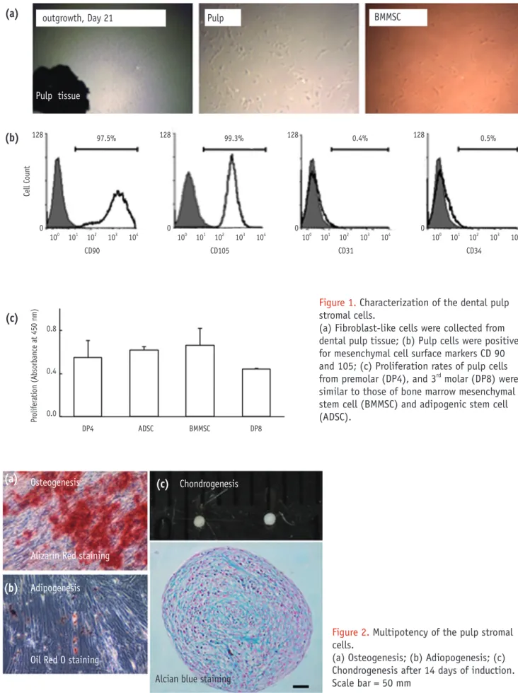
소구치로부터 치수조직을분리하여 outgrowth방법으로일차배양 한후약14일후부터일차배양한조직의경계로부터세포가자라나 와약21일경에배양기내에군집을이루며단층으로자란것을확인 할수있었다. 치수기질세포는골수유래줄기세포(BMMSC)와유사한 방추형의섬유모세포양의형태를보였다(Figure 1a).
표면표지자항체를이용한유세포분석결과, 치수기질세포에서중 간엽줄기세포표지자인 CD90, CD105이발현하였으며, 조혈세포와내
피세포의표지자인 CD31과 CD34에는미약한반응을보였다(Figure
1b).
소구치로부터 획득한 치수 기질세포(DPc4), 골수유래줄기세포
(BMMSC), 지방유래줄기세포(ADSC) 및제3대구치의치수기질세포
(DPc8)의자가증식능력을비교하였으며모든군에서유사한증식률을 관찰하였다(Figure 1c).
치수 세포의 다분화능력
분화유도를통해약14일후, Alizarin Red S 염색을통한석회화결 절, Oil-Red O 염색을통한지방조직및 glucosaminoglycan 기질침착 이관찰되었으며연골펠렛의형성을확인할수있었다(Figure 2).
분화 시기에 따른 펠렛의 형태학적 변화
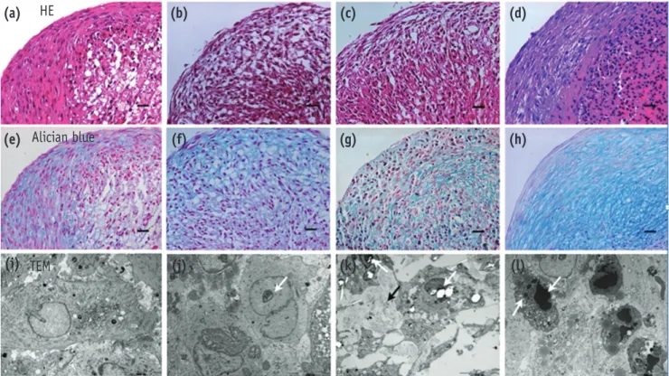
연골분화배지를이용하여치수세포를배양시작후이틀부터약1 mm3사이즈의백색펠렛이관찰되었다. 3, 7, 14, 21일의배양기간동 안크기나모양은유사하게유지되었다.
조직 구조를 살펴보면분화 3일 후에는 펠렛의 표면 형태와 평
행하게 나열되는납작한 spindle-like cell로 구성되는표층과크고
polygonal한세포와 세포간질이풍부한 심층으로구분되었다(Figure
3a). Glucosaminoglycan 기질침착은표층의세포간기질에서관찰되 었다(Figure 3e). 세포의초미세구조의경우, 세포기질내의미세구조
는 명확히구분되나핵소체가명확하게보이지는않았다(Figure 3i).
분화 7일에는 표면과평행하게 주행하는납작한세포로 구성된표
층과 polygonal한 세포로구성된심층사이에비대한 세포로이루어
진 중간층이관찰되었다(Figure 3b). Glucosaminoglycan 기질침착은 표층 및중간층의 세포간기질에서조금더 광범위하게관찰되었다
(Figure 3f). 전자현미경상에서도세포사이의간격이증가되며세포
핵내에서는 핵소체(흰화살표)가명확하게구분되어단백질합성등 세포의활성이높음을알수있었다(Figure 3j). 분화14일은7일과유 사하게 3개층으로이루어졌으나, 중간층의세포가조금더 응집되고 세포크기가작아지는양상이관찰되었다(Figure 3c). 이에반해세포 외기질의축적이증가하며펠렛내층까지분포하였다(Figure 3g). 세 포의형태는다소불규칙하였으나, 핵소체(흰화살표)는명확히구분 되었으며세포와세포사이사이의섬유성기질의침착이증가된양상 (검은화살표)을관찰할수있었다(Figure 3k). 분화21일째에는납작 한세포로구성된표층과동그란세포가응집되어있는심층이완전하 게구분되어2층구조를이루었다. 세포는7일, 14일에비해납작하거
나작아져비대한세포가거의존재하지않았으며 glucosaminoglycan
기질침착이보다광범위하고풍부하게관찰되었 (Figures 3d and 3h).
핵이농축되고(pyknosis) 및핵붕괴(karyorrhexis 흰화살표)를나타내 며괴사되는세포가다수발견되었다(Figure 3l).
세포 자멸사(apoptosis)의 변화
Apoptotic cell은 3일에는표층에서만매우드물게관찰되었으나7
일에는전층에걸쳐분포하며증가된양상이었다. 14일은전층에걸쳐 분포하였으나21일에는표층에서대부분관찰되었다(Figure 4).
Figure 1. Characterization of the dental pulp stromal cells.
(a) Fibroblast-like cells were collected from dental pulp tissue; (b) Pulp cells were positive for mesenchymal cell surface markers CD 90 and 105; (c) Proliferation rates of pulp cells from premolar (DP4), and 3rd molar (DP8) were similar to those of bone marrow mesenchymal stem cell (BMMSC) and adipogenic stem cell (ADSC).
Pulp tissue
outgrowth, Day 21 Pulp BMMSC
(a)
(b)
Cell Count
CD90 CD105 CD31 CD34
97.5% 99.3% 0.4% 0.5%
100 101 102 103 104 100 101 102 103 104 100 101 102 103 104 100 101 102 103 104 128
0
128
0
128
0
128
0
DP4 ADSC BMMSC DP8
(c)
Proliferation (Absorbance at 450 nm)
0.8
0.4
0.0
(a) (c)
(b)
Figure 2. Multipotency of the pulp stromal cells.
(a) Osteogenesis; (b) Adiopogenesis; (c) Chondrogenesis after 14 days of induction.
Scale bar = 50 mm Osteogenesis
Alizarin Red staining Adipogenesis
Oil Red O staining
Alcian blue staining Chondrogenesis
총괄 및 고안
본연구를통해성인의소구치의치수에서자라나온기질세포는줄 기세포의특징을보이며이를이용하여층구조를나타내는연골조직 을형성할수있음을알수있었다. 또한배양시기에따른형태학적인 분석을통해연골배양초기의펠렛이배양후기에비해세포의기질 형성능력이높고세포자멸사가적어향후재생치료에응용시유리 할수있음을알수있었다.
유치의 치수로부터로확인된줄기세포는 골수유래줄기세포나영 구치치수줄기세포에비해자가증식능력이유의하게뛰어나지만영 구치열기가시작된이후의성인환자에게서는 얻을수 없다는한계 가 있다.23,24발수된성인치수로부터도줄기세포를분리해낸바있으 며이는정상조직을제거하지않고도채취가용이하다는접근상의장 점이있는반면채취되는양이한정적이라는단점이있다.14본 연구 에서는교정치료를위해발치되는영구소구치를사용하였다. 소구치 는상대적으로발거가간단하며치료시양악소구치를모두발거하는 경우가많기때문에한환자에서한번의시술시획득할수있는치아 와그치아로부터얻을수있는세포의양이많다. 또한대부분기능하 고있던건전한치아로서온전한구조를가지고있다. 적정수준의기 계적장력은골수유래중간엽줄기세포증식에긍정적인조절인자이며 Figure 3. Morphologic evaluation of the chondrogenic pellets engineered from pulp cells.
HE (a, b, c, d) and Alcian blue (e, f, g, h) staining at 3, 7, 14 and 21 days of chondrogenic induction. Scale bar = 50 μm.
TEM (i, j, k, l) images of the cells within the pellets at 3, 7, 14 and 21 days of chondrogenic induction. White arrows, nucleolus; black arrow, extracellular matrix fibers, scale bar = 5 μm. HE, Hematoxylin-eosin; TEM, transmission eletron microscope.
(a)
(e)
(i)
(b)
(f)
(j)
(c)
(g)
(k)
(d)
(h)
(l) HE
Alician blue
TEM
Figure 4. Evaluation of apoptotic cells within the chon- drogenic pellets.
(a) At day 3, few Tunel positive cells were localized only within the superficial layer, but as induction period increased Tunel positive cells were scattered throughout the pellet; (b) at day 7; (c) at day 14; (d) at day 21.
Scale bar = 50 μm
(a) (b)
(c) (d)
인간의치수줄기세포의증식과분화, 기질생성에있어긍정적인영향 을끼칠수있다는결과도보고된바있어교합을이루어기능을하던 정상적인건전치아의세포는분열이나증식, 분화에있어매복치의세 포나염증조직으로부터유래된세포에비해더유리할것이라기대할 수있다.25,26
기존연구들의경우치아조직으로부터세포를배양할때크게두 가지방법을사용하고있다. 획득한조직을배양기바닥에붙여서조 직으로부터 기질세포가 자라나오도록 배양하는 outgrowth방법과 trypsin이나 collagenase 등의효소를이용하여조직을단일세포로분 해하여증식시키는 enzyme digestion의방법이다. Outgrowth방법에 비해 enzyme digestion방법의경우증식률과분화능력, 중간엽줄기 세포표지자의발현이상대적으로높아중간엽줄기세포특성의연구 에더적합하다고하였으나그술식이 복잡하고유래가다양한 효소 처리과정을 반복하기때문에임상적인응용을고려할때 단점이될 수있다.18,27따라서본연구에서는향후자가재생치료를목표로 out-
growth방법을선택하여치수조직의기질세포를효소처리없이얻어
사용하였다. 그결과영구치치수의기질세포는높은증식률과골, 지 방, 연골로분화되는다분화능력을가진간엽성줄기세포의특징을가 지고있음을알수있었다.
특히본연구의경우, 치수세포를이용해특별한지지체나원심분
리등의기술을동반하지않고기본적인배양기바닥에10-μl droplet
의세포를분주하고연골분화배지를사용하여배양하는방식으로비 교적간단하게펠렛형태의구형연골조직을얻을수있었다. 연골펠 렛의경우배양시기에따른크기의차이는크게관찰되지않았으나, 분화가진행됨에따라그내부미세구조및형태가변화는것을알수 있었다. 분화정도에따른형태학적변화는골수줄기세포, 활맥막줄기 세포, 연골세포를이용하여성생된연골펠렛의경우에도보고되고있 다.17골수줄기세포로분화시킨펠렛의경우활막, 연골세포와는달리 세포의형태에따라분화초기(1일)에는펠렛의표면형태와평행하게 나열되는납작한 spindle-like cell로구성되는표층과크고 polygonal 한세포와세포간질이풍부한 내층의2층으로구분되었다가, 중기(7 일)에는중간층에비대세포들이나타나면서3층으로구분되었다. 그 리고전자현미경관찰결과기질내의미세구조가명확히구분되고세 포와세포사이의간격이증가되면서그사이로섬유성기질의침착이 관찰되었다. 그러나분화후기(21일)로가면서골수줄기세포의경우 납작한세포로구성된표층과동그란세포가응집되어있는내층이완 전하게구분되어표층이명확한상태로남는2층구조로 관찰되었고 세포사멸이나타났다.17본연구결과치수기질세포는골수유래줄기 세포와같이간엽성줄기세포의표지자를나타내며다분화능력을가 지고있으며유사한정도의증식능력을나타내었다. 이를이용한연골 펠렛의형태및그변화또한골수유래줄기세포를이용한펠렛과가 장유사하였다.
Alcian blue 염색은연골조직에특이적으로나타나는 glycosamino-
glycan 기질을염색하는것으로분화기간전체에걸쳐서관찰되면그
양이증가하였다. 전자현미경을통해관찰한결과, 기질형성의측면 에서는관찰시기가진행됨에따라기질형성이증가하며연골분화가 유리하게진행됨을알수 있었다. 하지만 세포의경우, 7일경부터는
Tunel 양성의세포가나타내는 apoptosis가급격히증가하고분화후
기로갈수록핵이농축되어괴사되는세포가출현하였다. 비록7일경
을피크로 Tunel 양성세포양이줄어들기는하나여전히분화초기보
다는증가된양상을보이며이는골수줄기세포를이용한 연골분화의 경우와도유사하다. 연골펠렛의분화에따른세포의생존능력은분화 첫주에급감하고그후2 - 3주에는비교적적은양이감소한다고보 고되고있다.17반면, 전자현미경에의해관찰되는사멸세포는분화후 기로갈수록증가되었기때문에 apoptosis를경유하지않는세포사멸 또한공존하고있다고판단할수있다.
일반적으로 줄기세포를이용해 형성된연골조직은 골결손부또는 연골결손부의 재생치료에응용되며그효과또한긍적적으로보고되 고 있다.28따라서특수한효소처리없이분리, 배양한치수기질세포 유래의 연골조직을향후동물실험등을통해악관절영역의재생에 응용하기를기대할수있을것이다. 본연구에서보았듯이소구치발 거를통해서추출한기질세포가줄기세포의특징을보이며이를이용 하여층구조를나타내는연골조직으로분화될수있음을확인할수있 었다. 배양2일째생성된 펠렛의크기나모양이 전체배양기간동안 증가하지않는점, 배양3 - 7일동안표층과심층사이에비대한세포 로이루어진기질생성이왕성한중간층이두드러지는점, 7일을지나 면서사멸세포들이늘어나며14일이후후기에는세포형태나배열이 불규칙해지고세포괴사가일어나는점을토대로향후이를연골의재 생으로응용시에는배양초기의펠렛이배양후기에비해유리할것으 로사료된다.
결론
치수조직의기질세포에서중간엽줄기세포의특성을확인할수있 었다. 또한연골유도를시행할경우분리배양초기의연골조직이배 양 후기의조직에비해세포의기질형성능력이높고세포자멸사가 적어응용시유리할것으로판단된다.
References
1. Okeson JP. Management of temporomandibular disorder 5th edition. Philadelphia: Elsevier; 2003. p 15-22.
2. Arnett GW, Milam SB, Gottesman L. Progressive mandibular retrusion-idiopathic condylar resorption.
Part II. Am J Orthod Dentofacial Orthop 1996;110:117- 127.
3. Arnett GW, Milam SB, Gottesman L. Progressive mandibular retrusion-idiopathic condylar resorption.
Part I. Am J Orthod Dentofacial Orthop 1996;110:8-15.
4. Wolford LM, Cardenas L. Idiopathic condylar resorption:
diagnosis, treatment protocol, and outcomes. Am J Orthod Dentofacial Orthop 1999;116:667-677.
5. Crawford JG, Stoelinga PJ, Blijdorp PA, Brouns JJ.
Stability after reoperation for progressive condylar resorption after orthognathic surgery: report of seven cases. J Oral Maxillofac Surg 1994;52:460-466.
6. De Clercq CA, Neyt LF, Mommaerts MY, Abeloos JV, De Mot BM. Condylar resorption in orthognathic surgery:
a retrospective study. Int J Adult Orthodon Orthognath Surg 1994;9:233-240.
7. Merkx MA, Van Damme PA. Condylar resorption after orthognathic surgery. Evaluation of treatment in 8
patients. J Craniomaxillofac Surg 1994;22:53-58.
8. Mcllwraith CW, Frisbie DD, Rodkey WG, Kisiday JD, Werpy NM, Kawcak CE, Steadman JR. Evaluation of intra-articular mesenchymal stem cells to augment healing of microfractured chondral defects. Arthroscopy 2011;27:1552-1561.
9. Meyerrose T, Olson S, Pontow S, Kalomoiris S, Jung Y, Annett G, Bauer G, Nolta JA. Mesenchymal stem cells for the sustained in vivo delivery of bioactive factors.
Adv Drug Deliv Rev 2010;62:1167-1174.
10. van Buul GM, Kotek G, Wielopolski PA, Farrell E, Bos PK, Weinans H, Grohnert AU, Jahr H, Verhaar JA, Krestin GP, van Osch GJ, Bernsen MR. Clinically translatable cell tracking and quantification by MRI in cartilage repair using superparamagnetic iron oxides. PLoS One 2011;6:e17001.
11. Zhang J, Pan T, Im HJ, Fu FH, Wang JH. Differential properties of human ACL and MCL stem cells may be responsible for their differential healing capacity. BMC Med 2011;9:68.
12. Gronthos S, Mankani M, Brahim J, Robey PG, Shi S.
Postnatal human dental pulp stem cells (DPSCs) in vitro and in vivo. Proc Natl Acad Sci U S A 2000;97:13625- 13630.
13. Seo BM, Miura M, Gronthos S, Bartold PM, Batouli S, Brahim J, Young M, Robey PG, Wang CY, Shi S.
Investigation of multipotent postnatal stem cells from human periodontal ligament. Lancet 2004;364:149-155.
14. Huang AH, Chen YK, Chan AW, Shieh TY, Lin LM.
Isolation and characterization of human dental pulp stem/stromal cells from nonextracted crown-fractured teeth requiring root canal therapy. J Endod 2009;
35:673-681.
15. Pierdomenico L, Bonsi L, Calvitti M, Rondelli D, Arpinati M, Chirumbolo G, Becchetti E, Marchionni C, Alviano F, Fossati V, Staffolani N, Franchina M, Grossi A, Bagnara GP. Multipotent mesenchymal stem cells with immunosuppressive activity can be easily isolated from dental pulp. Transplantation 2005;80:836-842.
16. Struys T, Moreels M, Martens W, Donders R, Wolfs E, Lambrichts I. Ultrastructural and immunocytochemical analysis of multilineage differentiated human dental pulp- and umbilical cord-derived mesenchymal stem cells. Cells Tissues Organs 2011;193:366-378.
17. Ichinose S, Muneta T, Koga H, Segawa Y, Tagami M, Tsuji K, Sekiya I. Morphological differences during in vitro chondrogenesis of bone marrow-, synovium-MSCs, and chondrocytes. Lab Invest 2010;90:210-221.
18. Huang GT, Sonoyama W, Chen J, Park SH. In vitro characterization of human dental pulp cells: various isolation methods and culturing environments. Cell Tissue Res 2006;324:225-236.
19. Song SY, Jung JE, Jeon YR, Tark KC, Lew DH.
Determination of adipose-derived stem cell application on photo-aged fibroblasts, based on paracrine function.
Cytotherapy 2011;13:378-384.
20. Kim NR, Lee DH, Ahn SJ, Lee IS, Yang HC. The differentiation-inducing effect of conditioned media obtained from dental pulp cells. Oral Surg Oral Med Oral Pathol Oral Radiol Endod 2009;107:e54-59.
21. Dominici M, Le Blanc K, Mueller I, Slaper-Cortenbach I, Marini F, Krause D, Deans R, Keating A, Prockop Dj, Horwitz E. Minimal criteria for defining multipotent mesenchymal stromal cells. The International Society for Cellular Therapy position statement. Cytotherapy 2006;8:315-317.
22. Ahrens PB, Solursh M, Reiter RS. Stage-related capacity for limb chondrogenesis in cell culture. Dev Biol 1977;
60:69-82.
23. Miura M, Gronthos S, Zhao M, Lu B, Fisher LW, Robey PG, Shi S. SHED: stem cells from human exfoliated deciduous teeth. Proc Natl Acad Sci U S A 2003;100:5807-5812.
24. Nam H, Lee G. Identification of novel epithelial stem cell-like cells in human deciduous dental pulp. Biochem Biophys Res Commun 2009;386:135-139.
25. Choi KM, Seo YK, Yoon HH, Song KY, Kwon SY, Lee HS, Park JK. Effects of mechanical stimulation on the proliferation of bone marrow-derived human mesenchymal stem cells. Biotechnology and Bioprocess Engineering 2007;12:601-609.
26. Han MJ, Seo YK, Yoon HH, Song KY, Park JK. Effect of mechanical tension on the human dental pulp cells.
Biotechnology and Bioprocess Engineering 2008;13:410- 417.
27. Tanaka K, Iwasaki K, Feghali KE, Komaki M, Ishikawa I, Izumi Y. Comparison of characteristics of periodontal ligament cells obtained from outgrowth and enzyme- digested culture methods. Arch Oral Biol 2011;56:380- 388.
28. Mobasheri A, Csaki C, Clutterbuck AL, Rahmanzadeh M, Shakibaei M. Mesenchymal stem cells in connective tissue engineering and regenerative medicine:
applications in cartilage repair and osteoarthritis therapy. Histol Histopathol 2009;24:347-366.